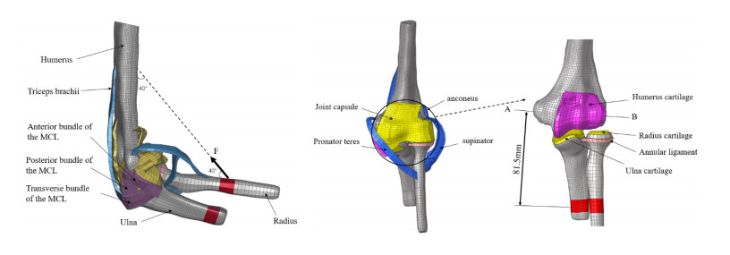
Dynamic orthoses have a significant effect on the treatment of elbow capsular contracture. Because of the lack of quantitative research on traction forces, determining the appropriate traction force to help stretch soft tissues and maintain the joint's range of motion is a challenge in the rehabilitation process. We developed a human elbow finite element (FE) model incorporating the activity behavior of the muscles and considering different capsular contracture locations, including total, anterior and posterior capsular contractures, to analyze the internal biomechanical responses of different capsular contracture models during flexion (30 to 80 degrees). Traction loads of 10, 20, 30 and 40 N were applied to the ulna and radius at the maximum flexion angle (80 degrees) to explore the appropriate traction loads at week 4 after a joint capsule injury. We observed a significant increase in posterior capsule stress with anterior capsular contracture (ACC), and the maximum peak stress was 1.3 times higher than that in the healthy model. During the fourth week after elbow capsule injury, the appropriate traction forces for total capsule contracture (TCC), ACC and posterior capsule contracture (PCC) were 20, 10 and 20 N, respectively; these forces maintained a stable biomechanical environment for the elbow joint and achieved a soft tissue pulling effect, thus increasing elbow mobility. The results can be used as a quantitative guide for the rehabilitation physicians to determine the traction load for a specific patient.
Citation: Fang Wang, Jiaming Wang, Mingxin Li, Jun Hu, Kehua Song, Jianguo Zhang, Yubo Fan. Biomechanical study of the effect of traction on elbow joint capsule contracture[J]. Mathematical Biosciences and Engineering, 2023, 20(12): 21451-21466. doi: 10.3934/mbe.2023949
Dynamic orthoses have a significant effect on the treatment of elbow capsular contracture. Because of the lack of quantitative research on traction forces, determining the appropriate traction force to help stretch soft tissues and maintain the joint's range of motion is a challenge in the rehabilitation process. We developed a human elbow finite element (FE) model incorporating the activity behavior of the muscles and considering different capsular contracture locations, including total, anterior and posterior capsular contractures, to analyze the internal biomechanical responses of different capsular contracture models during flexion (30 to 80 degrees). Traction loads of 10, 20, 30 and 40 N were applied to the ulna and radius at the maximum flexion angle (80 degrees) to explore the appropriate traction loads at week 4 after a joint capsule injury. We observed a significant increase in posterior capsule stress with anterior capsular contracture (ACC), and the maximum peak stress was 1.3 times higher than that in the healthy model. During the fourth week after elbow capsule injury, the appropriate traction forces for total capsule contracture (TCC), ACC and posterior capsule contracture (PCC) were 20, 10 and 20 N, respectively; these forces maintained a stable biomechanical environment for the elbow joint and achieved a soft tissue pulling effect, thus increasing elbow mobility. The results can be used as a quantitative guide for the rehabilitation physicians to determine the traction load for a specific patient.

| [1] |
J. W. Stoneback, B. D. Owens, J. Sykes, G. S. Athwal, L. Pointer, J. M. Wolf, Incidence of elbow dislocations in the United States population, J. Bone Jt. Surg., 94 (2012), 240–245. https://doi.org/10.2106/JBJS.J.01663 doi: 10.2106/JBJS.J.01663

|
| [2] |
J. N. Doornberg, T. Bosse, M. S. Cohen, J. B. Jupiter, D. Ring, P. Kloen, Temporary presence of myofibroblasts in human elbow capsule after trauma, J. Bone Jt. Surg., 96 (2014), e36. https://doi.org/10.2106/JBJS.M.00388 doi: 10.2106/JBJS.M.00388
|
| [3] |
A. L. C. Lindenhovius, J. B. Jupiter, The posttraumatic stiff elbow: A Review of the Literature, J. Hand Surg., 32 (2007), 1605–1623. https://doi.org/10.1016/j.jhsa.2007.09.015. doi: 10.1016/j.jhsa.2007.09.015
|
| [4] |
J. O. Sajbjerg, The stiff elbow: How I do it, Acta Orthop. Scand., 67 (1996), 626–631. https://doi.org/10.3109/17453679608997771 doi: 10.3109/17453679608997771
|
| [5] |
C. L. Dunham, R. M. Castile, N. Havlioglu, A. M. Chamberlain, S. P. Lake, Temporal patterns of motion in flexion-extension and pronation-supination in a rat model of posttraumatic elbow contracture, Clin. Orthop. Relat. Res., 476 (2018), 1878–1889. https://doi.org/10.1097/CORR.0000000000000388 doi: 10.1097/CORR.0000000000000388
|
| [6] |
L. E. Karbach, J. Elfar, Elbow Instability: Anatomy, biomechanics, diagnostic maneuvers, and testing, J. Hand Surg., 42 (2017), 118–126. https://doi.org/10.1016/j.jhsa.2016.11.025 doi: 10.1016/j.jhsa.2016.11.025
|
| [7] |
B. Attum, W. Obremskey, Posttraumatic elbow stiffness: A critical analysis review, JBJS Rev., 4 (2016). https://doi.org/10.2106/JBJS.RVW.15.00084 doi: 10.2106/JBJS.RVW.15.00084
|
| [8] |
D. Ring, J. B. Jupiter, Operative treatment of elbow stiffness, JBJS Essent. Surg. Tech., 1 (2011), e18. https://doi.org/10.2106/jbjs.st.k.00010 doi: 10.2106/jbjs.st.k.00010
|
| [9] |
K. A. Hildebrand, M. Zhang, A. D. Befus, P. T. Salo, D. A. Hart, A myofibroblast-mast cell-neuropeptide axis of fibrosis in post-traumatic joint contractures: An in vitro analysis of mechanistic components, J. Orthop. Res., 32 (2014), 1290–1296. https://doi.org/10.1002/jor.22676 doi: 10.1002/jor.22676
|
| [10] |
J. N. Doornberg, T. Bosse, M. S. Cohen, J. B. Jupiter, D. Ring, P. Kloen, Temporary presence of myofibroblasts in human elbow capsule after trauma, J. Bone Jt. Surg., 96 (2014), e36. https://doi.org/10.2106/JBJS.M.00388 doi: 10.2106/JBJS.M.00388
|
| [11] |
M. S. Cohen, D. R. Schimmel, K. Masuda, H. Hastings, C. Muehleman, Structural and biochemical evaluation of the elbow capsule after trauma, J. Shoulder Elbow Surg., 16 (2007), 484–490. https://doi.org/10.1016/j.jse.2006.06.018 doi: 10.1016/j.jse.2006.06.018
|
| [12] |
L. M. Reichel, O. A. Morales, Gross anatomy of the elbow capsule: A cadaveric study, J. Hand Surg., 38 (2013), 110–116. https://doi.org/10.1016/j.jhsa.2012.09.031 doi: 10.1016/j.jhsa.2012.09.031
|
| [13] |
B. F. Morrey, L. J. Askew, E. Y. Chao, A biomechanical study of normal functional elbow motion, J. Bone Jt. Surg., 63 (1981), 872–877. https://doi.org/10.2106/00004623-198163060-00002 doi: 10.2106/00004623-198163060-00002
|
| [14] |
G. Bhabra, C. S. Modi, T. Lawrence, Managing the stiff elbow, Orthop. Trauma, 30 (2016), 329–335. https://doi.org/10.1016/j.mporth.2016.04.005 doi: 10.1016/j.mporth.2016.04.005
|
| [15] |
C. C. L. Yau, J. Viveen, D. Eygendaal, B. The, Management of the stiff elbow, Orthop. Trauma, 34 (2020), 206–212. https://doi.org/10.1016/j.mporth.2020.05.003 doi: 10.1016/j.mporth.2020.05.003
|
| [16] |
R. Sivakumar, V. Somasheker, P. K. Shingi, T. Vinoth, M. Chidambaram, Treatment of stiff elbow in young patients with interpositional arthroplasty for mobility: Case Series, J. Orthop. Case Rep., 6 (2016), 49–52. https://doi.org/10.13107/jocr.2250-0685.566 doi: 10.13107/jocr.2250-0685.566
|
| [17] |
A. M. G. Cavalcanti, R. S. O. Filho, H. C. Gomes, A. B. S. Martins, E. B. Garcia, L. M. Ferreira, Review of articulated elbow orthotics for joint stiffness rehabilitation, Acta Ortop. Bras., 30 (2022), e254358. https://doi.org/10.1590/1413-785220223005e254358 doi: 10.1590/1413-785220223005e254358
|
| [18] |
P. M. Bonutti, J. E. Windau, B. A. Ables, B. G. Miller, Static progressive stretch to reestablish elbow range of motion, Clin. Orthop. Relat., 303 (1994), 128–134. https://doi.org/10.1097/00003086-199406000-00015 doi: 10.1097/00003086-199406000-00015
|
| [19] |
G. L. Gallucci, J. G. Boretto, M. A. Dávalos, A. Donndorff, V. A. Alfie, P. De Carli, Dynamic splint for the treatment of stiff elbow, Shoulder Elbow, 3 (2011), 52–55. https://doi.org/10.1111/j.1758-5740.2010.00096.x doi: 10.1111/j.1758-5740.2010.00096.x
|
| [20] |
G. Sim, J. Fleming, C. Glasgow, Mobilizing orthoses in the management of post-traumatic elbow contractures: A survey of Australian hand therapy practice, J. Hand Ther., 34 (2021), 90–99. https://doi.org/10.1016/j.jht.2019.12.014 doi: 10.1016/j.jht.2019.12.014
|
| [21] |
N. Young, N. Terrington, D. Francis, L. S. Robinson, Orthotic management of fixed flexion deformity of the proximal interphalangeal joint following traumatic injury: A systematic review, Hong Kong J. Occup. Ther., 31 (2018), 3–13. https://doi.org/10.1177/1569186118764067 doi: 10.1177/1569186118764067
|
| [22] |
M. J. Gluck, C. M. Beck, K. M. Sochol, D. A. London, M. R. Hausman, Comparative strength of elbow splint designs: a new splint design as a stronger alternative to posterior splints, J. Shoulder Elbow Surg., 28 (2019), e125–e130. https://doi.org/10.1016/j.jse.2018.10.015 doi: 10.1016/j.jse.2018.10.015
|
| [23] |
A. L. C. Lindenhovius, J. N. Doornberg, K. M. Brouwer, J. B. Jupiter, C. S. Mudgal, D. Ring, A prospective randomized controlled trial of dynamic versus static progressive elbow splinting for posttraumatic elbow stiffness, J. Bone Jt. Surg., 94 (2012), 694–700. https://doi.org/10.2106/JBJS.J.01761 doi: 10.2106/JBJS.J.01761
|
| [24] |
G. L. Gallucci, J. G. Boretto, M. A. Dávalos, A. Donndorff, V. A. Alfie, P. D. Carli, Dynamic splint for the treatment of stiff elbow, Shoulder Elbow, 3 (2011), 52–55. https://doi.org/10.1111/j.1758-5740.2010.00096.x doi: 10.1111/j.1758-5740.2010.00096.x
|
| [25] |
M. Li, M. S. Venäläinen, S. S. Chandra, R. Patel, J. Fripp, C. Engstrom, et al., Discrete element and finite element methods provide similar estimations for hip joint contact mechanics during walking gait, J. Biomech., 115 (2021), 110163. https://doi.org/10.1016/j.jbiomech.2020.110163 doi: 10.1016/j.jbiomech.2020.110163
|
| [26] |
M. E. Mononen, J. S. Jurvelin, R. K. Korhonen, Effects of radial tears and partial meniscectomy of lateral meniscus on the knee joint mechanics during the stance phase of the gait cycle-A 3D finite element study, J. Orthop. Res., 31 (2013), 1208–1217. https://doi.org/10.1002/jor.22358 doi: 10.1002/jor.22358
|
| [27] |
P. Xiang, K. M. Liew, Predicting buckling behavior of microtubules based on an atomistic-continuum model, Int. J. Solids Struct., 48 (2011), 1730–1737. https://doi.org/10.1016/j.ijsolstr.2011.02.022 doi: 10.1016/j.ijsolstr.2011.02.022
|
| [28] |
P. Xiang, K. M. Liew, Dynamic behaviors of long and curved microtubules based on an atomistic-continuum model, Comput. Methods Appl. Mech. Eng., (2012), 123–132. https://doi.org/10.1016/j.cma.2012.02.023 doi: 10.1016/j.cma.2012.02.023
|
| [29] |
P. Xiang, K. M. Liew, Free vibration analysis of microtubules based on an atomistic-continuum model, J. Sound Vib., 331 (2012), 213–230. https://doi.org/10.1016/j.jsv.2011.08.024 doi: 10.1016/j.jsv.2011.08.024
|
| [30] |
P. Xiang, K. M. Liew, A computational framework for transverse compression of microtubules based on a higher-order Cauchy-Born rule, Comput. Methods Appl. Mech. Eng., 254 (2013), 14–30. https://doi.org/10.1016/j.cma.2012.10.013 doi: 10.1016/j.cma.2012.10.013
|
| [31] |
P. Xiang, L. W. Zhang, K. M. Liew, Analysis of macromolecular microtubules using the potential-based matrix displacement method, Compos. Struct., 127 (2015), 224–230. https://doi.org/10.1016/j.compstruct.2015.03.004 doi: 10.1016/j.compstruct.2015.03.004
|
| [32] |
P. Xiang, L. W. Zhang, K. M. Liew, A mesh-free computational framework for predicting vibration behaviors of microtubules in an elastic medium, Compos. Struct., 149 (2016), 41–53. https://doi.org/10.1016/j.compstruct.2016.03.063 doi: 10.1016/j.compstruct.2016.03.063
|
| [33] |
K. Kamei, E. Sasaki, K. Fujisaki, Y. Harada, Y. Yamamoto, Y. Ishibashi, Ulnar collateral ligament dysfunction increases stress on the humeral capitellum: a finite element analysis, JSES Int., 5 (2021), 307–313. https://doi.org/10.1016/j.jseint.2020.10.022 doi: 10.1016/j.jseint.2020.10.022
|
| [34] |
A. Zarifian, A. A. Fough, D. Eygendaal, M. Rivlin, S. A. M. Shaegh, A. R. Kachooei, Length of plates and number of screws for the fixation of distal humerus fractures: A finite element biomechanical study, J. Hand Surg., 47 (2022), 690. https://doi.org/10.1016/j.jhsa.2021.07.010 doi: 10.1016/j.jhsa.2021.07.010
|
| [35] |
E. Akkurt, M. Yıldırım, F. Erenler, O. M. Tosun, M. F. Akkurt, B. Akkurt, et al., Mechanical evaluation for the finite element analysis of intramedullary nailing and plate screw system used in humerus transverse fractures, J. Orthop., 2023. https://doi.org/10.1016/j.jor.2023.10.024 doi: 10.1016/j.jor.2023.10.024
|
| [36] |
A. Schonning, B. Oommen, I. Ionescu, T. Conway, Hexahedral mesh development of free-formed geometry: The human femur exemplified, Comput. Aided Design, 41 (2009), 566–572. https://doi.org/10.1016/j.cad.2007.10.007 doi: 10.1016/j.cad.2007.10.007
|
| [37] |
M. Bendjaballah, A. Shirazi-Adl, D. Zukor, Finite element analysis of human knee joint in varus-valgus, Clin. Biomech., 12 (1997), 139–148. https://doi.org/10.1016/S0268-0033(97)00072-7 doi: 10.1016/S0268-0033(97)00072-7
|
| [38] |
J. G. Zhang, F. Wang, R. Zhou, Q. Xue, A three-dimensional finite element model of the cervical spine: an investigation of whiplash injury, Med. Biol. Eng. Comput., 49 (2011), 193–201. https://doi.org/10.1007/s11517-010-0708-9 doi: 10.1007/s11517-010-0708-9
|
| [39] |
P. Büchler, N. A. Ramaniraka, L. R. Rakotomanana, J. P. Iannotti, A. Farron, A finite element model of the shoulder: application to the comparison of normal and osteoarthritic joints, Clin. Biomech., 17 (2002), 630–639. https://doi.org/10.1016/S0268-0033(02)00106-7 doi: 10.1016/S0268-0033(02)00106-7
|
| [40] |
K. M. Quapp, J. A. Weiss, Material characterization of human medial collateral ligament, J. Biomech. Eng., 120 (1998), 757–763. https://doi.org/10.1115/1.2834890 doi: 10.1115/1.2834890
|
| [41] |
M. M. Schulz, T. Q. Lee, M. D. Sandusky, J. E. Tibone, P. J. McMahon, The healing effects on the biomechanical properties of joint capsular tissue treated with Ho:YAG laser: An in vivo rabbit study, Arthroscopy, 17 (2001), 342–347. https://doi.org/10.1053/jars.2001.19677 doi: 10.1053/jars.2001.19677
|
| [42] |
F. Li, H. Li, W. Hu, S. Su, B. Wang, Simulation of muscle activation with coupled nonlinear FE models, J. Mech. Med. Biol., 16 (2016), 1650082. https://doi.org/10.1142/S0219519416500822 doi: 10.1142/S0219519416500822
|
| [43] |
S. Hedenstierna, P. Halldin, K. Brolin, Evaluation of a combination of continuum and truss finite elements in a model of passive and active muscle tissue, Comput. Methods Biomech. Biomed. Eng., 11 (2008), 627–639. https://doi.org/10.1080/17474230802312516 doi: 10.1080/17474230802312516
|
| [44] |
B. S. Myers, C. A. V. Ee, D. L. A. Camacho, C. T. Woolley, T. M. Best, On the structural and material properties of mammalian skeletal muscle and its relevance to human, Cervical Impact Dyn., 104 (1995), 3095–3105. https://doi.org/10.4271/952723 doi: 10.4271/952723
|
| [45] |
B. S. Myers, C. T. Woolley, T. L. Slotter, W. E. Garrett, T. M. Best, The influence of strain rate on the passive and stimulated engineering stress-large strain behavior of the rabbit tibialis anterior muscle, J. Biomech. Eng., 120 (1998), 126–132. https://doi.org/10.1115/1.2834292 doi: 10.1115/1.2834292
|
| [46] |
K. Takatori, H. Hashizume, H. Wake, H. Inoue, N. Nagayama, Analysis of stress distribution in the humeroradial joint, J. Orthop. Sci., 7 (2002), 650–657. https://doi.org/10.1007/s007760200116 doi: 10.1007/s007760200116
|
| [47] |
F. Wang, S. Jia, M. Li, K. Pan, J. Zhang, Y. Fan, Effect of the medial collateral ligament and the lateral ulnar collateral ligament injury on elbow stability: a finite element analysis, Comput. Methods Biomech. Biomed. Eng., 24 (2021), 1517–1529. https://doi.org/10.1080/10255842.2021.1898601 doi: 10.1080/10255842.2021.1898601
|
| [48] |
L. Verstuyft, P. Caekebeke, R. van Riet, Postoperative rehabilitation in elbow surgery, J. Clin. Orthop. Trauma., 20 (2021), 101497. https://doi.org/10.1016/j.jcot.2021.101479 doi: 10.1016/j.jcot.2021.101479
|
| [49] | Z. Y. Sun, Development of a New Classification and Functional Score for Elbow Stiffness and Exploration of Long-Term Clinical Outcomes of Open Arthrolysis, Ph.D thesis, Shanghai Jiao Tong University, 2020. https://doi.org/10.27307/d.cnki.gsjtu.2020.000096 |
| [50] |
T. K. K. Koo, A. F. T. Mak, A neuromusculoskeletal model to simulate the constant angular velocity elbow extension test of spasticity, Med. Eng. Phys., 28 (2006), 60–69. https://doi.org/10.1016/j.medengphy.2005.03.012 doi: 10.1016/j.medengphy.2005.03.012
|
| [51] |
T. Kodek, M. Munih, An analysis of static and dynamic joint torques in elbow flexion-extension movements, Simul. Modell. Pract. Theory, 11 (2003), 297–311. https://doi.org/10.1016/S1569-190X(03)00063-7 doi: 10.1016/S1569-190X(03)00063-7
|
| [52] |
M. Hackl, K. Wegmann, S. L. Kahmann, N. Heinze, M. Staat, L. P. Müller, et al., Radial shortening osteotomy reduces radiocapitellar contact pressures while preserving valgus stability of the elbow, Knee Surg. Sport Traumatol. Arthroscopy, 25 (2017), 2280–2288. https://doi.org/10.1007/s00167-017-4468-z doi: 10.1007/s00167-017-4468-z
|
| [53] |
K. Takatori, H. Hashizume, H. Wake, H. Inoue, N. Nagayama, Analysis of stress distribution in the humeroradial joint, J. Orthop. Sci., 7 (2002), 650–657. https://doi.org/10.1007/s007760200116 doi: 10.1007/s007760200116
|
| [54] |
G. J. W. King, B. F. Morrey, K. N. An, Stabilizers of the elbow, J. Shoulder Elbow Surg., 2 (1993), 165–174. https://doi.org/10.1016/S1058-2746(09)80053-0 doi: 10.1016/S1058-2746(09)80053-0
|
| [55] |
S. H. Gallay, R. R. Richards, S. W. O'Driscoll, Intraarticular capacity and compliance of stiff and normal elbows, Arthroscopy, 9 (1993), 9–13. https://doi.org/10.1016/S0749-8063(05)80336-6 doi: 10.1016/S0749-8063(05)80336-6
|
| [56] |
K. Kimata, M. Yasui, H. Yokota, S. Hirai, M. Naito, T. Nakano, Transverse ligament of the elbow joint: an anatomic study of cadavers, J. Shoulder Elbow Surg., 28 (2019), 2253–2258. https://doi.org/10.1016/j.jse.2019.04.048 doi: 10.1016/j.jse.2019.04.048
|
| [57] |
G. F. Solitro, R. Fattori, K. Smidt, C. Nguyen, M. M. Morandi, R. S. Barton, Role of the transverse ligament of the ulnar collateral ligament of the elbow: a biomechanical study, JSES Int., 5 (2021), 549–553. https://doi.org/10.1016/j.jseint.2021.01.009 doi: 10.1016/j.jseint.2021.01.009
|
| [58] |
B. F. Morrey, K. N. An, Stability of the elbow: Osseous constraints, J. Shoulder Elbow Surg., 14 (2005), S174–S178. https://doi.org/10.1016/j.jse.2004.09.031 doi: 10.1016/j.jse.2004.09.031
|
| [59] |
C. L. B. Guglielmetti, M. E. C. Gracitelli, J. H. Assunção, F. B. Andrade-Silva, M. M. N. Pessa, A. A. Ferreira-Neto, et al., Randomized trial for the treatment of post-traumatic elbow stiffness: surgical release vs. rehabilitation, J. Shoulder Elbow Surg., 29 (2020), 1522–1529. https://doi.org/10.1016/j.jse.2020.03.023 doi: 10.1016/j.jse.2020.03.023
|
| [60] |
E. S. Veltman, J. N. Doornberg, D. Eygendaal, M. P. J. van den Bekerom, Static progressive versus dynamic splinting for posttraumatic elbow stiffness: a systematic review of 232 patients, Arch. Orthop. Trauma. Surg., 135 (2015), 613–617. https://doi.org/10.1007/s00402-015-2199-5 doi: 10.1007/s00402-015-2199-5
|
| [61] |
C. C. L. Yau, J. Viveen, D. Eygendaal, B. The, Management of the stiff elbow, Orthop. Trauma., 34 (2020), 206–212. https://doi.org/10.1016/j.mporth.2020.05.003 doi: 10.1016/j.mporth.2020.05.003
|
| [62] |
E. Mobedi, W. Kim, M. Leonori, N. G. Tsagarakis, A. Ajoudani, Design and control of an assistive device for elbow effort-compensation, IEEE/ASME Trans. Mechatron., 2023. https://doi.org/10.1109/TMECH.2023.3267681 doi: 10.1109/TMECH.2023.3267681
|
| [63] |
M. Ceccarelli, M. Riabtsev, A. Fort, M. Russo, M. A. Laribi, M. Urizar, Design and experimental characterization of l-CADEL v2, an assistive device for elbow motion, Sensors, 21 (2021), 1–15. https://doi.org/10.3390/s21155149 doi: 10.3390/s21155149
|
| [64] |
J. D. A. Rosero, D. C. B. Rosero, L. F. A. Realpe, A. F. S. Pino, E. R. González, Mechatronic Design of a prototype orthosis to support elbow joint rehabilitation, Bioengineering, 9 (2022), 287. https://doi.org/10.3390/bioengineering9070287 doi: 10.3390/bioengineering9070287
|
| [65] | R. Guachi, F. Napoleoni, F. Pipitone, M. Controzzi, Preliminary design and development of a selectable stiffness joint for elbow orthosis, in 2022 IEEE-RAS 21st International Conference on Humanoid Robots (Humanoids), (2022), 458–463. https://doi.org/10.1109/Humanoids53995.2022.10000188 |


Figures(4) / Tables(5)

Fang Wang, Jiaming Wang, Mingxin Li, Jun Hu, Kehua Song, Jianguo Zhang, Yubo Fan. Biomechanical study of the effect of traction on elbow joint capsule contracture[J]. Mathematical Biosciences and Engineering, 2023, 20(12): 21451-21466. doi: 10.3934/mbe.2023949


 DownLoad: