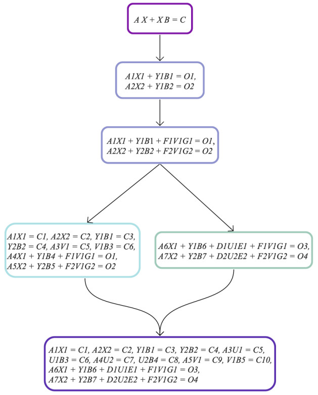
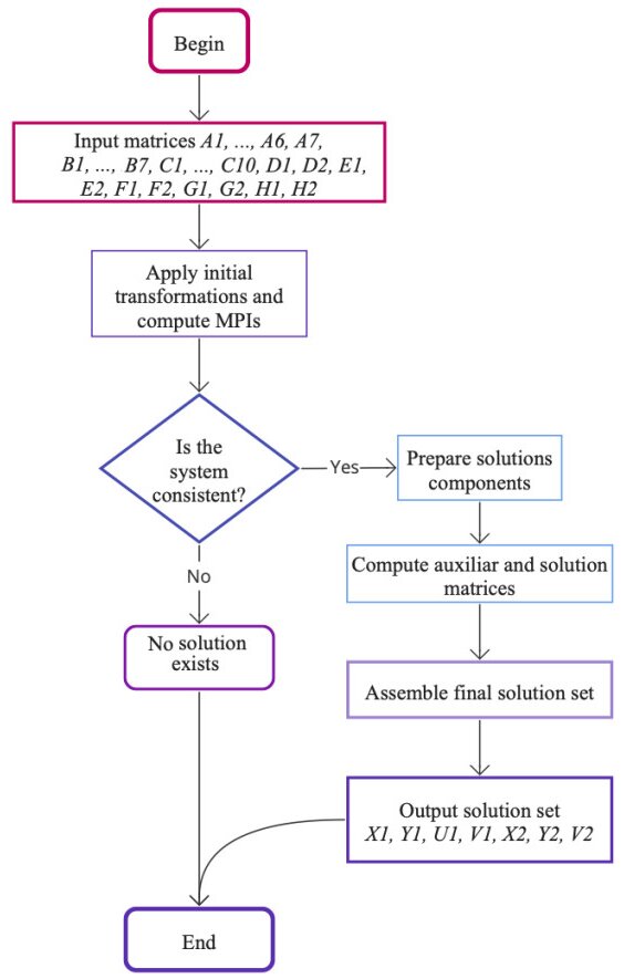
This article explores Sylvester quaternion matrix equations and potential applications, which are important in fields such as control theory, graphics, sensitivity analysis, and three-dimensional rotations. Recognizing that the determination of solutions and computational methods for these equations is evolving, our study contributes to the area by establishing solvability conditions and providing explicit solution formulations using generalized inverses. We also introduce an algorithm that utilizes representations of quaternion Moore-Penrose inverses to improve computational efficiency. This algorithm is validated with a numerical example, demonstrating its practical utility. Additionally, our findings offer a generalized framework in which various existing results in the area can be viewed as specific instances, showing the breadth and applicability of our approach. Acknowledging the challenges in handling large systems, we propose future research focused on further improving algorithmic efficiency and expanding the applications to diverse algebraic structures. Overall, our research establishes the theoretical foundations necessary for solving Sylvester-type quaternion matrix equations and introduces a novel algorithmic solution to address their computational challenges, enhancing both the theoretical understanding and practical implementation of these complex equations.
Citation: Abdur Rehman, Ivan Kyrchei, Muhammad Zia Ur Rahman, Víctor Leiva, Cecilia Castro. Solvability and algorithm for Sylvester-type quaternion matrix equations with potential applications[J]. AIMS Mathematics, 2024, 9(8): 19967-19996. doi: 10.3934/math.2024974
This article explores Sylvester quaternion matrix equations and potential applications, which are important in fields such as control theory, graphics, sensitivity analysis, and three-dimensional rotations. Recognizing that the determination of solutions and computational methods for these equations is evolving, our study contributes to the area by establishing solvability conditions and providing explicit solution formulations using generalized inverses. We also introduce an algorithm that utilizes representations of quaternion Moore-Penrose inverses to improve computational efficiency. This algorithm is validated with a numerical example, demonstrating its practical utility. Additionally, our findings offer a generalized framework in which various existing results in the area can be viewed as specific instances, showing the breadth and applicability of our approach. Acknowledging the challenges in handling large systems, we propose future research focused on further improving algorithmic efficiency and expanding the applications to diverse algebraic structures. Overall, our research establishes the theoretical foundations necessary for solving Sylvester-type quaternion matrix equations and introduces a novel algorithmic solution to address their computational challenges, enhancing both the theoretical understanding and practical implementation of these complex equations.

| [1] | R. A. Horn, C. R. Johnson, Matrix analysis, Cambridge: Cambridge University Press, 2012. https://doi.org/10.1017/CBO9780511810817 |
| [2] | K. Zhou, J. C. Doyle, K. Glover, Robust and optimal control, Upper Saddle River: Prentice Hall, 1996. Available from: https://dl.acm.org/doi/book/10.5555/225507 |
| [3] |
V. Simoncini, Computational methods for linear matrix equations, SIAM Rev., 58 (2016), 377–441. https://doi.org/10.1137/130912839 doi: 10.1137/130912839

|
| [4] |
V. L. Syrmos, F. L. Lewis, Coupled and constrained Sylvester equations in system design, Circuits Syst. Signal Process., 13 (1994), 663–694. https://doi.org/10.1007/BF02523122 doi: 10.1007/BF02523122
|
| [5] | K. R. Gavin, S. P. Bhattacharyya, Robust and well-conditioned eigenstructure assignment via Sylvester's equation, Proc. Amer. Control Conf., 1982. https://doi.org/10.1002/oca.4660040302 |
| [6] |
M. Darouach, Solution to Sylvester equation associated to linear descriptor systems, Syst. Control. Lett., 55 (2006), 835–838. https://doi.org/10.1016/j.sysconle.2006.04.004 doi: 10.1016/j.sysconle.2006.04.004
|
| [7] | G. H. Golub, C. F. V. Loan, Matrix computations, Baltimore: Johns Hopkins University Press, 2013. Available from: https://epubs.siam.org/doi/book/10.1137/1.9781421407944 |
| [8] |
K. Zuo, Y. Chen, L. Yuan, Further representations and computations of the generalized Moore-Penrose inverse, AIMS Math., 8 (2023), 23442–23458. https://doi.org/10.3934/math.20231191 doi: 10.3934/math.20231191
|
| [9] |
W. R. Hamilton, On quaternions, or on a new system of imaginaries in algebra, Philos. Mag., 25 (1844), 489–495. https://doi.org/10.1080/14786444408645047 doi: 10.1080/14786444408645047
|
| [10] |
S. D. Leo, G. Scolarici, Right eigenvalue equation in quaternionic quantum mechanics, J. Phys. A, 33 (2000), 2971–2995. http://doi.org/10.1088/0305-4470/33/15/306 doi: 10.1088/0305-4470/33/15/306
|
| [11] |
C. C. Took, D. P. Mandic, Augmented second-order statistics of quaternion random signals, Signal Process., 91 (2011), 214–224. https://doi.org/10.1016/j.sigpro.2010.06.024 doi: 10.1016/j.sigpro.2010.06.024
|
| [12] | S. L. Adler, Quaternionic quantum mechanics and quantum fields, New York: Oxford University Press, 1995. Available from: https://onlinelibrary.wiley.com/doi/abs/10.1002/qua.560600402 |
| [13] | J. B. Kuipers, Quaternions and rotation sequences, Princeton: Princeton University Press, 1999. |
| [14] |
A. Rehman, I. I. Kyrchei, I. Ali, M. Akram, A. Shakoor, The general solution of quaternion matrix equation having $\eta$-skew-Hermicity and its Cramer's rule, Math. Probl. Eng., 2019 (2019), 7939238. https://doi.org/10.1155/2019/7939238 doi: 10.1155/2019/7939238
|
| [15] |
A. Rehman, I. I. Kyrchei, I. Ali, M. Akram, A. Shakoor, Explicit formulas and determinantal representation for $\eta$-skew-Hermitian solution to a system of quaternion matrix equations, Filomat, 34 (2020), 2601–2627. https://doi.org/10.2298/FIL2008601R doi: 10.2298/FIL2008601R
|
| [16] |
A. Rehman, I. I. Kyrchei, Solving and algorithm to system of quaternion Sylvester-Type matrix equations with $*$-hermicity, Adv. Appl. Clifford Algebras, 32 (2022), 49. https://doi.org/10.1007/s00006-022-01222-2 doi: 10.1007/s00006-022-01222-2
|
| [17] |
Z. H. He, Q. W. Wang, Y. Zhang, A simultaneous decomposition for seven matrices with applications, J. Comput. Appl. Math., 349 (2019), 93–113. https://doi.org/10.1016/j.cam.2018.09.001 doi: 10.1016/j.cam.2018.09.001
|
| [18] |
S. W. Yu, Z. H. He, T. C. Qi, X. X. Wang, The equivalence canonical form of five quaternion matrices with applications to imaging and Sylvester-type equations, J. Comput. Appl. Math., 393 (2021), 113494. https://doi.org/10.1016/j.cam.2021.113494 doi: 10.1016/j.cam.2021.113494
|
| [19] |
E. K. W. Chu, L. Hou, D. B. Szyld, J. Zhou, Numerical solution of singular Sylvester equations, J. Comput. Appl. Math., 436 (2024), 115426. https://doi.org/10.1016/j.cam.2023.115426 doi: 10.1016/j.cam.2023.115426
|
| [20] |
X. Shao, Y. Wei, E. K. Chu, Numerical solutions of quaternionic Riccati equations, J. Appl. Math. Comput., 69 (2023), 2617–2639. https://doi.org/10.1007/s12190-023-01848-w doi: 10.1007/s12190-023-01848-w
|
| [21] |
L. S. Liu, S. Zhang, A coupled quaternion matrix equations with applications, J. Appl. Math. Comput., 69 (2023), 4069–4089. https://doi.org/10.1007/s12190-023-01916-1 doi: 10.1007/s12190-023-01916-1
|
| [22] |
Z. H. He, Some new results on a system of Sylvester-type quaternion matrix equations, Lin. Multilin. Algebra, 69 (2021), 3069–3091. https://doi.org/10.1080/03081087.2019.1704213 doi: 10.1080/03081087.2019.1704213
|
| [23] | Z. H. He, X. X. Wang, Y. F. Zhao, Eigenvalues of quaternion tensors with applications to color video processing, J. Sci. Comput., 94 (2023). https://doi.org/10.1007/s10915-022-02058-5 |
| [24] |
Z. H. He, C. Navasca, X. X. Wang, Decomposition for a quaternion tensor triplet with applications, Adv. Appl. Clifford Algebras, 32 (2022), 9. https://doi.org/10.1007/s00006-021-01195-8 doi: 10.1007/s00006-021-01195-8
|
| [25] | S. B. Aoun, N. Derbel, H. Jerbi, T. E. Simos, S. D. Mourtas, V. N. Katsikis, A quaternion Sylvester equation solver through noise-resilient zeroing neural networks with application to control the SFM chaotic system, AIMS Math., 8 (2023), 27376–27395. Available from: https://www.aimspress.com/article/doi/10.3934/math.20231401 |
| [26] |
M. Liu, H. Wu, Y. Shi, L. Jin, High-order robust discrete-time neural dynamics for time-varying multi-linear tensor equation with $\mathcal{M}$-tensor, IEEE Trans. Ind. Inform., 9 (2023), 9457–9467. http://dx.doi.org/ 10.1109/TII.2022.3228394 doi: 10.1109/TII.2022.3228394
|
| [27] | J. Respondek, Matrix black box algorithms-a survey, Bull. Pol. Acad. Sci. Tech. Sci., 2022, e140535. https://dx.doi.org/10.24425/bpasts.2022.140535 |
| [28] |
I. I. Kyrchei, Cramer's rule for quaternionic systems of linear equations, J. Math. Sci., 155 (2008), 839–858. https://doi.org/10.1007/s10958-008-9245-6 doi: 10.1007/s10958-008-9245-6
|
| [29] | I. I. Kyrchei, The theory of the column and row determinants in a quaternion linear algebra, Adv. Math. Resear., 15 (2012), 301–359. Available from: https://www.elibrary.ru/item.asp?id=29685532 |
| [30] | I. I. Kyrchei, Determinantal representations of the quaternion weighted Moore-Penrose inverse and its applications, Adv. Math. Resear., 23 (2017), 35–96. Available from: https://www.elibrary.ru/item.asp?id=35708733 |
| [31] | I. I. Kyrchei, Determinantal representations of the Drazin and W-weighted Drazin inverses over the quaternion skew field with applications, Quater. Theory Appl., 2017,201–275. Available from: https://www.elibrary.ru/item.asp?id = 38610582 |
| [32] |
I. I. Kyrchei, Cramer's Rules of $\eta$-(skew-)Hermitian solutions to the quaternion Sylvester-type matrix equations, Adv. Appl. Clifford Algebras, 29 (2019), 56. https://doi.org/10.1007/s00006-019-0972-1 doi: 10.1007/s00006-019-0972-1
|
| [33] |
I. I. Kyrchei, Determinantal representations of solutions to systems of two-sided quaternion matrix equations, Lin. Multilin. Algebra, 69 (2021), 648–672. https://doi.org/10.1080/03081087.2019.1614517 doi: 10.1080/03081087.2019.1614517
|
| [34] |
I. I. Kyrchei, Determinantal representations of general and (skew-)Hermitian solutions to the generalized Sylvester-type quaternion matrix equation, Abstr. Appl. Anal., 2019 (2019), 5926832. https://doi.org/10.1155/2019/5926832 doi: 10.1155/2019/5926832
|
| [35] |
O. Alshammari, M. Kchaou, H. Jerbi, S. B. Aoun, V. Leiva, A fuzzy design for a sliding mode observer-based control scheme of Takagi-Sugeno Markov jump systems under imperfect premise matching with bio-economic and industrial applications, Mathematics, 10 (2022), 3309. https://doi.org/10.3390/math10183309 doi: 10.3390/math10183309
|
| [36] |
P. B. Dhandapani, J. Thippan, C. Martin-Barreiro, V. Leiva, C. Chesneau, Numerical solutions of a differential system considering a pure hybrid fuzzy neutral delay theory, Electronics, 11 (2022), 1478. https://doi.org/10.3390/electronics11091478 doi: 10.3390/electronics11091478
|
| [37] | M. A. Akbar, V. Leiva, A new taxonomy of global software development best practices using prioritization based on a fuzzy system, J. Softw. Evol. Proc., 36 (2024). https://doi.org/10.1002/smr.2629 |
| [38] |
R. G. Aykroyd, V. Leiva, F. Ruggeri, Recent developments of control charts, identification of big data sources and future trends of current research, Technol. Forecast. Soc. Change, 144 (2019), 221–232. https://doi.org/10.1016/j.techfore.2019.01.005 doi: 10.1016/j.techfore.2019.01.005
|
| [39] |
A. Ghaffar, M. Z. Rahman, V. Leiva, C. Martin-Barreiro, X. Cabezas, C. Castro, Efficiency, optimality, and selection in a rigid actuation system with matching capabilities for an assistive robotic exoskeleton, Eng. Sci. Technol., 51 (2024), 101613. https://doi.org/10.1016/j.jestch.2023.101613 doi: 10.1016/j.jestch.2023.101613
|
| [40] |
A. Rehman, Q. W. Wang, Z. H. He, Solution to a system of real quaternion matrix equations encompassing $\eta$-Hermicity, Appl. Math. Comput., 265 (2015), 945–957. https://doi.org/10.1016/j.amc.2015.05.104 doi: 10.1016/j.amc.2015.05.104
|
| [41] |
A. Rehman, Q. W. Wang, I. Ali, M. Akram, M. O. Ahmad, A constraint system of generalized Sylvester quaternion matrix equations, Adv. Appl. Clifford Algebr., 3 (2017), 3183–3196. https://doi.org/10.1007/s00006-017-0803-1 doi: 10.1007/s00006-017-0803-1
|
| [42] |
A. Rehman, I. I. Kyrchei, I. Ali, M. Akram, A. Shakoor, Constraint solution of a classical system of quaternion matrix equations and its Cramer's rule, Iran J. Sci. Technol. Trans. Sci., 45 (2021), 1015–1024. https://doi.org/10.1007/s40995-021-01083-7 doi: 10.1007/s40995-021-01083-7
|
| [43] |
Z. Z. Bai, On Hermitian and skew-Hermitian splitting iteration methods for continuous Sylvester equations, J. Comput. Math., 29 (2011), 185–198. https://dx.doi.org/10.4208/jcm.1009-m3152 doi: 10.4208/jcm.1009-m3152
|
| [44] |
J. K. Baksalary, R. Kala, The matrix equation $AX-YB = C$, Linear Algebra Appl., 25 (1979), 41–43. https://doi.org/10.1016/0024-3795(79)90004-1 doi: 10.1016/0024-3795(79)90004-1
|
| [45] |
W. E. Roth, The equations $AX-YB = C$ and $AX-XB = C$ in matrices, Proc. Amer. Math. Soc., 3 (1952), 392–396. https://doi.org/10.2307/2031890 doi: 10.2307/2031890
|
| [46] |
L. Wang, Q. W. Wang, Z. H. He, The common solution of some matrix equations, Algebra Coll., 23 (2016), 71–81. https://doi.org/10.1142/S1005386716000092 doi: 10.1142/S1005386716000092
|
| [47] |
Q. W. Wang, Z. H. He, Solvability conditions and general solution for the mixed Sylvester equations, Automatica, 49 (2013), 2713–2719. https://doi.org/10.1016/j.automatica.2013.06.009 doi: 10.1016/j.automatica.2013.06.009
|
| [48] |
S. G. Lee, Q. P. Vu, Simultaneous solutions of matrix equations and simultaneous equivalence of matrices, Lin. Alg. Appl., 437 (2012), 2325–2339. https://doi.org/10.1016/j.laa.2012.06.004 doi: 10.1016/j.laa.2012.06.004
|
| [49] |
Y. Q. Lin, Y. M. Wei, Condition numbers of the generalized Sylvester equation, IEEE Trans. Automat. Control, 52 (2007), 2380–2385. http://doi.org/10.1109/TAC.2007.910727 doi: 10.1109/TAC.2007.910727
|
| [50] |
X. Zhang, A system of generalized Sylvester quaternion matrix equations and its applications, Appl. Math. Comput., 273 (2016), 74–81. https://doi.org/10.1016/j.amc.2015.09.074 doi: 10.1016/j.amc.2015.09.074
|
| [51] |
Z. H. He, Q. W. Wang, A pair of mixed generalized Sylvester matrix equations, J. Shanghai Univ. Nat. Sci., 20 (2014), 138–156. http://doi.org/10.3969/j.issn.1007-2861.2014.01.021 doi: 10.3969/j.issn.1007-2861.2014.01.021
|
| [52] |
Q. W. Wang, A. Rehman, Z. H. He, Y. Zhang, Constraint generalized Sylvester matrix equations, Automatica, 69 (2016), 60–64. https://doi.org/10.1016/j.automatica.2016.02.024 doi: 10.1016/j.automatica.2016.02.024
|
| [53] |
F. O. Farid, Z. H. He, Q. W. Wang, The consistency and the exact solutions to a system of matrix equations, Lin. Multilin. Algebra, 64 (2016), 2133–2158. https://doi.org/10.1080/03081087.2016.1140717 doi: 10.1080/03081087.2016.1140717
|
| [54] |
Z. H. He, Q. W. Wang, A system of periodic discrete-time coupled Sylvester quaternion matrix equations, Algebra Coll., 24 (2017), 169–180. https://doi.org/10.1142/S1005386717000104 doi: 10.1142/S1005386717000104
|
| [55] |
X. Liu, Z. H. He, $\eta$-Hermitian solution to a system of quaternion matrix equations, Bull. Malaysian Math. Sci. Soc., 43 (2020), 4007–4027. https://doi.org/10.1007/s40840-020-00907-w doi: 10.1007/s40840-020-00907-w
|
| [56] |
Q. W. Wang, Z. H. He, Systems of coupled generalized Sylvester matrix equations, Automatica, 50 (2014), 2840–2844. https://doi.org/10.1016/j.automatica.2014.10.033 doi: 10.1016/j.automatica.2014.10.033
|
| [57] |
Z. H. He, A system of coupled quaternion matrix equations with seven unknowns and its applications, Adv. Appl. Clifford Algebras, 29 (2019), 38. https://doi.org/10.1007/s00006-019-0955-2 doi: 10.1007/s00006-019-0955-2
|
| [58] |
V. L. Syrmos, F. L. Lewis, Output feedback eigenstructure assignment using two Sylvester equations, IEEE Trans. Autom. Cont., 38 (1993), 495–499. http://doi.org/10.1109/9.210155 doi: 10.1109/9.210155
|
| [59] |
R. C. Li, A bound on the solution to a structured Sylvester equation with an application to relative perturbation theory, SIAM J. Matrix Anal. Appl., 21 (1999), 440–445. https://doi.org/10.1137/S0895479898349586 doi: 10.1137/S0895479898349586
|
| [60] |
G. Marsaglia, G. P. H. Styan, Equalities and inequalities for ranks of matrices, Lin. Multilin. Algebra, 2 (1974), 269–292. https://doi.org/10.1080/03081087408817070 doi: 10.1080/03081087408817070
|
| [61] |
Q. W. Wang, Z. C. Wu, C. Y. Lin, Extremal ranks of a quaternion matrix expression subject to consistent systems of quaternion matrix equations with applications, Appl. Math. Comput., 182 (2006), 1755–1764. https://doi.org/10.1016/j.amc.2006.06.012 doi: 10.1016/j.amc.2006.06.012
|
| [62] |
Z. H. He, Q. W. Wang, The general solutions to some systems of matrix equations, Lin. Multilin. Algebra, 63 (2015), 2017–2032. https://doi.org/10.1080/03081087.2014.896361 doi: 10.1080/03081087.2014.896361
|
| [63] |
I. I. Kyrchei, Determinantal representations of the Moore-Penrose inverse over the quaternion skew field and corresponding Cramer's rules, Lin. Multilin. Algebra, 59 (2011), 413–431. https://doi.org/10.1080/03081081003586860 doi: 10.1080/03081081003586860
|
| [64] |
Y. Zhang, J. Zhang, J. Weng, Dynamic Moore-Penrose inversion with unknown derivatives: Gradient neural network approach, IEEE Trans. Neur. Net. Learn. Syst., 34 (2023), 10919–10929. http://doi.org/10.1109/TNNLS.2022.3171715 doi: 10.1109/TNNLS.2022.3171715
|
| [65] |
Y. Zhang, Improved GNN method with finite-time convergence for time-varying Lyapunov equation, Inform. Sci., 611 (2022), 494–503. https://doi.org/10.1016/j.ins.2022.08.061 doi: 10.1016/j.ins.2022.08.061
|
Figures(2)

Abdur Rehman, Ivan Kyrchei, Muhammad Zia Ur Rahman, Víctor Leiva, Cecilia Castro. Solvability and algorithm for Sylvester-type quaternion matrix equations with potential applications[J]. AIMS Mathematics, 2024, 9(8): 19967-19996. doi: 10.3934/math.2024974


 DownLoad: