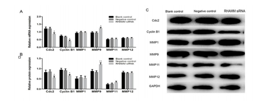
1.
Introduction
Lung cancer is one of the most common causes of cancer-related deaths worldwide [1] which can be divided into two types: small cell lung cancer (SCLC) and non-small cell lung cancer (NSCLC). Lung adenocarcinoma cancer (LUAD) is the predominantly common type of NSCLC accounting for over 40% of total incidence of lung cancer and has shown a significant increase in the past decades [2,3]. Due to the inconspicuous early symptoms and the lack of specific markers, the early diagnosis of LUAD is difficult, so the patients are likely to miss the best time for treatment [4,5,6]. Therefore, it is of great importance to study the genes related to tumor cell invasion and metastasis in LUAD patients and determine the relevant molecular mechanism for early diagnosis [7].
A series of genes have been found to play a role in the pathogenesis of NSCLC, such as RAS gene family, MYC gene family, ERBB family and p53 family [8]. RHAMM (hyaluronan mediated motility receptor), also known as CD168/HMMR/IHABP, has been widely studied as a protein receptor with multiple cell functions [9]. It was first identified as an important hyaluronic acid (HA) adhesion receptor. Antibodies against RHAMM can prevent HA from binding to lymphocytes, thus inhibiting the movement of lymphocytes. RHAMM plays an important role in tissue injury and wound healing [10,11] and can also be combined with calmodulin to analyze the differential phenotype of cells. During the interphase of cell division, RHAMM can be combined with GDP75 and GRP78 to microtubules, which maintains the stability of microtubules [12]. In vivo and in vitro experiments confirmed that RHAMM overexpression may regulate the protein expression of cell cycle dividing gene 2 (Cdc2) and CyclinB1, leading to the cell accumulation of G2/M phase [13]. The G2/M phase is one of the two checkpoints of the cell cycle and an important stage of cell proliferation, which is closely related to the instability of cell genome, the occurrence and treatment of tumors. The high expression of RHAMM at this stage suggests that RHAMM may control cell proliferation by regulating cell cycle. RHAMM is also related to a variety of kinases, and is distributed on the cell surface, cytoskeleton, mitochondria and nucleus which is transported to the outside of the cells through chaperone protein, and binds to the membranes by junction protein [13]. Current studies suggest that RHAMM may be an oncogenic factor, which is highly expressed in gastric cancer, colon cancer, breast cancer and other tumor cells, and regulates the migration and invasiveness of tumor cells [14].
RHAMM is a multifaceted molecule, and its various biological properties are known to be associated with malignant cell transformation and tumor development, while the more detailed molecular mechanism remains unclear [15]. The role of RHAMM in primary and metastatic NSCLC has been less studied than that in other tumors [16]. Wang et al. found that knockout RHAMM mediated by shRNA could inhibit the migration of LUAD cell lines H1975 and H3255 and predicted that RHAMM could be used as a prognostic factor for LUAD and as a therapeutic target to control the migration of NSCLC-like tumors [16]. Augustin et al. conducted a comprehensive analysis of differential expression profiles of 383 patients and found that RHAMM expression was associated with lower survival in large cell carcinomas (LCC) which indicated that RHAMM expression analysis was a valuable indicator to predict survival of LCC patients [17]. However, there is no research on the specific molecular mechanism of RHAMM affecting the proliferation, cell cycle and other characteristics of LUAD cells, and especially the effect caused by the induction of epithelial-mesenchymal transitions (EMT). Moreover, human LUAD A549 cell line is the most commonly used for researchers to study the occurrence and treatment of LUAD, and is used as the research object for the regulatory effect of various genes on LUAD. Xia et al. found that hypoxia could affect the protein expressions of p-glycoprotein and multidrug resistance protein in A549 cell line, thus affecting the growth of A549 cell line 18. Gao et al. reported that angelica inhibited the metastasis of A549 cell line by regulating MMPs/TIMPs and TGF-1, which had important reference value for the mechanism of angelica's regulation of LUAD 19. These results indicate that the study of RHAMM's function and mechanism in A549 cell line is of great significance to explore effects of RHAMM on LUAD.
In this study, we found that RHAMM expressed differentially in different clinical stages and the possible molecular mechanism of RHAMM overexpression inducing EMT of LUAD cells to promote cell proliferation and metastasis was studied.
2.
Materials and methods
2.1. Cells and main reagents
Human LUAD cell line A549 was obtained from the typical culture preservation center of Chinese Academy of Sciences (Shanghai, China). F-12k mixed culture medium (21127030) was purchased from Gibco (USA). MTT (tetrazolium blue, 298-93-1), DMSO (dimethyl sulfoxide, 2650) and trypsin (T2600000) were purchased from Sigma (USA). Inverted phase contrast microscope was purchased from OLYMPUS (USA). AccuriC6 flow cytometry and CellQuest software were purchased from BD company (USA). Micrometer was purchased from BIO-RAD (USA). Primary antibodies RHAMM (ab124729, 1:2000), Cdc2 (ab32094, 1:2000), CyclinB1 (ab32053, 1:300), MMP1 (ab137332, 1:1000), MMP9 (ab38898, 1:1000), MMP11 (ab53143, 1:1000), MMP12 (ab137444, 1:1000), E-cadherin (ab15148, 1:1000), Vimentin (ab137321, 1:500), GAPDH (ab9485, 1:2500), second antibody goat anti-rabbit IgG (ab205718) and enhanced chemiluminescence (ECL) kit (ab133406) were all purchased from abcam (China). SYBR Premix Ex Taq PCR kit (218076) was obtained from TaKaRa (Japan). Trizol (15596018), FBS (A3160910), Annexin V-FITC (bms500fi-100), BCA quantitative kit (23225) and cDNA synthesis kit (18091200) were obtained from Thermo Fisher Scientific (USA). Transfection kit LipoFiter was purchased from Hanbio (China). Primers of RHAMM, Cdc2, CyclinB1, MMP1, MMP9, MMP11, MMP12, E-cadherin, Vimentin and GAPDH were purchased from Ribobio Company (China).
2.2. Methods
2.2.1. Bioinformatic analysis
Four LUAD-related gene expression datasets (GSE10072, GSE32863, GSE43458, GSE118370) were downloaded from the Gene Expression Omnibus (GEO) database (https://www.ncbi.nlm.nih.gov/geo/), “RobustRankAgammaeg (RRA)” package of R software20 was utilized to integrate and analyze the four datasets to integrate differential expressed genes (DEGs) between normal tissue and cancerous tissue and set log|FC| > 1, padj < 0.05 as a threshold. At the same time, TCGA-LUAD mRNA expression and clinical data were downloaded from The Cancer Genome Atlas (TCGA) database (https://portal.gdc.cancer.gov/), and correlation analysis of clinical traits was carried out for the selected DEGs. KEGG pathway enrichment analysis and GO function annotation analysis were carried out using R package "clusterProfiler"21.
2.2.2. Cell culture and cell transfection
Human LUAD cell line A549 was cultured in F-12K medium containing 10% FBS and Kaighn's modification (1×) liquid (DMEM) at 37 ℃ with 5% CO2. Three pairs of 20 umol/L RHAMM-siRNA (RHAMM-siRNA1, RHAMM-siRNA2 and RHAMM-siRNA3) were designed and chemically synthesized by Guangzhou Ribobio Company using human RHAMM mRNA sequences provided by GenBank in NCBI as the template. According to the instructions of transfection kit of LipoFiter, A549 cell line was transfected with siRNA and control group.
2.2.3. qRT-PCR
Total RNA was extracted from cell samples using Trizol and 1 μg of total RNA was used to synthesize cDNA using cDNA synthesis kit. qRT-PCR was performed using SYBR Premix Ex Taq under the following thermal circulator conditions: 40 cycles of denaturation at 95 ℃ for 2 min, annealing at 95 ℃ for 5 s and extension at 60 ℃ for 30 s. Primers were shown in Table 1. Differences of relative mRNA expression of target genes in control group and experimental group were compared using 2-ΔΔCt method and the experiment was repeated three times.
2.2.4. Western blot
After 48 h transfection, the cells of each group were washed 3 times with cold PBS, and lysed on ice for 10 min after the whole protein lysate being added. The BCA quantitative kit was used for measuring protein concentrations, then 10 μL sample loading buffer was added to protein samples and the mixture was boiled for 10 min at 95 ℃. SDS-PAGE was performed at 100 V. After electrophoresis, proteins were transferred to NC membrane at 100 mA for 120 min and sealed with 5% BSA/TBST for 60 min. Then, the membrane was incubated with primary antibodies overnight at 4 ℃. After washing with 1×TBST solution three times for each of 5 min at room temperature, the membranes were incubated with secondary antibody at room temperature for 120 min. The membrane was washed with TBST 3 times for each of 20 min, the protein bands were observed using the ECL kit and the image was analyzed. Grayscale values of the bands were analyzed by Quantity One, and expressed by the ratio of grayscale values of the target bands and internal reference bands. The experiment was repeated three times.
2.2.5. MTT assay
Logarithmic phase cells of each group were seeded into 60 mm culture dish. After the cells had converged to 70-80%, they were digested with trypsin, resuspended in culture dish and counted. The diluted cell suspension was seeded in a 96-well plate at a density of 2×10³ and each group was made in sextuplicate. After being cultured at 37 ℃ with 5% CO2 in a incubator for 24 h, 48 h and 72 h respectively, each well was mixtured with 10 μL MTT solution (5 mg/mL) and cultured in the incubator. 4 hours later, the supernatant was absorbed and 150 μL DMSO solution was added. The absorbance value of each well at 490 nm was measured and recorded. The experiment was repeated three times and the mean value was taken.
2.2.6. Wound healing test
The 200 μL pipette tip was used to gently scrape the single layer through the center of the wells. The wells were washed twice briefly in the medium to remove the isolated cells. Then fresh medium was added, the cells were regrown for 24 h, and the cell migration was observed and photographed microscopically.
2.2.7. Transwell
The 24-well Transwell chamber (8 μm aperture) was used for Transwell invasion determination. The top chamber was coated with Matrigel matrix and about 2×10⁴ cells were plated in the upper chamber. The bottom chamber contained DMEM with 10% fetal bovine serum (FBS). After incubation at 37 ℃ for 24 h, the non-migratory cells side were cleared away with a cotton swab and the migratory cells on the lower side were stained with crystal violet. Finally, the cells on the bottom chamber were observed and counted under a microscope. The experiment was repeated three times.
2.2.8. Flow cytometry detected cell cycle and apoptosis
Cell cycle in A549 of each group was analyzed by propidium iodide (PI) staining. When logarithmic growth cells coverage reached 80%, the supernatant was moved, and the cells were washed once and digested with 0.25% trypsin. Then Cells were collected after low-speed centrifugation and each group was made in triplicate. The supernatant was discarded after centrifuging at 1200 rmp for 5 min. Cell precipitation was washed once with ice-cold PBS at 4 ℃, and centrifuged again at 1500 rmp for 5 min. Following fixation with ice-cold ethanol at 4 ℃ for 1 h. The fixed solution was removed by centrifugation of 1500 rmp for 5 min, the cells were washed by ice-cold PBS at 4 ℃ (PH = 7.2-7.4) for one time, and the cells were centrifuged at 1500 rmp for 5 min to collect the cells. According to the cell quantity, the cells were fixed with a certain volume of PI(1-1.5 mL) and stored in darkness at 4 for 30 min. Finally, the cell passing rate was 200-350 cells/s. The sample was filtered by a sieve (300 mesh) and placed in the flow pipe to test results.
Annexin V FITC/PI double staining was performed to detect cell apoptosis. Cells from each group were inoculated in a six-well plate with a density of 3.0×10⁵ cells/well overnight, and then resuspended in 100 μL of binding buffer containing 5 μL of Annexin V-FITC for 10 min at room temperature in the dark. Next the cells were added with 5 μL PI and incubated at room temperature for 5 min in the dark. Finally, cell apoptosis was detected using flow cytometry instrument, and CellQuest was used to analyze the data.
2.2.9. Statistical analysis
All data were processed by SPSS 22.0 statistical software, and exhibited as mean ± standard deviation. The differences between two groups were analyzed by t-test, and one-way ANOVA was used for comparison among multiple groups.
3.
Results
3.1. RHAMM gene is highly expressed in LUAD tissues and cells and is associated with cell cycle and ECM
In this study, 84 up-regulated genes and 146 down-regulated genes were identified by the combined analysis of four LUAD datasets (Figure 1A), among which RHAMM was highly expressed in LUAD tissues. The correlation analysis of the 230 genes screened in the TCGA-LUAD data set showed that only RHAMM expression level gradually increased from stage I-IV of LUAD, (Figure 1B), which indicated that RHAMM played a role in LUAD. Survival analysis results also showed that high RHAMM expression was significantly correlated with poor survival time (Figure 1C), which indicated that the expression of RHAMM had a significant impact on the prognosis of patients. GO enrichment analysis of up-regulated and down-regulated genes exhibited that RHAMM was involved in G2/M transition of cell mitosis cycle (Figure S1A), which was consistent with previous studies on RHAMM [13]. KEGG enrichment analysis of 230 DEGs revealed that RHAMM was involved in ECM-receptor interaction (Figure S1B). These results predicted that RHAMM was highly expressed in LUAD and might be associated with ECM.
3.2. RHAMM-siRNA1 transfected cells significantly down-regulate the expression of RHAMM in mRNA and protein levels
The interference efficiency of RHAMM was validated by qRT-PCR and Western blot, respectively. Compared with Blank control and Negative control groups, decreased mRNA and protein expression of RHAMM were observed in three siRNA groups (P < 0.01), among which the interference efficiency of siRNA1 was the best (Figure 2A-C). Based on the above results, RHAMM-siRNA1 was selected as the silent transfection group in the subsequent experiments. The transfection concentration gradient of RHAMM-siRNA1 was screened at 30, 50, 70 and 90 nmol/mL. As shown in Figure 2D, the expression level of RHAMM-siRNA1 was decreased with Blank control group and dose-dependent within a certain range. When the transfection concentration was 70 nmol/mL, the inhibitory effect was the most significant (P < 0.01). These results demonstrated that significant effect could be achieved by silencing RHAMM in A549 cell line.
3.3. Silencing RHAMM inhibits EMT, migration, and invasion
The results of qPCR and Western blot were shown in Figure 3A, compared with Blank and NC groups, the expression of E-cadherin after transfection with RHAMM-siRNA1was significantly up-regulated, while the expression of Vimentin was significantly down-regulated. EMT of tumor cells is closely related to tumor migration and invasion. Therefore, the migration ability of LUAD A549 cell line was detected by wound healing test. As displayed in Figure 3B, compared with Blank and NC groups, the cell migration rate of the RHAMM-siRNA group was significantly reduced (P < 0.05). The result of Transwell observed that compared with Blank and NC groups, the number of invasion cells in the RHAMM-siRNA group was significantly reduced (P < 0.05). These results illuminated that RHAMM significantly promoted the migration and invasion of A549 cell line, while inhibiting the expression of RHAMM significantly suppressed the migration and invasion of A549 cell line.
3.4. Silencing RHAMM inhibits cell proliferation, promotes apoptosis and changes cell cycle
To evaluated the effects of RHAMM on cell proliferation, MMT assays detected that (Figure 4A) silencing RHAMM inhibited cell viability in A549 cell line(P < 0.05) and Flow cytometry (FCM) results (Figure 4B) showed that the apoptosis rate of RHAMM-siRNA group was significantly increased (P < 0.01). FCM also detected cell cycle and the results (Figure 4C) demonstrated that the proportion of cells in RHAMM-siRNA group was increased significantly in G2/M phase, and decreased significantly in G0/G1 phase. Overall, the above results indicated that silencing RHAMM inhibited cell proliferation, promoted cell apoptosis, suppressed cell cycle in lung cells.
3.5. Silencing RHAMM inhibits the expression of Cdc2, CyclinB1 and promotes MMP9 protein expression
According to GO and KEGG analysis, RHAMM was found to be involved in G2/M transition of cell mitotic cycle (Figure S1A) and ECM-receptor interaction (Figure S1B). In order to demonstrate whether RHAMM can regulate the expression of Cdc2, CyclinB1 in mitotic cell cycle and MMPs degraded by ECM directly, RHAMM was silenced by RHAMM-siRNA1 to observe the expression of Cdc2 and CylclinB1 (Figure 1A) and the changes in mRNA and protein levels of four MMP (MMP1, MMP9, MMP11 and MMP12) with high correlation with RHAMM (Figure S1C). The results showed that (Figure 5A), the expression levels of Cdc2 and CyclinB1 were significantly down-regulated, and the expression levels of MMP9 were significantly increased (P < 0.05) after RHAMM gene was silenced. At the protein level (Figure 5B, C), Cdc2 and CyclinB1 expression were significantly down-regulated after RHAMM gene silencing (P < 0.05), while MMP9 expression was significantly increased (P < 0.01). However, there was no significant difference in mRNA and protein expression levels of MMP1, MMP11 and MMP12 after RHAMM gene was silenced. These results indicated that silencing RHAMM has a significant regulatory effect on the expressions of Cdc2, CyclinB1 and MMP9, and the close association of these genes with the ECM pathway indirectly suggested that RHAMM may influence the ECM pathway by regulating these genes.
4.
Discussion
RHAMM has been studied by many researchers as a marker gene in tumor prognosis. In past studies, RHAMM overexpression has been used as a marker of cancer invasiveness and agammaavation for many types of cancer [18,19,20]. The relationship between RHAMM and tumorigenesis development has also been reported by many studies. Mele et al. reported that silencing RHAMM inhibited the colorectal cancer cells migration and invasion and down-regulated expression of RHAMM gene in colorectal cancer was found to slow tumor growth and inhibit cancer cells metastasis to other organs in vivo [21]. It was also found that RHAMM expression level in ovarian surface epithelium, serum, urine samples and ovarian cell lysates of patients with ovarian cancer was increased significantly compared with normal people [22]. However, there are few studies on the role of RHAMM expression in LUAD. In this study, in order to directly determine the effect of RHAMM on the growth and development of LUAD, we selected A549 cell line for experiments. After silencing RHAMM, we found for the first time that RHAMM inhibited the proliferation, migration, invasion and EMT effect of A549 cell line. Our results were consistent with other reports. Wang et al. found that RHAMM was associated with poor prognosis and could promote the metastasis of NSCLC [16]. Song et al. demonstrated that CD44/RHAMM signaling pathway could promote cell proliferation of NSCLC [23]. The findings of our study to LUAD confirm once again that RHAMM can significantly promote the growth and development of cancer.
RHAMM gene, as a multifaceted molecule, has also been studied by many researchers on its molecular mechanism of regulating cancer. Kouvidi et al. demonstrated that cell proliferation of fibrosarcoma cell line HT1080 could be regulated through the β-catenin/c-myc signaling axis and abnormal expression of β-catenin/c-myc was an early event of cancer that played an important role in prognosis [24]. Researchers had found that the expression of HA family including RHAMM was positively correlated with the expression of β-catenin, Twist and Snail, while negatively correlated with the expression of E-cadherin which confirmed that RHAMM was involved in the regulation of EMT-related genes [25]. In our study, GO and KEGG analysis of screened genes revealed that RHAMM is involved in ECM and cell division cycle. It was observed that RHAMM gene could reduce the mRNA and protein expression levels of Cyc2 and cyclinB1 by silencing RHAMM gene. Moreover, studies have shown that the decreased expression levels of these two genes would lead to a large number of cell agammaegation in G2/M phase [13]. Other studies have shown that RHAMM may regulate the expression of Cyc2 and CyclinB1 by regulating their upstream genes [26]. These results suggested that RHAMM may further regulate the cell cycle of LUAD cells by regulating Cyc2 and CyclinB1. In the regulation of ECM pathway, CD44 gene, which also belongs to hyaluronic acid-binding protein with RHAMM, regulates ECM degradation by regulating MMP expression, and RHAMM is also related to ECM degradation [26,27]. Furthermore, it was revealed in our study that mRNA and protein expression levels of MMP9 were increased significantly. The results also prove once again that HA receptors such as RHAMM regulate ECM degradation by regulating MMP expression. Whether RHAMM regulates MMP protein secretion by regulating its upstream gene still needs to be confirmed.
In conclusion, RHAMM significantly promoted the proliferation, migration, invasion and EMT of LUAD cells. In this study, it was found that RHAMM may affect the EMT pathway, growth and development of LUAD cells by regulating the expression of Cdc2, CyclinB1, and MMP9.
Ethics approval and consent to participate
The authors have no ethical conflicts to disclose.
Acknowledgements
This study was supported by Science and Technology Project of Taizhou (No. 1802ky41).
Conflict of interest
The authors have no conflicts of interest to declare.