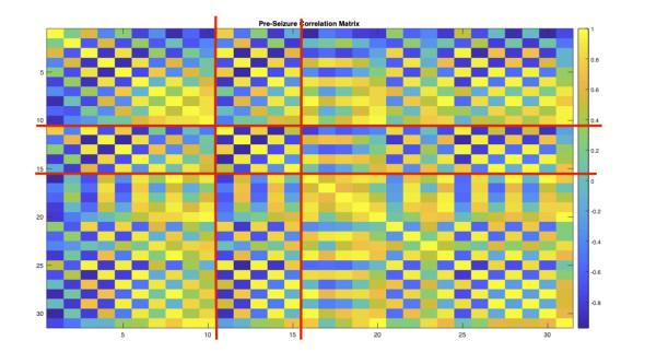
Epilepsy is a complex phenomena of a system of highly intensive and synchronized neurons simultaneously firing which can be traced to spatial and temporal patterns. Seizures are a well known physical feature for all types of epileptic disorders. The rhythms, patterns, and oscillatory dynamics explain the mechanistic nature of neurons especially in absence seizures. Previous models such as Wilson-Cowan (1973), introduced brain models showing the dynamics of a network of neurons consisting of excitatory and inhibitory neurons. Taylor et al. (2014) then adapted the Wilson-Cowan model to epileptic seizures using a thalamo-cortical based theory. Fan et al. (2018) projects that thalamic reticulus nuclei control spike wave discharges specifically in absence seizures. We identify brain activity patterns specific to Glucose (G1D) Transport Deficiency Epilepsy in a network model based on electroencephalogram device (EEG) data. Additionally, we study the EEG patterns to identify the plausible mechanism that causes G1D epileptic behavior. Our coupled thalamo-cortical model goes beyond a connection in a logical unidirectional pattern shown by Fan or in a bidirectional small world pattern. Our model is a network based on paired correlation of EEG signals more analogous to realistic seizure activity. Using our model, we are able to study stability analysis for equilibrium and periodic behavior. We also identify parameter values which cause synchronized activity or more stable activity. Lastly, we identify a synchronization index and sensitivity analysis regarding parameters that directly affect Spike Wave Discharges and other spiking behavior. We will show how our 32-unit data-driven network model reflects G1D seizure dynamics and discuss the limitations of the model.
Citation: Ariel Leslie, Jianzhong Su. Modeling and simulation of a network of neurons regarding Glucose Transporter Deficiency induced epileptic seizures[J]. Electronic Research Archive, 2022, 30(5): 1813-1835. doi: 10.3934/era.2022092
Epilepsy is a complex phenomena of a system of highly intensive and synchronized neurons simultaneously firing which can be traced to spatial and temporal patterns. Seizures are a well known physical feature for all types of epileptic disorders. The rhythms, patterns, and oscillatory dynamics explain the mechanistic nature of neurons especially in absence seizures. Previous models such as Wilson-Cowan (1973), introduced brain models showing the dynamics of a network of neurons consisting of excitatory and inhibitory neurons. Taylor et al. (2014) then adapted the Wilson-Cowan model to epileptic seizures using a thalamo-cortical based theory. Fan et al. (2018) projects that thalamic reticulus nuclei control spike wave discharges specifically in absence seizures. We identify brain activity patterns specific to Glucose (G1D) Transport Deficiency Epilepsy in a network model based on electroencephalogram device (EEG) data. Additionally, we study the EEG patterns to identify the plausible mechanism that causes G1D epileptic behavior. Our coupled thalamo-cortical model goes beyond a connection in a logical unidirectional pattern shown by Fan or in a bidirectional small world pattern. Our model is a network based on paired correlation of EEG signals more analogous to realistic seizure activity. Using our model, we are able to study stability analysis for equilibrium and periodic behavior. We also identify parameter values which cause synchronized activity or more stable activity. Lastly, we identify a synchronization index and sensitivity analysis regarding parameters that directly affect Spike Wave Discharges and other spiking behavior. We will show how our 32-unit data-driven network model reflects G1D seizure dynamics and discuss the limitations of the model.

| [1] |
G. Deco, A. Ponce-Alvarez, P. Hagmann, G. L. Romani, D. Mantini, M. Corbetta, How Local Excitation–Inhibition Ratio Impacts the Whole Brain Dynamics, J. Neurosci., 34 (2014), 7886–7898. https://doi.org/10.1523/JNEUROSCI.5068-13.2014 doi: 10.1523/JNEUROSCI.5068-13.2014

|
| [2] |
H. Zhang, J. Su, Q. Wang, Y. Liu, L. Good, J. Pascual, Predicting seizure by modeling synaptic plasticity based on EEG signals—a case study of inherited epilepsy, Commun. Nonlinear Sci. Numer. Simul., 56 (2018), 330–343. https://doi.org/10.1016/j.cnsns.2017.08.020 doi: 10.1016/j.cnsns.2017.08.020
|
| [3] | J. M. Bekkers, Pyramidal Neurons, Curr. Biol., 21 (2011), R975. https://doi.org/10.1016/j.cub.2011.10.037 |
| [4] |
R. B. Gonzales, C. J. DeLeon Galvan, Y. M. Rangel, B. J. Claiborne, Distribution of thorny excrescences on CA3 pyramidal neurons in the rat hippocampus, J. Comp. Neurol., 430 (2001), 357–368. https://doi.org/10.1002/1096-9861(20010212)430:3<357::AID-CNE1036>3.0.CO;2-K doi: 10.1002/1096-9861(20010212)430:3<357::AID-CNE1036>3.0.CO;2-K
|
| [5] |
S. B. Nelson, C. Hempel, K. Sugino, Probing the transcriptome of neuronal cellmtypes, Curr. Opin. Neurobiol., 16 (2006), 571–576. https://doi.org/10.1016/j.conb.2006.08.006 doi: 10.1016/j.conb.2006.08.006
|
| [6] | P. N. Taylor, G. Baier, S. S. Cash, J. Dauwels, J. Slotine, Y. Wang, A model of stimulus induced epileptic spike-wave discharges, 2013 IEEE Symposium on Computational Intelligence, Cognitive Algorithms, Mind, and Brain (CCMB), (2013), 53–59. https://doi.org/10.1109/CCMB.2013.6609165 |
| [7] | R. Swenson, The Thalamus - Thalamic Organization, Dartmouth Medical School, 2006, available from: www.dartmouth.edu/rswenson/NeuroSci/chapter_10.html. |
| [8] |
H. R. Wilson, J. D. Cowan, A mathematical theory of the functional dynamics of cortical and thalamic nervous tissue, Kybernetik, 13 (1973), 55–80. https://doi.org/10.1007/BF00288786 doi: 10.1007/BF00288786
|
| [9] | D. Golomb, Neuronal Synchrony Measures, Scholarpedia, 2 (2007), 1347. https://doi.org/10.4249/scholarpedia.1347 |
| [10] |
N. Kopell, G. B. Ermentrout, M. A. Whittington, R. D. Traub, Gamma Rhythms and Beta Rhythms Have Different Synchronization Properties, Proc. Natl. Acad. Sci. U.S.A., 97 (2000), 1867–1872. https://doi.org/10.1073/pnas.97.4.1867 doi: 10.1073/pnas.97.4.1867
|
| [11] |
P Suczynski, S Kalitzin, F. L. Da Silva, Dynamics of non-convulsive epileptic phenomena modeled by a bistable neuronal network, Neurosci., 12 (2004), 467–484. https://doi.org/10.1016/j.neuroscience.2004.03.014 doi: 10.1016/j.neuroscience.2004.03.014
|
| [12] | G. De Vries, A. Sherman, Channel sharing in pancreatic $\beta$-cell revisited: enhancement of emergent bursting by noise, J. Theor. Biol., 207 (2000), 513–530. https://doi.org/10.1006/jtbi.2000.2193 |
| [13] |
S. Intep, D. J. Higham, Zero, one and two-switch models of gene regulation, Discrete Contin. Dyn. Syst. B, 14 (2010), 495–513. https://doi.org/10.3934/dcdsb.2010.14.495 doi: 10.3934/dcdsb.2010.14.495
|
| [14] |
M. I. Freidlin, Quasi-deterministic approximation, metastability and stochastic resonance, Phys. D, 137 (2000), 333–352. https://doi.org/10.1016/S0167-2789(99)00191-8 doi: 10.1016/S0167-2789(99)00191-8
|
| [15] | L. Gammaitoni, P. Hanggi, P. Jung, F. Marchesoni, Stochastic Resonance, Rev. Mod. Phys., 70 (1998), 223–287. https://doi.org/10.1103/RevModPhys.70.223 |
| [16] |
T. R. Chay, H. S. Kang, Role of single-channel stochastic noise on bursting clusters of pancreatic_-cells, Biophys. J., 54 (1988), 427–435. https://doi.org/10.1016/S0006-3495(88)82976-X doi: 10.1016/S0006-3495(88)82976-X
|
| [17] |
A. Sherman, J. Rinzel, J. Keizer, Emergence of organized bursting in clusters of pancreatic beta-cells by channel sharing, Biophys. J., 54 (1988), 411–425. https://doi.org/10.1016/S0006-3495(88)82975-8 doi: 10.1016/S0006-3495(88)82975-8
|
| [18] |
M. Pedersen, M. Serensen, The effect of noise on $\beta$-cell burst period, SIAM J. Appl. Math., 67 (2007), 530–542. https://doi.org/10.1137/060655663 doi: 10.1137/060655663
|
| [19] |
J. Su, J. Rubin, D. Terman, Effects of noise on elliptic bursters, Nonlinearity, 17 (2004), 133–157. https://doi.org/10.1088/0951-7715/17/1/009 doi: 10.1088/0951-7715/17/1/009
|
| [20] |
P. N. Taylor, Y. Wang, M. Goodfellow, J. Dauwels, F. Moeller, U. Stephani, A computational study of stimulus driven epileptic seizure abatement, PloS One, 9 (2014), e114316. https://doi.org/10.1371/journal.pone.0114316 doi: 10.1371/journal.pone.0114316
|
| [21] |
R. D. Traub, D. Contreras, M. O. Cunningham, H. Murray, F. E. N. Lebeau, A. Roopun, et al., Single-column thalamocortical network model exhibiting gamma oscillations, sleep spindles, and epileptogenic bursts, J. Neurophysiol., 93 (2005), 2194–2232. https://doi.org/10.1152/jn.00983.2004 doi: 10.1152/jn.00983.2004
|
| [22] |
Z. Feng, A. Lubbe, Q. Lu, J. Su, On bursting solutions near chaotic regimes in a neuron model, Discrete Contin. Dyn. Syst. S, 7 (2014), 1363–1383. https://doi.org/10.3934/dcdss.2014.7.1363 doi: 10.3934/dcdss.2014.7.1363
|
| [23] |
P. N. Taylor, Y. Wang, M. Goodfellow, J. Dauwels, F. Moeller, U. Sephani, et al., A computational study of stimulus induced driven epileptic seizure abatement, PLoS One, 9 (2014), e114316. https://doi.org/10.1371/journal.pone.0114316 doi: 10.1371/journal.pone.0114316
|
| [24] |
R. D. Traub, R. K. Wong, R. Miles, H. Michelson, A Model of a CA3 Hippocampal Pyramidal Neuron Incorporating Voltage-Clamp Data on Intrinsic Conductances, J. Neurophysiol., 66 (1991), 635–650. https://doi.org/10.1152/jn.1991.66.2.635 doi: 10.1152/jn.1991.66.2.635
|
| [25] |
G. Baier, M. Goodfellow, P. N. Talor, Y. Wang, D. J. Garry, The Importance of Modeling Epileptic Seizure Dynamics as Spatio-Temporal Patterns, Front. Physiol., 3 (2012), 281. https://doi.org/10.3389/fphys.2012.00281 doi: 10.3389/fphys.2012.00281
|
| [26] |
Q. Wang, X. Shi, G. Chen, Delay-induced synchronization transition in small-world Hodgkin-Huxley neuronal networks with channel blocking, Discrete Contin. Dyn. Syst. B, 16 (2011), 607–621. https://doi.org/10.3934/dcdsb.2011.16.607 doi: 10.3934/dcdsb.2011.16.607
|
| [27] |
A. Daci, A. Bozalija, F. Jashari, S. Krasniqi, Individualizing Treatment Approaches for Epileptic Patients with Glucose Transporter TYPE1 (GLUT-1) Deficiency, Int. J. Mol. Sci., 19 (2018), 122. https://doi.org/10.3390/ijms19010122 doi: 10.3390/ijms19010122
|
| [28] |
D. Fan, S. Liu, Q. Wang, Stimulus-Induced Epileptic Spike-Wave Discharges in Thalamocortical Model with Disinhibition, Sci. Rep., 6 (2016), 1–21. https://doi.org/10.1038/srep37703 doi: 10.1038/srep37703
|
| [29] | D. Fan, J. Su, A. Bowman, Rich Dynamics Induced by Synchronization Varieties in the Coupled Thalamocortical Circuitry Model, In Brain Informatics: International Conference, BI 2018, Springer, 2018, 78–84. https://doi.org/10.1007/978-3-030-05587-5_8 |
| [30] |
D. Fan, Q. Wang, J. Su, H. Xi, Stimulus-Induced Transitions between Spike-Wave Discharges and Spindles with the Modulation of Thalamic Reticular Nucleus, J. Comput. Neurosci., 43 (2017), 203–225. https://doi.org/10.1007/s10827-017-0658-4 doi: 10.1007/s10827-017-0658-4
|
| [31] |
R. Kuske, S. M. Baer, Asymptotic analysis of noise sensitivity in a neuronal burster, Bull. Math. Biol., 64 (2002), 447–481. https://doi.org/10.1006/bulm.2002.0279 doi: 10.1006/bulm.2002.0279
|
| [32] |
H. Zhang, J. Jacobs, Traveling Theta Waves in the Human Hippocampus, J. Neurosci., 35 (2015), 12477–12487. https://doi.org/10.1523/jneurosci.5102-14.2015 doi: 10.1523/jneurosci.5102-14.2015
|


Figures(13) / Tables(4)
Ariel Leslie, Jianzhong Su. Modeling and simulation of a network of neurons regarding Glucose Transporter Deficiency induced epileptic seizures[J]. Electronic Research Archive, 2022, 30(5): 1813-1835. doi: 10.3934/era.2022092


 DownLoad: