Delineation of retinal vessels in fundus images is essential for detecting a range of eye disorders. An automated technique for vessel segmentation can assist clinicians and enhance the efficiency of the diagnostic process. Traditional methods fail to extract multiscale information, discard unnecessary information, and delineate thin vessels. In this paper, a novel residual U-Net architecture that incorporates multi-scale feature learning and effective attention is proposed to delineate the retinal vessels precisely. Since drop block regularization performs better than drop out in preventing overfitting, drop block was used in this study. A multi-scale feature learning module was added instead of a skip connection to learn multi-scale features. A novel effective attention block was proposed and integrated with the decoder block to obtain precise spatial and channel information. Experimental findings indicated that the proposed model exhibited outstanding performance in retinal vessel delineation. The sensitivities achieved for DRIVE, STARE, and CHASE_DB datasets were 0.8293, 0.8151 and 0.8084, respectively.
Citation: G. Prethija, Jeevaa Katiravan. EAMR-Net: A multiscale effective spatial and cross-channel attention network for retinal vessel segmentation[J]. Mathematical Biosciences and Engineering, 2024, 21(3): 4742-4761. doi: 10.3934/mbe.2024208
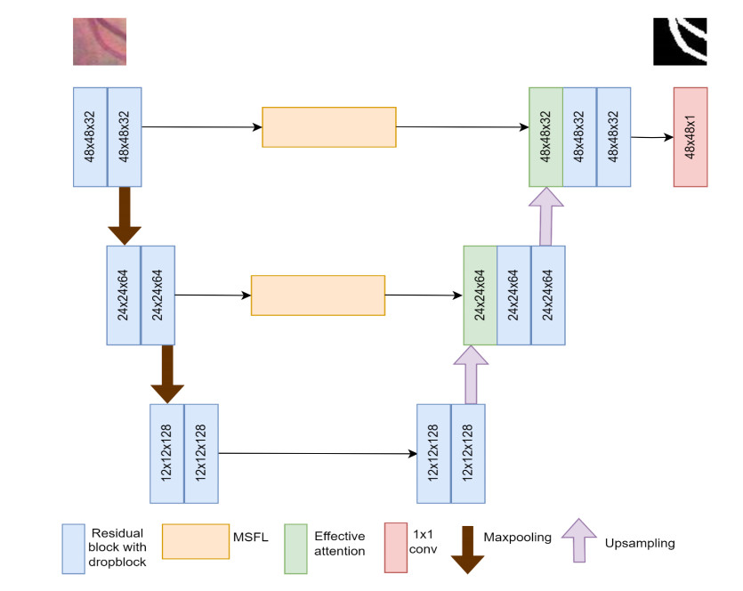
Delineation of retinal vessels in fundus images is essential for detecting a range of eye disorders. An automated technique for vessel segmentation can assist clinicians and enhance the efficiency of the diagnostic process. Traditional methods fail to extract multiscale information, discard unnecessary information, and delineate thin vessels. In this paper, a novel residual U-Net architecture that incorporates multi-scale feature learning and effective attention is proposed to delineate the retinal vessels precisely. Since drop block regularization performs better than drop out in preventing overfitting, drop block was used in this study. A multi-scale feature learning module was added instead of a skip connection to learn multi-scale features. A novel effective attention block was proposed and integrated with the decoder block to obtain precise spatial and channel information. Experimental findings indicated that the proposed model exhibited outstanding performance in retinal vessel delineation. The sensitivities achieved for DRIVE, STARE, and CHASE_DB datasets were 0.8293, 0.8151 and 0.8084, respectively.

| [1] | Centers for Disease Control and Prevention, National diabetes statistics report, 2020: estimates of diabetes and its burden in the United States, CDC, 2020. Available from: https://stacks.cdc.gov/view/cdc/85309 |
| [2] | Eye Complications, ADA, 2021. Available from: https://diabetes.org/about-diabetes/complications/eye-complication. |
| [3] |
M. M. Fraz, P. Remagnino, A. Hoppe, B. Uyyanonvara, A. R. Rudnicka, C. G. Owen, et al., Blood vessel segmentation methodologies in retinal images—a survey, Comput. Methods Programs Biomed., 108 (2012), 407–433. https://doi.org/10.1016/j.cmpb.2012.03.009 doi: 10.1016/j.cmpb.2012.03.009

|
| [4] |
S. Dash, S. Verma, Kavita, S. Bevinakoppa, M. Wozniak, J. Shafi, et al., Guidance image-based enhanced matched filter with modified thresholding for blood vessel extraction, Symmetry, 14 (2022), 194. https://doi.org/10.3390/sym14020194 doi: 10.3390/sym14020194
|
| [5] |
S. Chatterjee, A. Suman, R. Gaurav, S. Banerjee, A. K. Singh, B. K. Ghosh, et al., Retinal blood vessel segmentation using edge detection method, J. Phys. Conf. Ser., 1717 (2021), 012008. https://doi.org/10.1088/1742-6596/1717/1/012008 doi: 10.1088/1742-6596/1717/1/012008
|
| [6] | P. Kuppusamy, M. M. Basha, C. L. Hung, Retinal blood vessel segmentation using random forest with Gabor and Canny edge features, in 2022 International Conference on Smart Technologies and Systems for Next Generation Computing (ICSTSN), Villupuram, India, (2022), 1–4. https://doi.org/10.1109/ICSTSN53084.2022.9761339 |
| [7] |
S. Roychowdhury, D. D. Koozekanani, K. K. Parhi, Blood vessel segmentation of fundus images by major vessel extraction and subimage classification, IEEE J. Biomed. Health Inf., 19 (2014) 1118–1128. https://doi.org/10.1109/JBHI.2014.2335617 doi: 10.1109/JBHI.2014.2335617
|
| [8] | E. Chakour, Y. Mrad, A. Mansouri, Y. Elloumi, M. H. Bedoui, I. B. Andaloussi, et al., Blood vessel segmentation of retinal fundus images using dynamic preprocessing and mathematical morphology, in 2022 8th International Conference on Control, Decision and Information Technologies (CoDIT), Istanbul, Turkey, (2022), 1473–1478. https://doi.org/10.1109/CoDIT55151.2022.9804004 |
| [9] | P. R. Wankhede, K. B. Khanchandani, Retinal blood vessel segmentation using graph cut analysis, in 2015 International Conference on Industrial Instrumentation and Control (ICIC), Pune, India, (2015), 1429–1432. https://doi.org/10.1109/IIC.2015.7150973 |
| [10] |
M. R. K. Mookiah, S. Hogg, T. J. MacGillivray, V. Prathiba, R. Pradeepa, V. Mohan, et al., A review of machine learning methods for retinal blood vessel segmentation and artery/vein classification, Med. Image Anal., 68 (2021), 101905. https://doi.org/10.1016/j.media.2020.101905 doi: 10.1016/j.media.2020.101905
|
| [11] |
O. O. Sule, A survey of deep learning for retinal blood vessel segmentation methods: Taxonomy, trends, challenges and future directions, IEEE Access, 10 (2022), 38202–38236. https://doi.org/10.1109/ACCESS.2022.3163247 doi: 10.1109/ACCESS.2022.3163247
|
| [12] |
T. J. Jebaseeli, C. A. D. Durai, J. D. Peter, Retinal blood vessel segmentation from diabetic retinopathy images using tandem PCNN model and deep learning based SVM, Optik, 199 (2019), 163328. https://doi.org/10.1016/j.ijleo.2019.163328 doi: 10.1016/j.ijleo.2019.163328
|
| [13] |
X. Yang, Z. Li, Y. Guo, D. Zhou, Retinal vessel segmentation based on an improved deep forest, Int. J. Imaging Syst. Technol., 31 (2021), 1792–1802. https://doi.org/10.1002/ima.22610 doi: 10.1002/ima.22610
|
| [14] |
D. Yang, G. Liu, M. Ren, B. Xu, J. Wang, A multi-scale feature fusion method based on U-net for retinal vessel segmentation, Entropy, 22 (2020), 811. https://doi.org/10.3390/E22080811 doi: 10.3390/E22080811
|
| [15] |
M. Padmapriya, S. Pasupathy, V. Punitha, Early diagnosis of diabetic retinopathy using unsupervised learning, Soft Comput., 27 (2023), 9093–9104. https://doi.org/10.1007/s00500-023-08418-z doi: 10.1007/s00500-023-08418-z
|
| [16] |
N. Muzammil, S. A. A. Shah, A. Shahzad, M. A. Khan, R. M. Ghoniem, Multifilters-based unsupervised method for retinal blood vessel segmentation, Appl. Sci., 12 (2022), 6393. https://doi.org/10.3390/app12136393 doi: 10.3390/app12136393
|
| [17] | Z. Qaiser, W. Ahmad, M. Y. Umair, Z. Mahmood, Unsupervised vessel segmentation method in retinal images, in 2022 International Conference on Frontiers of Information Technology (FIT), Islamabad, Pakistan, (2022), 65–70. https://doi.org/10.1109/FIT57066.2022.00022 |
| [18] |
K. Upadhyay, M. Agrawal, P. Vashist, Unsupervised multiscale retinal blood vessel segmentation using fundus images, IET Image Proc., 14 (2020) 2616–2625. https://doi.org/10.1049/iet-ipr.2019.0969 doi: 10.1049/iet-ipr.2019.0969
|
| [19] | O. Ronneberger, P. Fischer, T. Brox, U-net: Convolutional networks for biomedical image segmentation, in Medical Image Computing and Computer-Assisted Intervention–MICCAI 2015: 18th International Conference, Munich, Germany, (2015), 234–241. https://doi.org/10.1007/978-3-319-24574-4_28 |
| [20] | T. Laibacher, S. Jalali, M2u-net: Effective and efficient retinal vessel segmentation for real-world applications, in Proceedings of the IEEE/CVF conference on computer vision and pattern recognition workshops, Long Beach, CA, USA, 2019. https://doi.org/10.1109/CVPRW.2019.00020 |
| [21] |
H. Boudegga, Y. Elloumi, M. Akil, M. H. Bedoui, R. Kachouri, A. B. Abdallah, Fast and efficient retinal blood vessel segmentation method based on deep learning network, Comput. Med. Imaging Graphics, 90 (2021). https://doi.org/10.1016/j.compmedimag.2021.101902 doi: 10.1016/j.compmedimag.2021.101902
|
| [22] |
X. Yang, Z. Li, Y. Guo, D. Zhou, DCU-net: a deformable convolutional neural network based on cascade U-net for retinal vessel segmentation, Multimed. Tools Appl., 81 (2022), 15593–15607. https://doi.org/10.1007/s11042-022-12418-w doi: 10.1007/s11042-022-12418-w
|
| [23] |
H. Wang, G. Xu, X. Pan, Z. Liu, N. Tang, R. Lan, et al., Attention-inception-based U-Net for retinal vessel segmentation with advanced residual, Comput. Electr. Eng., 98 (2022), 107670. https://doi.org/10.1016/j.compeleceng.2021.107670 doi: 10.1016/j.compeleceng.2021.107670
|
| [24] |
X. Wang, X. Jiang, J. Ren, Blood vessel segmentation from fundus image by a cascade classification framework, Pattern Recognit., 88 (2019), 331–341. https://doi.org/10.1016/j.patcog.2018.11.030 doi: 10.1016/j.patcog.2018.11.030
|
| [25] |
F. Dong, D. Wu, C. Guo, S. Zhang, B. Yang, X. Gong, CRAUNet: A cascaded residual attention U-Net for retinal vessel segmentation, Comput. Biol. Med., 147 (2022), 105651. https://doi.org/10.1016/j.compbiomed.2022.105651 doi: 10.1016/j.compbiomed.2022.105651
|
| [26] |
Y. Liu, J. Shen, L. Yang, G. Bian, H. Yu, ResDO-UNet: A deep residual network for accurate retinal vessel segmentation from fundus images, Biomed. Signal Process. Control, 79 (2023), 104087. https://doi.org/10.1016/j.bspc.2022.104087 doi: 10.1016/j.bspc.2022.104087
|
| [27] |
K. Ren, L. Chang, M. Wan, G. Gu, Q. Chen, An improved U-net based retinal vessel image segmentation method, Heliyon, 8 (2022), e11187. https://doi.org/10.1016/j.heliyon.2022.e11187 doi: 10.1016/j.heliyon.2022.e11187
|
| [28] |
J. Li, G. Gao, L. Yang, Y. Liu, GDF-Net: A multi-task symmetrical network for retinal vessel segmentation, Biomed. Signal Process. Control, 81 (2023), 104426. https://doi.org/10.1016/j.bspc.2022.104426 doi: 10.1016/j.bspc.2022.104426
|
| [29] |
Y. Liu, J. Shen, L. Yang, H. Yu, G. Bian, Wave-Net: A lightweight deep network for retinal vessel segmentation from fundus images, Comput. Biol. Med., 152 (2023), 106341. https://doi.org/10.1016/j.compbiomed.2022.106341 doi: 10.1016/j.compbiomed.2022.106341
|
| [30] |
S. Yi, Y. Wei, G. Zhang, T. Wang, F. She, X. Yang, Segmentation of retinal vessels based on MRANet, Heliyon, 9 (2023). https://doi.org/10.1016/j.heliyon.2022.e12361 doi: 10.1016/j.heliyon.2022.e12361
|
| [31] |
A. Kumar, R. K. Agrawal, L. Joseph, IterMiUnet: A lightweight architecture for automatic blood vessel segmentation, Multimedia Tools Appl., 82 (2023), 1–25. https://doi.org/10.1007/s11042-023-15433-7 doi: 10.1007/s11042-023-15433-7
|
| [32] |
R. Liu, T. Wang, X. Zhang, X. Zhou, DA-Res2UNet: Explainable blood vessel segmentation from fundus images, Alexandria Eng. J., 68 (2023) 539–549. https://doi.org/10.1016/j.aej.2023.01.049 doi: 10.1016/j.aej.2023.01.049
|
| [33] |
K. Sun, Y. Chen, Y. Chao, J. Geng, Y. Chen, A retinal vessel segmentation method based improved U-Net model, Biomed. Signal Process. Control, 82 (2023), 104574. https://doi.org/10.1016/j.bspc.2023.104574 doi: 10.1016/j.bspc.2023.104574
|
| [34] |
J. Li, G. Gao, Y. Liu, L. Yang, MAGF-Net: A multiscale attention-guided fusion network for retinal vessel segmentation, Measurement, 206 (2023), 112316. https://doi.org/10.1016/j.measurement.2022.112316 doi: 10.1016/j.measurement.2022.112316
|
| [35] | K. He, X. Zhang, S. Ren, J. Sun, Deep residual learning for image recognition, in Proceedings of the IEEE Conference on Computer Vision and Pattern Recognition (CVPR), Las Vegas, NV, USA, (2016), 770–778. https://doi.org/10.1007/s11042-023-15433-7 |
| [36] | G. V. Ghiasi, T. Y. Lin, Q. Le, DropBlock: A regularization method for convolutional networks, Adv. Neural Inf. Process. Syst., 31 (2018). |
| [37] |
L. C. Chen, G. Papandreou, S. Member, I. Kokkinos, K. Murphy, A. L. Yuille, Deeplab: Semantic image segmentation with deep convolutional nets, atrous convolution, and fully connected crfs, IEEE Trans. Pattern Anal. Mach. Intell., 40 (2017), 834–848. https://doi.org/10.1109/TPAMI.2017.2699184 doi: 10.1109/TPAMI.2017.2699184
|
| [38] | L. C. Chen, G. Papandreou, F. Schroff, H. Adam, Rethinking atrous convolution for semantic image segmentation, preprint, arXiv: 1706.05587. https://doi.org/10.48550/arXiv.1706.05587 |
| [39] | F. Yu, V. Koltun, Multi-scale context aggregation by dilated convolutions, preprint arXiv: 1511.07122. https://doi.org/10.48550/arXiv.1511.07122 |
| [40] |
R. Liu, F. Tao, X. Liu, J. Na, H. Leng, J. Wu, et al., RAANet: a residual ASPP with attention framework for semantic segmentation of high-resolution remote sensing images, Remote Sens., 14 (2022), 3109. https://doi.org/10.3390/rs14133109 doi: 10.3390/rs14133109
|
| [41] |
Y. Qiu, Y. Liu, Y. Chen, J. Zhang, J. Zhu, J. Xu, A2SPPNet: Attentive atrous spatial pyramid pooling network for salient object detection, IEEE Trans. Multimedia, 25 (2023) 1991–2006. https://doi.org/10.1109/TMM.2022.3141933 doi: 10.1109/TMM.2022.3141933
|
| [42] | G. Cao, S. Luo, Multimodal perception for dexterous manipulation, in Tactile Sensing, Skill Learning, and Robotic Dexterous Manipulation, Academic Press, (2022), 45–58. https://doi.org/10.1016/B978-0-32-390445-2.00010-6 |
| [43] | J. Hu, Squeeze-and-Excitation networks, in Proceedings of the IEEE conference on computer vision and pattern recognition, Salt Lake City, UT, USA, (2018), 7132–7141. |
| [44] | Q. Wang, B. Wu, P. Zhu, P. Li, W. Zuo, Q. Hu, ECA-Net: Efficient channel attention for deep convolutional neural networks, in Proceedings of the IEEE/CVF conference on computer vision and pattern recognition, Seattle, WA, USA, (2020), 11534–11542. https://doi.org/10.1109/CVPR42600.2020.01155 |
| [45] |
M. Z. Alom, C. Yakopcic, M. Hasan, T. M. Taha, V. K. Asari, Recurrent residual U-Net for medical image segmentation, J. Med. Imaging, 6 (2019), 014006. https://doi.org/10.1117/1.JMI.6.1.014006 doi: 10.1117/1.JMI.6.1.014006
|
| [46] |
W. Ding, Y. Sun, J. Huang, H. Ju, C. Zhang, G. Yang, et al., RCAR-UNet: Retinal vessel segmentation network algorithm via novel rough attention mechanism, Inf. Sci., 657 (2024), 120007. https://doi.org/10.1016/j.ins.2023.120007 doi: 10.1016/j.ins.2023.120007
|


Figures(8) / Tables(4)

G. Prethija, Jeevaa Katiravan. EAMR-Net: A multiscale effective spatial and cross-channel attention network for retinal vessel segmentation[J]. Mathematical Biosciences and Engineering, 2024, 21(3): 4742-4761. doi: 10.3934/mbe.2024208


 DownLoad: