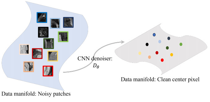
The two-dimensional (2D) cine cardiovascular magnetic resonance (CMR) technique is the reference standard for assessing cardiac function. However, one challenge with 2D cine is that the acquisition time for the whole cine stack is long and requires multiple breath holds, which may not be feasible for pediatric or ill patients. Though single breath-hold multi-slice cine may address the issue, it can only acquire low-resolution images, and hence, affect the accuracy of cardiac function assessment. To address these challenges, a Ferumoxytol-enhanced, free breathing, isotropic high-resolution 3D cine technique was developed. The method produces high-contrast cine images with short acquisition times by using compressed sensing together with a manifold-based method for image denoising. This study included fifteen patients (9.1 $ \pm $ 5.6 yrs.) who were referred for clinical cardiovascular magnetic resonance imaging (MRI) with Ferumoxytol contrast and were prescribed the 3D cine sequence. The data was acquired on a 1.5T scanner. Statistical analysis shows that the manifold-based denoised 3D cine can accurately measure ventricular function with no significant differences when compared to the conventional 2D breath-hold (BH) cine. The multiplanar reconstructed images of the proposed 3D cine method are visually comparable to the golden standard 2D BH cine method in terms of clarity, contrast, and anatomical precision. The proposed method eliminated the need for breath holds, reduced scan times, enabled multiplanar reconstruction within an isotropic data set, and has the potential to be used as an effective tool to access cardiovascular conditions.
Citation: Anna Andrews, Pezad Doctor, Lasya Gaur, F. Gerald Greil, Tarique Hussain, Qing Zou. Manifold-based denoising for Ferumoxytol-enhanced 3D cardiac cine MRI[J]. Mathematical Biosciences and Engineering, 2024, 21(3): 3695-3712. doi: 10.3934/mbe.2024163
The two-dimensional (2D) cine cardiovascular magnetic resonance (CMR) technique is the reference standard for assessing cardiac function. However, one challenge with 2D cine is that the acquisition time for the whole cine stack is long and requires multiple breath holds, which may not be feasible for pediatric or ill patients. Though single breath-hold multi-slice cine may address the issue, it can only acquire low-resolution images, and hence, affect the accuracy of cardiac function assessment. To address these challenges, a Ferumoxytol-enhanced, free breathing, isotropic high-resolution 3D cine technique was developed. The method produces high-contrast cine images with short acquisition times by using compressed sensing together with a manifold-based method for image denoising. This study included fifteen patients (9.1 $ \pm $ 5.6 yrs.) who were referred for clinical cardiovascular magnetic resonance imaging (MRI) with Ferumoxytol contrast and were prescribed the 3D cine sequence. The data was acquired on a 1.5T scanner. Statistical analysis shows that the manifold-based denoised 3D cine can accurately measure ventricular function with no significant differences when compared to the conventional 2D breath-hold (BH) cine. The multiplanar reconstructed images of the proposed 3D cine method are visually comparable to the golden standard 2D BH cine method in terms of clarity, contrast, and anatomical precision. The proposed method eliminated the need for breath holds, reduced scan times, enabled multiplanar reconstruction within an isotropic data set, and has the potential to be used as an effective tool to access cardiovascular conditions.

| [1] |
S. V. Raman, M. Markl, A. R. Patel, J. Bryant, B. D. Allen, S. Plein, et al., 30-minute cmr for common clinical indications: a society for cardiovascular magnetic resonance white paper, J. Cardiovasc. Magn. Reson., 24 (2022), 13. https://doi.org/10.1186/s12968-022-00844-6 doi: 10.1186/s12968-022-00844-6

|
| [2] | M. A. Syed, S. V. Raman, O. P. Simonetti, Basic principles of cardiovascular MRI: physics and imaging techniques, Springer, 2015. |
| [3] |
R. Menchón-Lara, F. Simmross-Wattenberg, P. Higuera, M. Martín-Fernández, C. Alberola-López, Reconstruction techniques for cardiac cine mri, Insights Imag., 10 (2019), 1–16. https://doi.org/10.1186/s13244-019-0754-2 doi: 10.1186/s13244-019-0754-2
|
| [4] |
C. M. Kramer, J. Barkhausen, C. Bucciarelli-Ducci, S. D. Flamm, R. J. Kim, E. Nagel, Standardized cardiovascular magnetic resonance imaging (cmr) protocols: 2020 update, J. Cardiovasc. Magn. Reson., 22 (2020), 1–18. https://doi.org/10.1186/s12968-020-00607-1 doi: 10.1186/s12968-020-00607-1
|
| [5] |
J. Liu, P. Spincemaille, N. C. F. Codella, T. D. Nguyen, M. R. Prince, Y. Wang, Respiratory and cardiac self-gated free-breathing cardiac cine imaging with multiecho 3d hybrid radial ssfp acquisition, Magn. Reson. Med., 63 (2010), 1230–1237. https://doi.org/10.1002/mrm.22306 doi: 10.1002/mrm.22306
|
| [6] |
Q. Lyu, H. Shan, Y. Xie, A. C. Kwan, Y. Otaki, K. Kuronuma, et al., Cine cardiac mri motion artifact reduction using a recurrent neural network, IEEE Trans. Med. Imaging, 40 (2021), 2170–2181. https://doi.org/10.1109/TMI.2021.3073381 doi: 10.1109/TMI.2021.3073381
|
| [7] |
Q. Zou, A. H. Ahmed, P. Nagpal, S. Kruger, M. Jacob, Dynamic imaging using a deep generative storm (gen-storm) model, IEEE Trans. Med. Imaging, 40 (2021), 3102–3112. https://doi.org/10.1109/TMI.2021.3065948 doi: 10.1109/TMI.2021.3065948
|
| [8] |
Q. Zou, A. H. Ahmed, S. Dzelebdzic, T. Hussain, Free-breathing and ungated cardiac mri reconstruction using a deep kernel representation, Appl. Sci., 13 (2023), 2281. https://doi.org/10.3390/app13042281 doi: 10.3390/app13042281
|
| [9] |
D. C. Peters, R. Nezafat, H. Eggers, C. Stehning, W. J. Manning, 2d free-breathing dual navigator-gated cardiac function validated against the 2d breath-hold acquisition, J. Magn. Reson. Imaging, 28 (2008), 773–777. https://doi.org/10.1002/jmri.21417 doi: 10.1002/jmri.21417
|
| [10] |
M. H. Moghari, A. Barthur, M. E. Amaral, T. Geva, A. J. Powell, Free-breathing whole-heart 3d cine magnetic resonance imaging with prospective respiratory motion compensation, Magn. Reson. Med., 80 (2018), 181–189. https://doi.org/10.1002/mrm.27021 doi: 10.1002/mrm.27021
|
| [11] |
M. Usman, B. Ruijsink, M. S. Nazir, G. Cruz, C. Prieto, Free breathing whole-heart 3d cine mri with self-gated cartesian trajectory, Magn. Reson. Med., 38 (2017), 129–137. https://doi.org/10.1016/j.mri.2016.12.021 doi: 10.1016/j.mri.2016.12.021
|
| [12] |
J. Liu, L. Feng, H. -W. Shen, C. Zhu, Y. Wang, K. Mukai, et al., Highly-accelerated self-gated free-breathing 3d cardiac cine mri: Validation in assessment of left ventricular function, Magn. Reson. Mater. Phys. Biol. Med., 30 (2017), 337–346. https://doi.org/10.1007/s10334-017-0607-2 doi: 10.1007/s10334-017-0607-2
|
| [13] |
T. Küstner, A. Bustin, O. Jaubert, R. Hajhosseiny, P. G. Masci, R. Neji, et al., Isotropic 3d cartesian single breath-hold cine mri with multi-bin patch-based low-rank reconstruction, Magn. Reson. Med., 84 (2020), 2018–2033. https://doi.org/10.1002/mrm.28267 doi: 10.1002/mrm.28267
|
| [14] |
H. Y. Carr, Steady-state free precession in nuclear magnetic resonance, Phys. Rev., 112 (1958), 1693. https://doi.org/10.1103/PhysRev.112.1693 doi: 10.1103/PhysRev.112.1693
|
| [15] |
J. Hamilton, D. Franson, N. Seiberlich, Recent advances in parallel imaging for mri, Prog. Nucl. Magn. Reson. Spectrosc., 101 (2017), 71–95. https://doi.org/10.1016/j.pnmrs.2017.04.002 doi: 10.1016/j.pnmrs.2017.04.002
|
| [16] |
M. H. Moghari, T. Geva, A. J. Powell, Prospective heart tracking for whole-heart magnetic resonance angiography, Magn. Reson. Med, 77 (2017), 759–765. https://doi.org/10.1002/mrm.26117 doi: 10.1002/mrm.26117
|
| [17] |
M. Lustig, D. L. Donoho, J. M. Santos, J. M. Pauly, Compressed sensing mri, IEEE Signal Process. Mag., 25 (2008), 72–82. https://doi.org/10.1109/MSP.2007.914728 doi: 10.1109/MSP.2007.914728
|
| [18] |
M. R. Bashir, L. Bhatti, D. Marin, R. C. Nelson, Emerging applications for ferumoxytol as a contrast agent in mri, J. Magn. Reson. Imaging, 41 (2015), 884–898. https://doi.org/10.1002/jmri.24691 doi: 10.1002/jmri.24691
|
| [19] |
Y. Zhang, H. Lin, Y. Li, H. Ma, A patch based denoising method using deep convolutional neural network for seismic image, IEEE Access, 7 (2019), 156883–156894. https://doi.org/10.1109/ACCESS.2019.2949774 doi: 10.1109/ACCESS.2019.2949774
|
| [20] |
J. He, L. Ding, L. Jiang, Z. Li, Q. Hu, Intrinsic dimensionality estimation based on manifold assumption, J. Vis. Commun. Image Represent., 25 (2014), 740–747. https://doi.org/10.1016/j.jvcir.2014.01.006 doi: 10.1016/j.jvcir.2014.01.006
|
| [21] | T. Kessler, G. Dorian, J. H. Mack, Application of a rectified linear unit (relu) based artificial neural network to cetane number predictions, in Internal Combustion Engine Division Fall Technical Conference, American Society of Mechanical Engineers, 58318 (2017), V001T02A006. https://doi.org/10.1115/ICEF2017-3614 |
| [22] |
T. K. Kim, T test as a parametric statistic, Korean J. Anesthesiol., 68 (2015), 540–546. https://doi.org/10.4097/kjae.2015.68.6.540 doi: 10.4097/kjae.2015.68.6.540
|
| [23] | G. B. Barrett, The coefficient of determination: Understanding r squared and r squared, Math. Teacher, 93 (2000), 230–234. |
| [24] |
D. Giavarina, Understanding bland altman analysis, Biochem. Med., 25 (2015), 141–151. https://doi.org/10.11613/BM.2015.015 doi: 10.11613/BM.2015.015
|


Figures(7) / Tables(3)

Anna Andrews, Pezad Doctor, Lasya Gaur, F. Gerald Greil, Tarique Hussain, Qing Zou. Manifold-based denoising for Ferumoxytol-enhanced 3D cardiac cine MRI[J]. Mathematical Biosciences and Engineering, 2024, 21(3): 3695-3712. doi: 10.3934/mbe.2024163


 DownLoad: