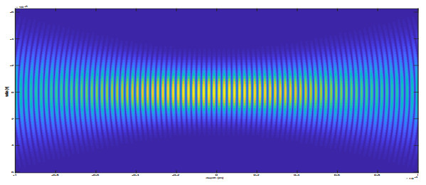
We review mathematical models describing how Optical Coherence Tomography works. Hereby, we focus on models based on Maxwell's equations and their simplifications. We highlight especially the effects of different modeling assumptions for the incident illumination, the medium, the light propagation, and the measurement setup and illustrate the qualitatively differing behavior in numerical simulations of the OCT data and compare them with real data from OCT measurements.
Citation: Peter Elbau, Leonidas Mindrinos, Leopold Veselka. Development of mathematical models for quantitative OCT: A review[J]. AIMS Mathematics, 2023, 8(2): 2508-2531. doi: 10.3934/math.2023130
We review mathematical models describing how Optical Coherence Tomography works. Hereby, we focus on models based on Maxwell's equations and their simplifications. We highlight especially the effects of different modeling assumptions for the incident illumination, the medium, the light propagation, and the measurement setup and illustrate the qualitatively differing behavior in numerical simulations of the OCT data and compare them with real data from OCT measurements.

| [1] |
S. G. Adie, T. R. Hillman, D. D. Sampson, Detection of multiple scattering in optical coherence tomography using the spatial distribution of stokes vectors, Opt. express, 15 (2007), 18033–18049. https://doi.org/10.1364/OE.15.018033 doi: 10.1364/OE.15.018033

|
| [2] |
P. E. Andersen, T. M. Jørgensen, L. Thrane, A. Tycho, H. T. Yura, Modeling light–tissue interaction in optical coherence tomography systems, Optical Coherence Tomography, (2008), 73–115. https://doi.org/10.1007/978-3-540-77550-8_3 doi: 10.1007/978-3-540-77550-8_3
|
| [3] |
P. E. Andersen, L. Thrane, H. T. Yura, A. Tycho, T. M. Jørgensen, M. H. Frosz, Advanced modelling of optical coherence tomography systems, Phys. Med. Biol., 49 (2004), 1307–1327. https://doi.org/10.1088/0031-9155/49/7/017 doi: 10.1088/0031-9155/49/7/017
|
| [4] | M. Born, E. Wolf, Principles of Optics, Cambridge University Press, Cambridge, 7 ed., 1999. |
| [5] |
T. Brenner, D. Reitzle, A. Kienle, Optical coherence tomography images simulated with an analytical solution of maxwell's equations for cylinder scattering, J. Biomed. Opt., 21 (2016), 045001. https://doi.org/10.1117/1.JBO.21.4.045001 doi: 10.1117/1.JBO.21.4.045001
|
| [6] |
O. Bruno, J. Chaubell, Inverse scattering problem for optical coherence tomography, Opt. Lett., 28 (2003), 2049–2051. https://doi.org/10.1364/OL.28.002049 doi: 10.1364/OL.28.002049
|
| [7] |
O. Bruno, J. Chaubell, One-dimensional inverse scattering problem for optical coherence tomography, Inverse Probl., 21 (2005), 499–524. https://doi.org/10.1088/0266-5611/21/2/006 doi: 10.1088/0266-5611/21/2/006
|
| [8] | D. Colton, R. Kress, Inverse Acoustic and Electromagnetic Scattering Theory, no. 93 in Applied Mathematical Sciences, Springer, Berlin, 3 ed., 2013. https://doi.org/10.1007/978-1-4614-4942-3 |
| [9] | P. Elbau, L. Mindrinos, O. Scherzer, Mathematical methods of optical coherence tomography, in Handbook of Mathematical Methods in Imaging, O. Scherzer, ed., Springer New York, 2015. https://doi.org/10.1007/978-1-4939-0790-8_44 |
| [10] |
P. Elbau, L. Mindrinos, O. Scherzer, Inverse problems of combined photoacoustic and optical coherence tomography, Math. Method. Appl. Sci., 40 (2017), 505–522. https://doi.org/10.1002/mma.3915 doi: 10.1002/mma.3915
|
| [11] |
P. Elbau, L. Mindrinos, O. Scherzer, The inverse scattering problem for orthotropic media in polarization-sensitive optical coherence tomography, GEM Int. J. Geomathema., 9 (2018), 145–165. https://doi.org/10.1007/s13137-017-0102-y doi: 10.1007/s13137-017-0102-y
|
| [12] | P. Elbau, L. Mindrinos, L. Veselka, Reconstructing the optical parameters of a layered medium with optical coherence elastography, in Mathematical and Numerical Approaches for Multi-Wave Inverse Problems, L. Beilina, M. Bergounioux, M. Christofol, A. Da Silva, and A. Litman, eds., no. 328 in Springer Proceedings in Mathematics & Statistics, Springer, 2020. https://doi.org/10.1007/978-3-030-48634-1_8 |
| [13] | P. Elbau, L. Mindrinos, L. Veselka, Quantitative oct reconstructions for dispersive media, in Time-dependent Problems in Imaging and Parameter Identification, B. Kaltenbacher, T. Schuster, and A. Wald, eds., Springer, Cham, 2021. https://doi.org/10.1007/978-3-030-57784-1_8 |
| [14] |
A. F. Fercher, Optical coherence tomography, J. Biomed. Opt., 1 (1996), 157–173. https://doi.org/10.1117/12.231361 doi: 10.1117/12.231361
|
| [15] |
A. F. Fercher, Optical coherence tomography - development, principles, applications, Z. Med. Phys., 20 (2010), 251–276. https://doi.org/10.1016/j.zemedi.2009.11.002 doi: 10.1016/j.zemedi.2009.11.002
|
| [16] |
A. F. Fercher, W. Drexler, C. K. Hitzenberger, T. Lasser, Optical coherence tomography - principles and applications, Rep. Prog. Phys., 66 (2003), 239–303. https://doi.org/10.1088/0034-4885/66/2/204 doi: 10.1088/0034-4885/66/2/204
|
| [17] | A. F. Fercher, C. K. Hitzenberger, Optical coherence tomography, in Progress in optics, vol. 44, Elsevier, 2002. https://doi.org/10.1016/S0079-6638(02)80017-8 |
| [18] |
A. F. Fercher, C. K. Hitzenberger, M. Sticker, R. Zawadzki, B. Karamata, T. Lasser, Numerical dispersion compensation for partial coherence interferometry and optical coherence tomography, Opt. express, 9 (2001), 610–615. https://doi.org/10.1364/OE.9.000610 doi: 10.1364/OE.9.000610
|
| [19] |
A. F. Fercher, C. K. Hitzenberger, M. Sticker, R. Zawadzki, B. Karamata, T. Lasser, Dispersion compensation for optical coherence tomography depth-scan signals by a numerical technique, Opt. Commun., 204 (2002), 67–74. https://doi.org/10.1016/S0030-4018(02)01137-9 doi: 10.1016/S0030-4018(02)01137-9
|
| [20] |
G. V. Gelikonov, L. S. Dolin, E. A. Sergeeva, I. V. Turchin, Multiple backscattering effects in optical coherence tomography images of layered turbid media, Radiophysics and quantum electronics, 46 (2003), 565–576. https://doi.org/10.1023/B:RAQE.0000019871.67609.3f doi: 10.1023/B:RAQE.0000019871.67609.3f
|
| [21] |
U. Haberland, V. Blazek, H. J. Schmitt, Chirp optical coherence tomography of layered scattering media, J. Biomed. Opt., 3 (1998), 259–266. https://doi.org/10.1117/1.429889 doi: 10.1117/1.429889
|
| [22] |
M. R. Hee, D. Huang, E. A. Swanson, J. G. Fujimoto, Polarization-sensitive low-coherence reflectometer for birefringence characterization and ranging, J. Opt. Soc. Am. B, 9 (1992), 903–908. https://doi.org/10.1364/JOSAB.9.000903 doi: 10.1364/JOSAB.9.000903
|
| [23] |
D. Huang, E. A. Swanson, C. P. Lin, J. S. Schuman, G. Stinson, W. Chang, et al., Optical coherence tomography, Science, 254 (1991), 1178–1181. https://doi.org/10.1126/science.1957169 doi: 10.1126/science.1957169
|
| [24] | J. A. Izatt, M. A. Choma, Theory of optical coherence tomography, in Optical coherence tomography, W. Drexler and J. G. Fujimoto, eds., Springer, 2008. https://doi.org/10.1007/978-3-540-77550-8_2 |
| [25] | J. D. Jackson, Classical Electrodynamics, Wiley, 3 ed., 1998. |
| [26] | J. Kalkman, Fourier-domain optical coherence tomography signal analysis and numerical modeling, International Journal of Optics, 2017. https://doi.org/10.1155/2017/9586067 |
| [27] |
B. Karamata, M. Laubscher, M. Leutenegger, S. Bourquin, T. Lasser, P. Lambelet, Multiple scattering in optical coherence tomography. i. investigation and modeling, JOSA A, 22 (2005), 1369–1379. https://doi.org/10.1364/JOSAA.22.001369 doi: 10.1364/JOSAA.22.001369
|
| [28] |
B. Karamata, M. Leutenegger, M. Laubscher, S. Bourquin, T. Lasser, P. Lambelet, Multiple scattering in optical coherence tomography. ii. experimental and theoretical investigation of cross talk in wide-field optical coherence tomography, JOSA A, 22 (2005), 1380–1388. https://doi.org/10.1364/JOSAA.22.001380 doi: 10.1364/JOSAA.22.001380
|
| [29] |
J. U. Kim, H. Choi, Y. Park, J. Shin, Finite-difference time-domain analysis of increased penetration depth in optical coherence tomography by wavefront shaping, Biomed. Opt. Express, 9 (2018), 3883–3897. https://doi.org/10.1364/BOE.9.003883 doi: 10.1364/BOE.9.003883
|
| [30] |
A. Knüttel, M. Boehlau-Godau, Spatially confined and temporally resolved refractive index and scattering evaluation in human skin performed with optical coherence tomography, J. Biomed. Opt., 5 (2000), 83–92. https://doi.org/10.1117/1.429972 doi: 10.1117/1.429972
|
| [31] |
R. F. Lutomirski, H. T. Yura, Propagation of a finite optical beam in an inhomogeneous medium, Appl. Optics, 10 (1971), 1652–1658. https://doi.org/10.1364/AO.10.001652 doi: 10.1364/AO.10.001652
|
| [32] | L. Mandel, E. Wolf, Optical coherence and quantum optics, Cambridge University Press, Cambridge, England, 1995. https://doi.org/10.1017/CBO9781139644105 |
| [33] |
D. L. Marks, B. J. Davis, S. A. Boppart, P. S. Carney, Partially coherent illumination in full-field interferometric synthetic aperture microscopy, JOSA A, 26 (2009), 376–386. https://doi.org/10.1364/JOSAA.26.000376 doi: 10.1364/JOSAA.26.000376
|
| [34] |
D. L. Marks, T. S. Ralston, S. A. Boppart, P. S. Carney, Inverse scattering for frequency-scanned full-field optical coherence tomography, JOSA A, 24 (2007), 1034–1041. https://doi.org/10.1364/JOSAA.24.001034 doi: 10.1364/JOSAA.24.001034
|
| [35] |
P. R. T. Munro, Three-dimensional full wave model of image formation in optical coherence tomography, Opt. express, 24 (2016), 27016–27031. https://doi.org/10.1364/OE.24.027016 doi: 10.1364/OE.24.027016
|
| [36] |
V. D. Nguyen, D. J. Faber, E. van der Pol, T. G. van Leeuwen, J. Kalkman, Dependent and multiple scattering in transmission and backscattering optical coherence tomography, Opt. Express, 21 (2013), 29145–29156. https://doi.org/10.1364/OE.21.029145 doi: 10.1364/OE.21.029145
|
| [37] |
P. Rajai, H. Schriemer, A. Amjadi, R. Munger, Simultaneous measurement of refractive index and thickness of multilayer systems using fourier domain optical coherence tomography, part 1: theory, J. Biomed. Opt., 22 (2017), 015002. https://doi.org/10.1117/1.JBO.22.1.015002 doi: 10.1117/1.JBO.22.1.015002
|
| [38] |
T. S. Ralston, D. L. Marks, P. S. Carney, S. A. Boppart, Inverse scattering for optical coherence tomography, Journal of the Optical Society of America A 23 (2006), 1027–1037. https://doi.org/10.1364/JOSAA.23.001027 doi: 10.1364/JOSAA.23.001027
|
| [39] | T. S. Ralston, D. L. Marks, P. S. Carney, S. A. Boppart, Phase stability technique for inverse scattering in optical coherence tomography, in 3rd IEEE International Symposium on Biomedical Imaging: Nano to Macro, 2006. |
| [40] |
T. S. Ralston, D. L. Marks, P. S. Carney, S. A. Boppart, Interferometric synthetic aperture microscopy, Nat. phys., 3 (2007), 129–134. https://doi.org/10.1038/nphys514 doi: 10.1038/nphys514
|
| [41] |
T. S. Ralston, S. G. Adie, D. L. Marks, S. A. Boppart, P. S. Carney, Cross-validation of interferometric synthetic aperture microscopy and optical coherence tomography, Opt. Lett., 35 (2010), 1683–1685. https://doi.org/10.1364/OL.35.001683 doi: 10.1364/OL.35.001683
|
| [42] | M. Santos, A. Araújo, S. Barbeiro, F. Caramelo, A. Correia, M. I. Marques, et al.,, Simulation of cellular changes on optical coherence tomography of human retina, in 2015 37th Annual International Conference of the IEEE Engineering in Medicine and Biology Society (EMBC), IEEE, 2015. https://doi.org/10.1109/EMBC.2015.7320285 |
| [43] |
J. M. Schmitt, Optical coherence tomography (OCT): A review, IEEE J. Sel. Top. Quant., 5 (1999), 1205–1215. https://doi.org/10.1109/2944.796348 doi: 10.1109/2944.796348
|
| [44] |
J. M. Schmitt, A. Knüttel, Model of optical coherence tomography of heterogeneous tissue, JOSA A, 14 (1997), 1231–1242. https://doi.org/10.1364/JOSAA.14.001231 doi: 10.1364/JOSAA.14.001231
|
| [45] |
J. M. Schmitt, A. Knüttel, R. F. Bonner, Measurement of optical properties of biological tissues by low-coherence reflectometry, Appl. Optics, 32 (1993), 6032–6042. https://doi.org/10.1364/AO.32.006032 doi: 10.1364/AO.32.006032
|
| [46] |
J. M. Schmitt, S. H. Xiang, K. M. Yung, Speckle in optical coherence tomography, J. Biomed. Opt., 4 (1999), 95–105. https://doi.org/10.1117/1.429925 doi: 10.1117/1.429925
|
| [47] |
C. S. Seelamantula, S. Mulleti, Super-resolution reconstruction in frequency-domain optical-coherence tomography using the finite-rate-of-innovation principle, IEEE T. Signal Proces., 62 (2014), 5020–5029. https://doi.org/10.1109/TSP.2014.2340811 doi: 10.1109/TSP.2014.2340811
|
| [48] | A. S. F. C. Silva, A. L. Correia, From optical coherence tomography to maxwell's equations, IEEE 3rd Portuguese Meeting in Bioengineering, 2013. |
| [49] | O. Svelto, Principles of Lasers, Springer Verlag, 5 ed., 2010. https://doi.org/10.1007/978-1-4419-1302-9 |
| [50] |
G. J. Tearney, M. E. Brezinski, J. F. Southern, B. E. Bouma, M. R. Hee, J. G. Fujimoto, Determination of the refractive index of highly scattering human tissue by optical coherence tomography, Opt. Lett., 20 (1995), 2258–2260. https://doi.org/10.1364/OL.20.002258 doi: 10.1364/OL.20.002258
|
| [51] |
L. Thrane, H. T. Yura, P. E. Andersen, Analysis of optical coherence tomography systems based on the extended huygens - fresnel principle, JOSA A, 17 (2000), 484–490. https://doi.org/10.1364/JOSAA.17.000484 doi: 10.1364/JOSAA.17.000484
|
| [52] |
P. H. Tomlins, R. K. Wang, Theory, developments and applications of optical coherence tomography, J. Phys. D Appl. Phys., 38 (2005), 2519. https://doi.org/10.1088/0022-3727/38/15/002 doi: 10.1088/0022-3727/38/15/002
|
| [53] |
P. H. Tomlins, R. K. Wang, Matrix approach to quantitative refractive index analysis by fourier domain optical coherence tomography, JOSA A, 23 (2006), 1897–1907. https://doi.org/10.1364/JOSAA.23.001897 doi: 10.1364/JOSAA.23.001897
|
| [54] |
U. Tricoli, R. Carminati, Modeling of full-field optical coherence tomography in scattering media, JOSA A, 36 (2019), C122–C129. https://doi.org/10.1364/JOSAA.36.00C122 doi: 10.1364/JOSAA.36.00C122
|
| [55] |
I. V. Turchin, E. A. Sergeeva, L. S. Dolin, N. M. Shakhova, R. R. Richards-Kortum, Novel algorithm of processing optical coherence tomography images for differentiation of biological tissue pathologies, J. Biomed. Opt., 10 (2005), 064024. https://doi.org/10.1117/1.2137670 doi: 10.1117/1.2137670
|
| [56] |
T. G. van Leeuwen, D. J. Faber, M. C. Aalders, Measurement of the axial point spread function in scattering media using single-mode fiber-based optical coherence tomography, IEEE J. Sel. Top. Quant., 9 (2003), 227–233. https://doi.org/10.1109/JSTQE.2003.813299 doi: 10.1109/JSTQE.2003.813299
|
| [57] |
L. Veselka, L. Krainz, L. Mindrinos, W. Drexler, P. Elbau, A quantitative model for optical coherence tomography, Sensors, 21 (2021), 6864. https://doi.org/10.3390/s21206864 doi: 10.3390/s21206864
|
| [58] |
M. J. Yadlowsky, J. M. Schmitt, R. F. Bonner, Multiple scattering in optical coherence microscopy, Appl. Optics, 34 (1995), 5699–5707. https://doi.org/10.1364/AO.34.005699 doi: 10.1364/AO.34.005699
|
| [59] |
J. Yi, J. Gong, X. Li, Analyzing absorption and scattering spectra of micro-scale structures with spectroscopic optical coherence tomography, Opt. Express, 17 (2009), 13157–13167. https://doi.org/10.1364/OE.17.013157 doi: 10.1364/OE.17.013157
|
| [60] |
K. M. Yung, S. L. Lee, J. M. Schmitt, Phase-domain processing of optical coherence tomography images, J. Biomed. Opt., 4 (1999), 125–136. https://doi.org/10.1117/1.429942 doi: 10.1117/1.429942
|
| [61] |
H. T. Yura, S. G. Hanson, Optical beam wave propagation through complex optical systems, JOSA A, 4 (1987), 1931–1948. https://doi.org/10.1364/JOSAA.4.001931 doi: 10.1364/JOSAA.4.001931
|


Figures(7) / Tables(2)

Peter Elbau, Leonidas Mindrinos, Leopold Veselka. Development of mathematical models for quantitative OCT: A review[J]. AIMS Mathematics, 2023, 8(2): 2508-2531. doi: 10.3934/math.2023130


 DownLoad: