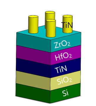
Ferroelectricity is demonstrated for the first time in Si(100)/SiO2/TiN/HfO2-ZrO2/TiN stack using pulsed laser deposition (PLD) and the effects of temperatures, partial oxygen pressures, and thickness for the stabilization of the ferroelectric phase were mapped. Thin films deposited at a higher temperature and a higher oxygen partial pressure have a higher thickness, demonstrating a better ferroelectric response with ~12 μC/cm2 remnant polarization, a leakage current of 10−7 A (at 8 V) and endurance > 1011 cycles indicative of an orthorhombic crystal phase. In contrast, thin films deposited at lower temperatures and pressures does not exhibit ferroelectric behavior. These films can be attributed to having a dominant monoclinic phase, having lower grain size and increased leakage current. Finally, the effects of ZrO2 as top and bottom layer were also investigated which showed that ZrO2 as the top layer provided better mechanical confinement for stabilizing the orthorhombic phase instead of as the bottom layer.
Citation: Sree Sourav Das, Zach Fox, Md Dalim Mia, Brian C Samuels, Rony Saha, Ravi Droopad. Demonstration of ferroelectricity in PLD grown HfO2-ZrO2 nanolaminates[J]. AIMS Materials Science, 2023, 10(2): 342-355. doi: 10.3934/matersci.2023018
Ferroelectricity is demonstrated for the first time in Si(100)/SiO2/TiN/HfO2-ZrO2/TiN stack using pulsed laser deposition (PLD) and the effects of temperatures, partial oxygen pressures, and thickness for the stabilization of the ferroelectric phase were mapped. Thin films deposited at a higher temperature and a higher oxygen partial pressure have a higher thickness, demonstrating a better ferroelectric response with ~12 μC/cm2 remnant polarization, a leakage current of 10−7 A (at 8 V) and endurance > 1011 cycles indicative of an orthorhombic crystal phase. In contrast, thin films deposited at lower temperatures and pressures does not exhibit ferroelectric behavior. These films can be attributed to having a dominant monoclinic phase, having lower grain size and increased leakage current. Finally, the effects of ZrO2 as top and bottom layer were also investigated which showed that ZrO2 as the top layer provided better mechanical confinement for stabilizing the orthorhombic phase instead of as the bottom layer.

| [1] |
Zhang Z, Wang Z, Shi T, et al. (2020) Memory materials and devices: From concept to application. InfoMat 2: 261–90. https://doi.org/10.1002/inf2.12077. doi: 10.1002/inf2.12077

|
| [2] |
Bouaziz J, Rojo Romeo P, Baboux N, et al. (2019) Characterization of ferroelectric hafnium/zirconium oxide solid solutions deposited by reactive magnetron sputtering. J Vac Sci Technol B 37: 021203. https://doi.org/10.1116/1.5060643. doi: 10.1116/1.5060643
|
| [3] |
Weeks SL, Pal A, Narasimhan VK, et al. (2017) Engineering of ferroelectric HfO2-ZrO2 nanolaminates. ACS Appl Mater Interfaces 9: 13440–13447. https://doi.org/10.1021/acsami.7b00776. doi: 10.1021/acsami.7b00776
|
| [4] |
Fan Z, Chen J, Wang J (2016) Ferroelectric HfO2-based materials for next-generation ferroelectric memories. J Adv Dielectr 6: 1630003. https://doi.org/10.1142/S2010135X16300036 doi: 10.1142/S2010135X16300036
|
| [5] |
Lu J, Luo W, Feng J, et al. (2018) Unusual ferroelectricity in two-dimensional perovskite oxide thin films. Nano Lett 18: 595–601. https://doi.org/10.1021/acs.nanolett.7b04797 doi: 10.1021/acs.nanolett.7b04797
|
| [6] |
Sai N, Kolpak AM, Rappe AM (2005) Ferroelectricity in ultrathin perovskite films. Phys Rev B 72: 20101. https://doi.org/10.1103/PhysRevB.72.020101 doi: 10.1103/PhysRevB.72.020101
|
| [7] |
Cohen RE (1992) Origin of ferroelectricity in perovskite oxides. Nature 358: 136–138. https://doi.org/10.1038/358136a0 doi: 10.1038/358136a0
|
| [8] |
Kim K, Lee S (2006) Integration of lead zirconium titanate thin films for high density ferroelectric random access memory. J Appl Phys 100: 51604. https://doi.org/10.1063/1.2337361 doi: 10.1063/1.2337361
|
| [9] |
Martin LW, Rappe AM (2016) Thin-film ferroelectric materials and their applications. Nat Rev Mater 2: 1–14. https://doi.org/10.1038/natrevmats.2016.87 doi: 10.1038/natrevmats.2016.87
|
| [10] |
De Araujo C-P, Cuchiaro JD, McMillan LD, et al. (1995) Fatigue-free ferroelectric capacitors with platinum electrodes. Nature 374: 627–629. https://doi.org/10.1038/374627a0 doi: 10.1038/374627a0
|
| [11] |
Park BH, Kang BS, Bu SD, et al. (1999) Lanthanum-substituted bismuth titanate for use in non-volatile memories. Nature 401: 682–684. https://doi.org/10.1038/44352 doi: 10.1038/44352
|
| [12] |
Ishiwara H (2012) Ferroelectric random access memories. J Nanosci Nanotechnol 12: 7619–7627. https://doi.org/10.1166/jnn.2012.6651 doi: 10.1166/jnn.2012.6651
|
| [13] |
Park MH, Lee YH, Mikolajick T, et al. (2018) Review and perspective on ferroelectric HfO2-based thin films for memory applications. MRS Commun 8: 795–808. https://doi.org/10.1557/mrc.2018.175. doi: 10.1557/mrc.2018.175
|
| [14] |
Jiang H, Gomez-Abal RI, Rinke P, et al. (2010) Electronic band structure of zirconia and hafnia polymorphs from the GW perspective. Phys Rev B 81: 85119. https://doi.org/10.1103/PhysRevB.81.085119. doi: 10.1103/PhysRevB.81.085119
|
| [15] |
Martin D, Yurchuk E, Müller S, et al. (2013) Downscaling ferroelectric field effect transistors by using ferroelectric Si-doped HfO2. Solid State Electron 88: 65–68. https://doi.org/10.1016/j.sse.2013.04.013 doi: 10.1016/j.sse.2013.04.013
|
| [16] |
Müller J, Polakowski P, Mueller S, et al. (2015) Ferroelectric hafnium oxide based materials and devices: Assessment of current status and future prospects. ECS J Solid State Sci Technol 4: N30–N35. https://doi.org/10.1149/2.0081505jss. doi: 10.1149/2.0081505jss
|
| [17] |
Sang X, Grimley ED, Schenk T, et al. (2015) On the structural origins of ferroelectricity in HfO2 thin films. Appl Phys Lett 106: 162905. https://doi.org/10.1063/1.4919135 doi: 10.1063/1.4919135
|
| [18] |
Shimizu T, Katayama K, Kiguchi T, et al. (2016) The demonstration of significant ferroelectricity in epitaxial Y-doped HfO2 film. Sci Rep 6: 32931. https://doi.org/10.1038/srep32931 doi: 10.1038/srep32931
|
| [19] |
Park MH, Schenk T, Fancher CM, et al. (2017) A comprehensive study on the structural evolution of HfO2 thin films doped with various dopants. J Mater Chem C 5: 4677–90. https://doi.org/10.1039/C7TC01200D. doi: 10.1039/C7TC01200D
|
| [20] |
Shandalov M, Mcintyre P (2009) Size-dependent polymorphism in HfO2 nanotubes and nanoscale thin films. J Appl Phys 106: 84322. https://doi.org/10.1063/1.3243077 doi: 10.1063/1.3243077
|
| [21] |
Muller J, Boscke TS, Schroder U, et al. (2012) Ferroelectricity in simple binary ZrO2 and HfO2. Nano Lett 12: 4318–23. https://doi.org/10.1021/nl302049k doi: 10.1021/nl302049k
|
| [22] |
Lomenzo PD, Zhao P, Takmeel Q, et al. (2014) Ferroelectric phenomena in Si-doped HfO2 thin films with TiN and Ir electrodes. J Vac Sci Technol B, Nanotechnol Microelectron Mater Process Meas Phenom 32: 03D123. https://doi.org/10.1116/1.4873323 doi: 10.1116/1.4873323
|
| [23] |
Chernikova AG, Kozodaev MG, Negrov DV, et al. (2018) Improved ferroelectric switching endurance of La-doped Hf0.5Zr0.5O2 thin films. ACS Appl Mater Interfaces 10: 2701–8. https://doi.org/10.1021/acsami.7b15110 doi: 10.1021/acsami.7b15110
|
| [24] |
Hoffmann M, Schroeder U, Schenk T, et al. (2015) Stabilizing the ferroelectric phase in doped hafnium oxide. J Appl Phys 118: 72006. https://doi.org/10.1063/1.4927805 doi: 10.1063/1.4927805
|
| [25] |
Lee YH, Kim HJ, Moon T, et al. (2017) Preparation and characterization of ferroelectric Hf0.5Zr0.5O2 thin films grown by reactive sputtering. Nanotechnology 28: 305703. https://doi.org/10.1088/1361-6528/aa7624 doi: 10.1088/1361-6528/aa7624
|
| [26] | Lyu J, Fina I, Bachelet R, et al. (2019) Enhanced ferroelectricity in epitaxial Hf0.5Zr0.5O2 thin films integrated with Si (001) using SrTiO3 templates. Appl Phys Lett 114: 222901. https://doi.org/10.1063/1.5096002 |
| [27] |
Lu YW, Shieh J, Tsai FY (2016) Induction of ferroelectricity in nanoscale ZrO2/HfO2 bilayer thin films on Pt/Ti/SiO2/Si substrates. Acta Mater 115: 68–75. https://doi.org/10.1016/j.actamat.2016.05.029 doi: 10.1016/j.actamat.2016.05.029
|
| [28] |
Park MH, Kim HJ, Lee G, et al. (2019) A comprehensive study on the mechanism of ferroelectric phase formation in hafnia-zirconia nanolaminates and superlattices. Appl Phys Rev 6: 041403. https://doi.org/10.1063/1.5118737. doi: 10.1063/1.5118737
|
| [29] |
Polakowski P, Müller J (2015) Ferroelectricity in undoped hafnium oxide. Appl Phys Lett 106: 232905. https://doi.org/10.1063/1.4922272 doi: 10.1063/1.4922272
|
| [30] |
Cho HW, Pujar P, Choi M, et al. (2021) Direct growth of orthorhombic Hf0.5Zr0.5O2 thin films for hysteresis-free MoS2 negative capacitance field-effect transistors. Npj 2D Mater Appl 5: 46. https://doi.org/10.1038/s41699-021-00229-w. doi: 10.1038/s41699-021-00229-w
|
| [31] |
Nukala P, Antoja-Lleonart J, Wei Y, et al. (2019) Direct epitaxial growth of polar (1-X)HfO2-(x)ZrO2 ultrathin films on silicon. ACS Appl Electron Mater 1: 2585–2593. https://doi.org/10.1021/acsaelm.9b00585. doi: 10.1021/acsaelm.9b00585
|
| [32] | Krebs H-U, Weisheit M, Faupel J, et al. (2003) Pulsed laser deposition (PLD)—A versatile thin film technique, In: Kramer B, Advances in Solid State Physics, Springer Berlin, Heidelberg, 505–518. https://doi.org/10.1007/978-3-540-44838-9_36. |
| [33] | Enkelmann V (1998) The oligomeric approach, In: Mullen K, Wegner G, 2 Eds., Electronic Materials, Wiley: Chichester, 295. |
| [34] |
Lyu J, Fina I, Solanas R, et al. (2019) Growth window of ferroelectric epitaxial Hf0.5Zr0.5O2 thin films. ACS Appl Electron Mater 1: 220–228. https://doi.org/10.1021/acsaelm.8b00065 doi: 10.1021/acsaelm.8b00065
|
| [35] | Gunst T, Stradi D, Blom A (2020) Identification of zirconia and hafnia crystalline phases by optical spectroscopy from first-principles, Nanoengineering: Fabrication, Properties, Optics, Thin Films, and Devices XVII, 11467: 1146708. https://doi.org/10.1117/12.2568807. |
| [36] |
Gao L, Yalon E, Chew AR, et al. (2017) Effect of oxygen vacancies and strain on the phonon spectrum of HfO2 thin films. J Appl Phys 121: 224101. https://doi.org/10.1063/1.4984833. doi: 10.1063/1.4984833
|
| [37] |
Tan T, Liu Z, Lu H, et al. (2010) Structure and optical properties of HfO2 thin films on silicon after rapid thermal annealing. Opt Mater (Amst) 32: 432–435. https://doi.org/10.1016/j.optmat.2009.10.003. doi: 10.1016/j.optmat.2009.10.003
|
| [38] |
Böscke TS, Müller J, Bräuhaus D, et al. (2011) Ferroelectricity in hafnium oxide thin films. Appl Phys Lett 99: 102903. https://doi.org/10.1063/1.3634052 doi: 10.1063/1.3634052
|
| [39] |
Park JH, Kim BK, Park JG, et al. (1999) Dielectric hysteresis measurement in lossy ferroelectrics. Ferroelectrics 230: 151–156. https://doi.org/10.1080/00150199908214911. doi: 10.1080/00150199908214911
|
| [40] |
Pandian MS, Ramasamy P, Kumar B (2012) A comparative study of ferroelectric triglycine sulfate (TGS) crystals grown by conventional slow evaporation and unidirectional method. Mater Res Bull 47: 1587–1597. https://doi.org/10.1016/j.materresbull.2012.01.030 doi: 10.1016/j.materresbull.2012.01.030
|
 matersci-10-02-018-s01.docx matersci-10-02-018-s01.docx |
 |


Figures(9)

Sree Sourav Das, Zach Fox, Md Dalim Mia, Brian C Samuels, Rony Saha, Ravi Droopad. Demonstration of ferroelectricity in PLD grown HfO2-ZrO2 nanolaminates[J]. AIMS Materials Science, 2023, 10(2): 342-355. doi: 10.3934/matersci.2023018


 DownLoad: