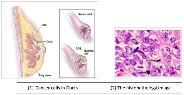
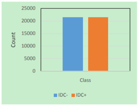
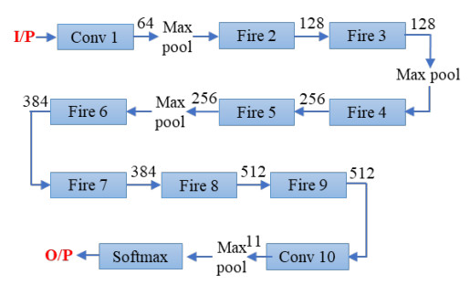
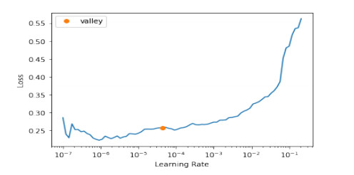
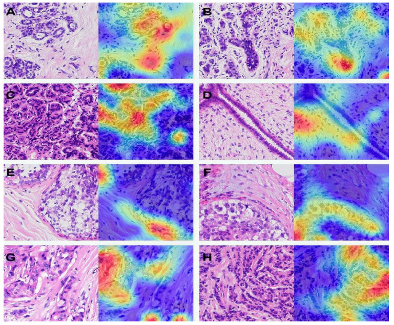
One of the most effective approaches for identifying breast cancer is histology, which is the meticulous inspection of tissues under a microscope. The kind of cancer cells, or whether they are cancerous (malignant) or non-cancerous, is typically determined by the type of tissue that is analyzed by the test performed by the technician (benign). The goal of this study was to automate IDC classification within breast cancer histology samples using a transfer learning technique. To improve our outcomes, we combined a Gradient Color Activation Mapping (Grad CAM) and image coloring mechanism with a discriminative fine-tuning methodology employing a one-cycle strategy using FastAI techniques. There have been lots of research studies related to deep transfer learning which use the same mechanism, but this report uses a transfer learning mechanism based on lightweight Squeeze Net architecture, a variant of CNN (Convolution neural network). This strategy demonstrates that fine-tuning on Squeeze Net makes it possible to achieve satisfactory results when transitioning generic features from natural images to medical images.
Citation: Sushovan Chaudhury, Kartik Sau, Muhammad Attique Khan, Mohammad Shabaz. Deep transfer learning for IDC breast cancer detection using fast AI technique and Sqeezenet architecture[J]. Mathematical Biosciences and Engineering, 2023, 20(6): 10404-10427. doi: 10.3934/mbe.2023457
One of the most effective approaches for identifying breast cancer is histology, which is the meticulous inspection of tissues under a microscope. The kind of cancer cells, or whether they are cancerous (malignant) or non-cancerous, is typically determined by the type of tissue that is analyzed by the test performed by the technician (benign). The goal of this study was to automate IDC classification within breast cancer histology samples using a transfer learning technique. To improve our outcomes, we combined a Gradient Color Activation Mapping (Grad CAM) and image coloring mechanism with a discriminative fine-tuning methodology employing a one-cycle strategy using FastAI techniques. There have been lots of research studies related to deep transfer learning which use the same mechanism, but this report uses a transfer learning mechanism based on lightweight Squeeze Net architecture, a variant of CNN (Convolution neural network). This strategy demonstrates that fine-tuning on Squeeze Net makes it possible to achieve satisfactory results when transitioning generic features from natural images to medical images.

| [1] |
A. Vulli, P. N. Srinivasu, M. S. K. Sashank, J. Shafi, J. Choi, M. F. Ijaz, Fine-Tuned DenseNet-169 for breast cancer metastasis prediction using FastAI and 1-cycle policy, Sensors, 22 (2022), 2988. https://doi.org/10.3390/s22082988 doi: 10.3390/s22082988

|
| [2] |
L. Benning, A. Peintner, L. Peintner, Advances in and the applicability of machine learning-based screening and early detection approaches for cancer: A primer, Cancers (Basel), 14 (2022), 1–15. https://doi.org/10.3390/cancers14030623 doi: 10.3390/cancers14030623
|
| [3] |
T. M. C. Pereira, R. C. Conceição, V. Sencadas, R. Sebastião, Biometric recognition: A systematic review on electrocardiogram data acquisition methods, Sensors, 23 (2023), 1507. https://doi.org/10.3390/s23031507 doi: 10.3390/s23031507
|
| [4] |
T. Thakur, I. Batra, M. Luthra, S. Vimal, G. Dhiman, A. Malik, et al., Gene Expression-Assisted Cancer Prediction Techniques, J. Healthcare Eng., 2021 (2021), 1–9. https://doi.org/10.1155/2021/4242646 doi: 10.1155/2021/4242646
|
| [5] | G. K. Saini, H. Chouhan, S. Kori, A. Gupta, M. Shabaz, V. Jagota, et al., Recognition of human sentiment from image using machine learning, Ann. RSCB, (2021), 1802–1808. https://www.annalsofrscb.ro/index.php/journal/article/view/4703 |
| [6] |
S. N. H. Bukhari, A. Jain, E. Haq, M. A. Khder, R. Neware, J. Bhola, Machine learning-based ensemble model for Zika virus T-cell epitope prediction, J. Healthcare Eng., 2021 (2021), 1–10. https://doi.org/10.1155/2021/9591670 doi: 10.1155/2021/9591670
|
| [7] |
S. Chaudhury, N. Shelke, K. Sau, B. Prasanalakshmi, M. Shabaz, A novel approach to classifying breast cancer histopathology biopsy images using bilateral knowledge distillation and label smoothing regularization, Comput. Math. Methods Med., 2021 (2021), 1–11. https://doi.org/10.1155/2021/4019358 doi: 10.1155/2021/4019358
|
| [8] |
G. Kaur, S. Bhushan, D. Singh, Fusion in multimodal biometric system: A review, Indian J. Sci. Technol., 10 (2017), 1–10. https://doi.org/10.17485/ijst/2017/v10i19/114382 doi: 10.17485/ijst/2017/v10i19/114382
|
| [9] | R. L. Siegel, K. D. Miller, H. E. Fuchs, A. Jemal, Cancer statistics, 2022, CA Cancer J. Clin., 72 (2022), 7–33. https://doi.org/10.3322/caac.21708 |
| [10] |
H. J. Hoffman, A. Khan, K. M. Ajmera, L. Zolfaghari, J. R. Schenfeld, P. H. Levine, Initial response to chemotherapy, not delay in diagnosis, predicts overall survival in inflammatory breast cancer cases, Am. J. Clin. Oncol., 37 (2014), 315–321. https://doi.org/10.1097/COC.0b013e318271b34b doi: 10.1097/COC.0b013e318271b34b
|
| [11] |
S. L. Bangare, G. Pradeepini, S. T. Patil, Regenerative pixel mode and tumour locus algorithm development for brain tumour analysis: A new computational technique for precise medical imaging, Int. J. Biomed. Eng. Technol., 27 (2018), 76. https://doi.org/10.1504/IJBET.2018.093087 doi: 10.1504/IJBET.2018.093087
|
| [12] |
R. Kumar, D. Shringi, K. N. Bairwa, Enhancing the tribological behavior of hybrid Al6061 metal matrix composites through the incorporation of nickel and chromium nanoparticles, Int. J. Adv. Eng. Manag. Sci., 6 (2020), 414–420. https://doi.org/10.22161/ijaems.69.1 doi: 10.22161/ijaems.69.1
|
| [13] |
R. F. Mustapa, R. Rifin, M. E. Mahadan, A. Zainuddin, Interactive water level control system simulator based on OMRON CX-Programmer and CX-Designer, Int. J. Emerg. Technol. Adv. Eng., 11 (2021), 91–99. https://doi.org/10.46338/ijetae0921_11 doi: 10.46338/ijetae0921_11
|
| [14] | A. Singh, Mathematical modeling language/tool with disciplinary as a solution strategy in the study of worldwide subjects, J. Positive Psychol. Wellbeing, 6 (2022), 1690–1698. https://www.journalppw.com/index.php/jppw/article/view/2630 |
| [15] | V. Jagota, V. Bhatia, L. Vives, A. B. Prasad, ML-PASD, Adv. Med. Diagn. Treatment Care. (2021), 82–93. https://doi.org/10.4018/978-1-7998-7460-7.ch006 |
| [16] |
Y. Yari, T. V. Nguyen, H. T. Nguyen, Deep learning applied for histological diagnosis of breast cancer, IEEE Access, 8 (2020), 162432–162448. https://doi.org/10.1109/access.2020.3021557 doi: 10.1109/access.2020.3021557
|
| [17] |
A. Mehbodniya, I. Alam, S. Pande, R. Neware, K. P. Rane, M. Shabaz, M. V. Madhavan, Financial fraud detection in healthcare using machine learning and deep learning techniques, Secur. Commun. Networks, 2021 (2021), 1–8. https://doi.org/10.1155/2021/9293877 doi: 10.1155/2021/9293877
|
| [18] |
G. S. Sajja, Machine learning based detection of depression and anxiety, Int. J. Computer Appl., 183 (2021), 20–23. https://doi.org/10.5120/ijca2021921856 doi: 10.5120/ijca2021921856
|
| [19] | R. Godasu, D. Zeng, K. Sutrave, Transfer learning in medical image classification: Challenges and opportunities, MWAIS 2020 Proceedings, (2020), 5–28. https://aisel.aisnet.org/mwais2020/18 |
| [20] |
A. Osareh, B. Shadgar, Machine learning techniques to diagnose breast cancer, 2010 5th Int. Symp. Heal. Informatics Bioinformatics, 2010 (2010), 114–120. https://doi.org/10.1109/HIBIT.2010.5478895 doi: 10.1109/HIBIT.2010.5478895
|
| [21] | N. Bayramoglu, J. Kannala, J. Heikkila, Deep learning for magnification independent breast cancer histopathology image classification, 23rd International Conference on Pattern Recognition (ICPR), (2016). https://doi.org/10.1109/icpr.2016.7900002 |
| [22] | C. Pearce, Convolutional Neural Networks and the Analysis of Cancer Imagery, (2017). http://cs231n.stanford.edu/reports/2017/pdfs/25.pdf |
| [23] | A. Pillai, A. Nizam, M. Joshee, A. Pinto, S. Chavan, Breast cancer detection in mammograms using deep learning, Adv. Intell. Syst. Comput., 1354 (2021), 121–127. https://doi.org/10.1007/978-981-16-2008-9_11 |
| [24] | Y. S. Vang, Z. Chen, X. Xie, Deep learning framework for multi-class breast cancer histology image classification, Lecture Notes Computer Sci., (2018), 914–922. https://doi.org/10.1007/978-3-319-93000-8_104 |
| [25] | E. Deniz, A. Şengür, Z. Kadiroğlu, Y. Guo, V. Bajaj, Ü. Budak, Transfer learning based histopathologic image classification for breast cancer detection, Health Inform. Sci. Syst., 6 (2018). https://doi.org/10.1007/s13755-018-0057-x |
| [26] |
S. Chaudhury, N. Shelke, K. Sau, B. Prasanalakshmi, M. Shabaz, A novel approach to classifying breast cancer histopathology biopsy images using bilateral knowledge distillation and label smoothing regularization, Comput. Math. Methods Med., 2021 (2021), 1–11. https://doi.org/10.1155/2021/4019358 doi: 10.1155/2021/4019358
|
| [27] |
S. Chaudhury, M. Rakhra, N. Memon, K. Sau, M. T. Ayana, Breast cancer calcifications: Identification using a novel segmentation approach, Comput. Math. Methods Med., 2021 (2021), 1–13. https://doi.org/10.1155/2021/9905808 doi: 10.1155/2021/9905808
|
| [28] |
S. Chaudhury, A. N. Krishna, S. Gupta, K. S. Sankaran, S. Khan, K. Sau, A. Raghuvanshi, et al., Effective image processing and segmentation-based machine learning techniques for diagnosis of breast cancer, Comput. Math. Methods Med., 2022 (2022), 1–6. https://doi.org/10.1155/2022/6841334 doi: 10.1155/2022/6841334
|
| [29] | A. Patil, D. Tamboli, S. Meena, D. Anand, A. Sethi, Breast cancer histopathology image classification and localization using multiple instance learning, 2019 IEEE International WIE Conference on Electrical and Computer Engineering (WIECON-ECE), (2019), 1–4. https://doi.org/10.1109/WIECON-ECE48653.2019.9019916 |
| [30] |
F. A. Spanhol, L. S. Oliveira, C. Petitjean, L. Heutte, A dataset for breast cancer histopathological image classification, IEEE Transact. Biomed. Eng., 63 (2016), 1455–1462. https://doi.org/10.1109/TBME.2015.2496264 doi: 10.1109/TBME.2015.2496264
|
| [31] | F. A. Spanhol, L. S. Oliveira, C. Petitjean, L. Heutte, Breast cancer histopathological image classification using Convolutional Neural Networks, 2016 International Joint Conference on Neural Networks (IJCNN), (2016), 2560–2567. https://doi.org/10.1109/IJCNN.2016.7727519 |
| [32] |
S.-C. B. Lo, S.-L. A. Lou, Jyh-Shyan Lin, M. T. Freedman, M. V. Chien, S. K. Mun, Artificial convolution neural network techniques and applications for lung nodule detection, IEEE Transact. Med. Imag., 14 (1995), 711–718. https://doi.org/10.1109/42.476112 doi: 10.1109/42.476112
|
| [33] |
B. E. Bejnordi, M. Mullooly, R. M. Pfeiffer, S. Q. Fan, P. M. Vacek, D. L. Weaver, et al., Using deep convolutional neural networks to identify and classify tumor-associated stroma in diagnostic breast biopsies, Modern Pathol., 31 (2018), 1502–1512. https://doi.org/10.1038/s41379-018-0073-z doi: 10.1038/s41379-018-0073-z
|
| [34] |
J. Howard, S. Gugger, Fastai: A layered API for deep learning, Information, 11 (2020), 108. https://doi.org/10.3390/info11020108 doi: 10.3390/info11020108
|
| [35] |
A. Cruz-Roa, A. Basavanhally, F. González, H. Gilmore, M. Feldman, S. Ganesan, et al., Automatic detection of invasive ductal carcinoma in whole slide images with convolutional neural networks, SPIE Proceed., 9041 (2014), 904103. https://doi.org/10.1117/12.2043872 doi: 10.1117/12.2043872
|
| [36] | M. Romano, A. A. Hernandez, Enhanced deep learning approach for predicting invasive ductal carcinoma from histopathology images, 2019 2nd International Conference on Artificial Intelligence and Big Data (ICAIBD), (2019), 142–148. https://doi.org/10.1109/ICAIBD.2019.8837044 |
| [37] |
S. J. Pan, Q. Yang, A survey on transfer learning, IEEE Transact. Knowled. Data Eng., 22 (2010), 1345–1359. https://doi.org/10.1109/TKDE.2009.191 doi: 10.1109/TKDE.2009.191
|
| [38] |
A. M. Dawud, K. Yurtkan, H. Oztoprak, Application of deep learning in neuroradiology: Brain haemorrhage classification using transfer learning, Comput. Intell. Neurosci., 2019 (2019), 1–12. https://doi.org/10.1155/2019/4629859 doi: 10.1155/2019/4629859
|
| [39] | F. N. Iandola, S. Han, M. W. Moskewicz, K. Ashraf, W. J. Dally, K. Keutzer, SqueezeNet: AlexNet-level accuracy with 50x fewer parameters and < 0.5MB model size, (2016), 1–13. http://arXiv.org/abs/1602.07360. |
| [40] |
E. H. Houssein, M. Dirar, L. Abualigah, W. M. Mohamed, An efficient equilibrium optimizer with support vector regression for stock market prediction, Neural Comput. Appl., 34 (2021), 3165–3200. https://doi.org/10.1007/s00521-021-06580-9 doi: 10.1007/s00521-021-06580-9
|
| [41] |
A. Krizhevsky, I. Sutskever, G. E. Hinton, ImageNet classification with deep convolutional neural networks, Commun. ACM, 60 (2017), 84–90. https://doi.org/10.1145/3065386 doi: 10.1145/3065386
|
| [42] |
R. Ranjbarzadeh, S. Dorosti, S. J. Ghoushchi, A. Caputo, E. B. Tirkolaee, S. S. Ali, et al., Breast tumor localization and segmentation using machine learning techniques: Overview of datasets, findings, and methods, Comput. Biol. Med., 152 (2023), 106443. https://doi.org/10.1016/j.compbiomed.2022.106443 doi: 10.1016/j.compbiomed.2022.106443
|
| [43] | R. Ranjbarzadeh, N. T. Sarshar, S. J. Ghoushchi, M. S. Esfahani, M. Parhizkar, Y. Pourasad, et al., MRFE-CNN: Multi-route feature extraction model for breast tumor segmentation in Mammograms using a convolutional neural network, Ann. Operat. Res., (2022). https://doi.org/10.1007/s10479-022-04755-8 |
| [44] |
R. Ranjbarzadeh, A. B. Kasgari, S. J. Ghoushchi, S. Anari, M. Naseri, M. Bendechache, Brain tumor segmentation based on deep learning and an attention mechanism using MRI multi-modalities brain images, Sci. Rep., 11 (2021), 10930. https://doi.org/10.1038/s41598-021-90428-8 doi: 10.1038/s41598-021-90428-8
|
| [45] | R. Ranjbarzadeh, S. J. Ghoushchi, S. Anari, S. Safavi, N. T. Sarshar, E. B. Tirkolaee, et al., A deep learning approach for robust, multi-oriented, and curved text detection, Cognit. Comput., (2022). https://doi.org/10.1007/s12559-022-10072-w |
| [46] |
R. Ranjbarzadeh, A. Caputo, E. B. Tirkolaee, S. J. Ghoushchi, M. Bendechache, Brain tumor segmentation of MRI images: A comprehensive review on the application of artificial intelligence tools, Comput. Biol. Med., 152 (2023), 106405. https://doi.org/10.1016/j.compbiomed.2022.106405 doi: 10.1016/j.compbiomed.2022.106405
|
| [47] |
R. Ranjbarzadeh, S. Dorosti, S. J. Ghoushchi, S. Safavi, N. Razmjooy, N. T. Sarshar, et al., Nerve optic segmentation in CT images using a deep learning model and a texture descriptor, Complex Intell. Syst., 8 (2022), 3543–3557. https://doi.org/10.1007/s40747-022-00694-w doi: 10.1007/s40747-022-00694-w
|
| [48] |
A. Aghamohammadi, R. Ranjbarzadeh, F. Naiemi, M. Mogharrebi, S. Dorosti, M. Bendechache, TPCNN: Two-path convolutional neural network for tumor and liver segmentation in CT images using a novel encoding approach, Expert Syst. Appl., 183 (2021), 115406. https://doi.org/10.1016/j.eswa.2021.115406 doi: 10.1016/j.eswa.2021.115406
|
| [49] |
S. Anari, N. T. Sarshar, N. Mahjoori, S. Dorosti, A. Rezaie, Review of deep learning approaches for thyroid cancer diagnosis, Math. Problems Eng., 2022 (2022), 1–8. https://doi.org/10.1155/2022/5052435 doi: 10.1155/2022/5052435
|
| [50] |
S. Chaudhury, K. Sau, A BERT encoding with recurrent neural network and long-short term memory for breast cancer image classification, Decision Anal. J., 6 (2023), 100177. https://doi.org/10.1016/j.dajour.2023.100177 doi: 10.1016/j.dajour.2023.100177
|
| [51] |
D. Singh, J. P. Choudhury, M. De, A comparative study of meta heuristic model to assess the type of breast cancer disease, IETE J. Res., 68 (2022), 3683–3694. https://doi.org/10.1080/03772063.2020.1775139 doi: 10.1080/03772063.2020.1775139
|
Figures(12) / Tables(4)

Sushovan Chaudhury, Kartik Sau, Muhammad Attique Khan, Mohammad Shabaz. Deep transfer learning for IDC breast cancer detection using fast AI technique and Sqeezenet architecture[J]. Mathematical Biosciences and Engineering, 2023, 20(6): 10404-10427. doi: 10.3934/mbe.2023457


 DownLoad: