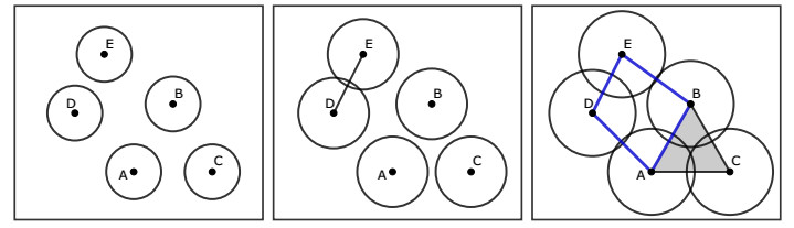
Filament-motor interactions inside cells play essential roles in many developmental as well as other biological processes. For instance, actin-myosin interactions drive the emergence or closure of ring channel structures during wound healing or dorsal closure. These dynamic protein interactions and the resulting protein organization lead to rich time-series data generated by using fluorescence imaging experiments or by simulating realistic stochastic models. We propose methods based on topological data analysis to track topological features through time in cell biology data consisting of point clouds or binary images. The framework proposed here is based on computing the persistent homology of the data at each time point and on connecting topological features through time using established distance metrics between topological summaries. The methods retain aspects of monomer identity when analyzing significant features in filamentous structure data, and capture the overall closure dynamics when assessing the organization of multiple ring structures through time. Using applications of these techniques to experimental data, we show that the proposed methods can describe features of the emergent dynamics and quantitatively distinguish between control and perturbation experiments.
Citation: Madeleine Dawson, Carson Dudley, Sasamon Omoma, Hwai-Ray Tung, Maria-Veronica Ciocanel. Characterizing emerging features in cell dynamics using topological data analysis methods[J]. Mathematical Biosciences and Engineering, 2023, 20(2): 3023-3046. doi: 10.3934/mbe.2023143
Filament-motor interactions inside cells play essential roles in many developmental as well as other biological processes. For instance, actin-myosin interactions drive the emergence or closure of ring channel structures during wound healing or dorsal closure. These dynamic protein interactions and the resulting protein organization lead to rich time-series data generated by using fluorescence imaging experiments or by simulating realistic stochastic models. We propose methods based on topological data analysis to track topological features through time in cell biology data consisting of point clouds or binary images. The framework proposed here is based on computing the persistent homology of the data at each time point and on connecting topological features through time using established distance metrics between topological summaries. The methods retain aspects of monomer identity when analyzing significant features in filamentous structure data, and capture the overall closure dynamics when assessing the organization of multiple ring structures through time. Using applications of these techniques to experimental data, we show that the proposed methods can describe features of the emergent dynamics and quantitatively distinguish between control and perturbation experiments.

| [1] |
H. Edelsbrunner, J. Harer, Persistent homology-a survey, Contemp. Math., 453 (2008), 257–282. https://doi.org/10.1090/conm/453/08802 doi: 10.1090/conm/453/08802

|
| [2] |
L. Wasserman, Topological data analysis, Annu. Rev. Stat. Appl., 5 (2018), 501–532. https://doi.org/10.1146/annurev-statistics-031017-100045 doi: 10.1146/annurev-statistics-031017-100045
|
| [3] |
G. Carlsson, Topological methods for data modelling, Nat. Rev. Phys., 2 (2020), 697–708. https://doi.org/10.1038/s42254-020-00249-3 doi: 10.1038/s42254-020-00249-3
|
| [4] |
E. J. Amézquita, M. Y. Quigley, T. Ophelders, E. Munch, D. H. Chitwood, The shape of things to come: Topological data analysis and biology, from molecules to organisms, Dev. Dyn., 249 (2020), 816–833. https://doi.org/10.1002/dvdy.175 doi: 10.1002/dvdy.175
|
| [5] |
A. Bukkuri, N. Andor, I. K. Darcy, Applications of topological data analysis in oncology, Front. Artif. Intell., 4 (2021), 38. https://doi.org/10.3389/frai.2021.659037 doi: 10.3389/frai.2021.659037
|
| [6] |
Y. Skaf, R. Laubenbacher, Topological data analysis in biomedicine: A review, J. Biomed. Inform., 130 (2022), 104082. https://doi.org/10.1016/j.jbi.2022.104082 doi: 10.1016/j.jbi.2022.104082
|
| [7] |
K. Garside, R. Henderson, I. Makarenko, C. Masoller, Topological data analysis of high resolution diabetic retinopathy images, PLOS ONE, 14 (2019), e0217413. https://doi.org/10.1371/journal.pone.0217413 doi: 10.1371/journal.pone.0217413
|
| [8] |
C. Ellis, M. Lesnick, G. Henselman-Petrusek, B. Keller, J. Cohen, Feasibility of topological data analysis for event-related fMRI, Network Neurosci., 3 (2019), 695–706. https://doi.org/10.1162/netn_a_00095 doi: 10.1162/netn_a_00095
|
| [9] |
M. McGuirl, A. Volkening, B. Sandstede, Topological data analysis of zebrafish patterns, PNAS, 117 (2020), 5113–5124. https://doi.org/10.1073/pnas.1917763117 doi: 10.1073/pnas.1917763117
|
| [10] | V. Maroulas, C. P. Micucci, F. Nasrin, Bayesian topological learning for classifying the structure of biological networks, preprint, arXiv: 2009.11974. |
| [11] |
D. Cohen-Steiner, H. Edelsbrunner, J. Harer, Stability of persistence diagrams, Discrete Comput. Geom., 37 (2007), 103–120. https://doi.org/10.1007/s00454-006-1276-5 doi: 10.1007/s00454-006-1276-5
|
| [12] | M. J. Jimenez, M. Rucco, P. Vicente-Munuera, P. Gómez-Gálvez, L. M. Escudero, Topological data analysis for self-organization of biological tissues, in International Workshop on Combinatorial Image Analysis, Springer, 2017,229–242. |
| [13] |
L. L. Bonilla, A. Carpio, C. Trenado, Tracking collective cell motion by topological data analysis, PLOS Comput. Biol., 16 (2020), e1008407. https://doi.org/10.1371/journal.pcbi.1008407 doi: 10.1371/journal.pcbi.1008407
|
| [14] | B. Lin, Topological data analysis in time series: Temporal filtration and application to single-cell genomics, preprint, arXiv: 2204.14048. |
| [15] | D. Cohen-Steiner, H. Edelsbrunner, D. Morozov, Vines and vineyards by updating persistence in linear time, in Proceedings of the twenty-second annual symposium on Computational geometry, ACM, (2006), 119–126. |
| [16] | A. Hickok, D. Needell, M. A. Porter, Analysis of spatiotemporal anomalies using persistent homology: case studies with COVID-19 data, preprint, arXiv: 2107.09188. |
| [17] |
C. M. Topaz, L. Ziegelmeier, T. Halverson, Topological data analysis of biological aggregation models, PloS ONE, 10 (2015), e0126383. https://doi.org/10.1371/journal.pone.0126383 doi: 10.1371/journal.pone.0126383
|
| [18] |
M. Ulmer, L. Ziegelmeier, C. M. Topaz, A topological approach to selecting models of biological experiments, PloS ONE, 14 (2019), e0213679. https://doi.org/10.1371/journal.pone.0213679 doi: 10.1371/journal.pone.0213679
|
| [19] |
M. V. Ciocanel, R. Juenemann, A. T. Dawes, S. A. McKinley, Topological data analysis approaches to uncovering the timing of ring structure onset in filamentous networks, Bull. Math. Biol., 83 (2021), 1–25. https://doi.org/10.1007/s11538-020-00847-3 doi: 10.1007/s11538-020-00847-3
|
| [20] |
K. Popov, J. Komianos, G. A. Papoian, MEDYAN: mechanochemical simulations of contraction and polarity alignment in actomyosin networks, PLoS Comput. Biol., 12 (2016), e1004877. https://doi.org/10.1371/journal.pcbi.1004877 doi: 10.1371/journal.pcbi.1004877
|
| [21] |
C. A. Mandato, W. M. Bement, Contraction and polymerization cooperate to assemble and close actomyosin rings around Xenopus oocyte wounds, J. Cell Biol., 154 (2001), 785–798. https://doi.org/10.1083/jcb.200103105 doi: 10.1083/jcb.200103105
|
| [22] |
R. D. Mortensen, R. P. Moore, S. M. Fogerson, H. Y. Chiou, C. V. Obinero, N. K. Prabhu, et al., Identifying genetic players in cell sheet morphogenesis using a Drosophila deficiency screen for genes on chromosome 2R involved in dorsal closure, G3 Genes Genomes Genetics, 8 (2018), 2361–2387. https://doi.org/10.1534/g3.118.200233 doi: 10.1534/g3.118.200233
|
| [23] |
M. V. Ciocanel, A. Chandrasekaran, C. Mager, Q. Ni, G. A. Papoian, A. Dawes, Simulated actin reorganization mediated by motor proteins, PLoS Comput. Biol., 18 (2022), e1010026. https://doi.org/10.1371/journal.pcbi.1010026 doi: 10.1371/journal.pcbi.1010026
|
| [24] |
H. A. Benink, W. M. Bement, Concentric zones of active Rhoa and Cdc42 around single cell wounds, J. Cell Biol., 168 (2005), 429–439. https://doi.org/10.1083/jcb.200411109 doi: 10.1083/jcb.200411109
|
| [25] |
R. D. Mortensen, R. P. Moore, S. M. Fogerson, H. Y. Chiou, C. V. Obinero, N. K. Prabhu, et al., Supplemental material for Mortensen et al., 2018, GSA J., 2018. https://doi.org/10.25387/g3.6207470.v2 doi: 10.25387/g3.6207470.v2
|
| [26] |
D. Legland, I. Arganda-Carreras, P. Andrey, MorphoLibJ: integrated library and plugins for mathematical morphology with ImageJ, Bioinformatics, 32 (2016), 3532–3534, https://doi.org/10.1093/bioinformatics/btw413 doi: 10.1093/bioinformatics/btw413
|
| [27] | A. Clark, Pillow (pil fork) documentation, 2015, Available from: https://pillow.readthedocs.io/en/stable/. |
| [28] |
C. Tralie, N. Saul, R. Bar-On, Ripser.py: A lean persistent homology library for Python, J. Open Source Software, 3 (2018), 925, https://doi.org/10.21105/joss.00925 doi: 10.21105/joss.00925
|
| [29] |
U. Bauer, Ripser: efficient computation of Vietoris-Rips persistence barcodes, J. Appl. Comput. Topol., 5 (2021), 391–423, https://doi.org/10.1007/s41468-021-00071-5 doi: 10.1007/s41468-021-00071-5
|
| [30] |
J. T. Nardini, B. J. Stolz, K. B. Flores, H. A. Harrington, H. M. Byrne, Topological data analysis distinguishes parameter regimes in the Anderson-Chaplain model of angiogenesis, PLOS Comput. Biol., 17 (2021), e1009094. https://doi.org/10.1371/journal.pcbi.1009094 doi: 10.1371/journal.pcbi.1009094
|
| [31] | J. J. Berwald, J. M. Gottlieb, E. Munch, Computing Wasserstein distance for persistence diagrams on a quantum computer, preprint, arXiv: 1809.06433. |
| [32] |
R. Ghrist, Barcodes: the persistent topology of data, Bull. Am. Math. Soc., 45 (2008), 61–75. https://doi.org/10.1090/S0273-0979-07-01191-3 doi: 10.1090/S0273-0979-07-01191-3
|
| [33] | GitHub, Code for "Topological data analysis distinguishes parameter regimes in the Anderson-Chaplain model of angiogenesis", Available from: https://github.com/johnnardini/Angio_TDA. |
| [34] |
D. Cohen-Steiner, H. Edelsbrunner, J. Harer, Y. Mileyko, Lipschitz functions have $l_{p}$-stable persistence, Found. Comput. Math., 10 (2010), 127–139. https://doi.org/10.1007/s10208-010-9060-6 doi: 10.1007/s10208-010-9060-6
|
| [35] | C. Tralie, Persim Package in Python, 2021. Available from: https://persim.scikit-tda.org/en/latest/reference/index.html. |
| [36] | GitHub, Sample code for image analysis and construction of significant topological paths corresponding to the time evolution of 1-dimensional holes (actin-myosin ring channels) in point cloud or binary image datasets, 2022. Available from: https://github.com/veronica-ciocanel/TDA_actomyosin/. |
| [37] |
B. Stolz, H. Harrington, M. Porter, Persistent homology of time-dependent functional networks constructed from coupled time series, Chaos, 27 (2017), 047410. https://doi.org/10.1063/1.4978997 doi: 10.1063/1.4978997
|
| [38] |
M. Feng, M. A. Porter, Persistent homology of geospatial data: A case study with voting, SIAM Rev., 63 (2021), 67–99. https://doi.org/10.1137/19M1241519 doi: 10.1137/19M1241519
|
| [39] |
B. T. Fasy, F. Lecci, A. Rinaldo, L. Wasserman, S. Balakrishnan, A. Singh, Confidence sets for persistence diagrams, Ann. Stat., 42 (2014), 2301–2339. https://doi.org/10.1214/14-AOS1252 doi: 10.1214/14-AOS1252
|
| [40] | F. Chazal, B. T. Fasy, F. Lecci, A. Rinaldo, A. Singh, L. Wasserman, On the bootstrap for persistence diagrams and landscapes, preprint, arXiv: 1311.0376. |
| [41] |
F. Chazal, B. Fasy, F. Lecci, B. Michel, A. Rinaldo, A. Rinaldo, et al., Robust topological inference: Distance to a measure and kernel distance, J. Mach. Learn. Res., 18 (2017), 5845–5884. https://doi.org/10.48550/arXiv.1412.7197 doi: 10.48550/arXiv.1412.7197
|
| [42] |
O. Bobrowski, M. Kahle, P. Skraba, Maximally persistent cycles in random geometric complexes, Ann. Appl. Probab., 27 (2017), 2032–2060. https://doi.org/10.1214/16-AAP1232 doi: 10.1214/16-AAP1232
|
| [43] |
O. Bobrowski, M. Kahle, Topology of random geometric complexes: a survey, J. Appl. Comput. Topol., 1 (2018), 331–364. https://doi.org/10.1007/s41468-017-0010-0 doi: 10.1007/s41468-017-0010-0
|
| [44] |
N. Chenavier, C. Hirsch, Extremal lifetimes of persistent cycles, Extremes, 25 (2022), 299–330. https://doi.org/10.1007/s10687-021-00430-6 doi: 10.1007/s10687-021-00430-6
|
| [45] |
C. Schwayer, M. Sikora, J. Slováková, R. Kardos, C. P. Heisenberg, Actin rings of power, Dev. Cell, 37 (2016), 493–506. https://doi.org/10.1016/j.devcel.2016.05.024 doi: 10.1016/j.devcel.2016.05.024
|
| [46] |
R. P. Moore, S. M. Fogerson, U. S. Tulu, J. W. Yu, A. H. Cox, M. A. Sican, et al., Super-resolution microscopy reveals actomyosin dynamics in medioapical arrays, Mol. Biol. Cell., 11 (2022), ar94. https://doi.org/10.1091/mbc.E21-11-0537 doi: 10.1091/mbc.E21-11-0537
|
| [47] |
Z. Zhang, Y. Nishimura, P. Kanchanawong, Extracting microtubule networks from superresolution single-molecule localization microscopy data, Mol. Biol. Cell., 28 (2017), 333–345. https://doi.org/10.1091/mbc.E16-06-0421 doi: 10.1091/mbc.E16-06-0421
|
| [48] |
D. A. Flormann, M. Schu, E. Terriac, D. Thalla, L. Kainka, M. Koch, et al., A novel universal algorithm for filament network tracing and cytoskeleton analysis, FASEB J., 35 (2021), e21582. https://doi.org/10.1096/fj.202100048R doi: 10.1096/fj.202100048R
|
| [49] |
D. Haertter, X. Wang, S. M. Fogerson, N. Ramkumar, J. M. Crawford, K. D. Poss, et al., DeepProjection: Rapid and structure-specific projections of tissue sheets embedded in 3D microscopy stacks using deep learning, bioRxiv, 2021. https://doi.org/10.1101/2021.11.17.468809 doi: 10.1101/2021.11.17.468809
|


Figures(8)

Madeleine Dawson, Carson Dudley, Sasamon Omoma, Hwai-Ray Tung, Maria-Veronica Ciocanel. Characterizing emerging features in cell dynamics using topological data analysis methods[J]. Mathematical Biosciences and Engineering, 2023, 20(2): 3023-3046. doi: 10.3934/mbe.2023143


 DownLoad: