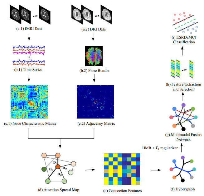
The structure and function of brain networks (BN) may be altered in patients with end-stage renal disease (ESRD). However, there are relatively few attentions on ESRD associated with mild cognitive impairment (ESRDaMCI). Most studies focus on the pairwise relationships between brain regions, without taking into account the complementary information of functional connectivity (FC) and structural connectivity (SC). To address the problem, a hypergraph representation method is proposed to construct a multimodal BN for ESRDaMCI. First, the activity of nodes is determined by connection features extracted from functional magnetic resonance imaging (fMRI) (i.e., FC), and the presence of edges is determined by physical connections of nerve fibers extracted from diffusion kurtosis imaging (DKI) (i.e., SC). Then, the connection features are generated through bilinear pooling and transformed into an optimization model. Next, a hypergraph is constructed according to the generated node representation and connection features, and the node degree and edge degree of the hypergraph are calculated to obtain the hypergraph manifold regularization (HMR) term. The HMR and L1 norm regularization terms are introduced into the optimization model to achieve the final hypergraph representation of multimodal BN (HRMBN). Experimental results show that the classification performance of HRMBN is significantly better than that of several state-of-the-art multimodal BN construction methods. Its best classification accuracy is 91.0891%, at least 4.3452% higher than that of other methods, verifying the effectiveness of our method. The HRMBN not only achieves better results in ESRDaMCI classification, but also identifies the discriminative brain regions of ESRDaMCI, which provides a reference for the auxiliary diagnosis of ESRD.
Citation: Zhengtao Xi, Tongqiang Liu, Haifeng Shi, Zhuqing Jiao. Hypergraph representation of multimodal brain networks for patients with end-stage renal disease associated with mild cognitive impairment[J]. Mathematical Biosciences and Engineering, 2023, 20(2): 1882-1902. doi: 10.3934/mbe.2023086
The structure and function of brain networks (BN) may be altered in patients with end-stage renal disease (ESRD). However, there are relatively few attentions on ESRD associated with mild cognitive impairment (ESRDaMCI). Most studies focus on the pairwise relationships between brain regions, without taking into account the complementary information of functional connectivity (FC) and structural connectivity (SC). To address the problem, a hypergraph representation method is proposed to construct a multimodal BN for ESRDaMCI. First, the activity of nodes is determined by connection features extracted from functional magnetic resonance imaging (fMRI) (i.e., FC), and the presence of edges is determined by physical connections of nerve fibers extracted from diffusion kurtosis imaging (DKI) (i.e., SC). Then, the connection features are generated through bilinear pooling and transformed into an optimization model. Next, a hypergraph is constructed according to the generated node representation and connection features, and the node degree and edge degree of the hypergraph are calculated to obtain the hypergraph manifold regularization (HMR) term. The HMR and L1 norm regularization terms are introduced into the optimization model to achieve the final hypergraph representation of multimodal BN (HRMBN). Experimental results show that the classification performance of HRMBN is significantly better than that of several state-of-the-art multimodal BN construction methods. Its best classification accuracy is 91.0891%, at least 4.3452% higher than that of other methods, verifying the effectiveness of our method. The HRMBN not only achieves better results in ESRDaMCI classification, but also identifies the discriminative brain regions of ESRDaMCI, which provides a reference for the auxiliary diagnosis of ESRD.

| [1] |
S. M. Li, X. F. Ma, R. W. Huang, M. Li, J. Z. Tian, H. Wen, et al., Abnormal degree centrality in neurologically asymptomatic patients with end-stage renal disease: A resting-state fMRI study, Clin. Neurophysiol., 127 (2016), 602–609. https://doi.org/10.1016/j.clinph.2015.06.022 doi: 10.1016/j.clinph.2015.06.022

|
| [2] |
X. F. Ma, G. H. Jiang, S. M. Li, J. H. Wang, W. F. Zhan, S. Q. Zeng, et al., Aberrant functional connectome in neurologically asymptomatic patients with end-stage renal disease, PloS One, 10 (2015), e0121085. https://doi.org/10.1371/journal.pone.0121085 doi: 10.1371/journal.pone.0121085
|
| [3] |
E. O'Lone, M. Connors, P. Masson, S. Wu, P. J. Kelly, D. Gillespie, et al., Cognition in people with end-stage kidney disease treated with hemodialysis: A systematic review and Meta-analysis, Am. J. Kidney Dis., 67 (2016), 925–935. https://doi.org/10.1053/j.ajkd.2015.12.028 doi: 10.1053/j.ajkd.2015.12.028
|
| [4] |
K. Karunaratne, D. Taube, N. Khalil, R. Perry, P. A. Malhotra, Neurological complications of renal dialysis and transplantation, Pract. Neurol., 18 (2018), 115–125. https://doi.org/10.1136/practneurol-2017-001657 doi: 10.1136/practneurol-2017-001657
|
| [5] |
S. H. Wang, Y. Zhang, Y. J. Li, W. J. Jia, F. Y. Liu, M. M. Yang, et al., Single slice based detection for Alzheimer's disease via wavelet entropy and multilayer perceptron trained by biogeography-based optimization, Multimed. Tools Appl., 77 (2018), 10393–10417. https://doi.org/10.1007/s11042-016-4222-4 doi: 10.1007/s11042-016-4222-4
|
| [6] |
S. H. Wang, Y. D. Zhang, G. Liu, P. Preetha, T. F. Yuan, Detection of Alzheimer's disease by three-dimensional displacement field estimation in structural magnetic resonance imaging, Int. J. Alzheimers Dis., 50 (2016), 233–248. https://doi.org/10.3233/JAD-150848 doi: 10.3233/JAD-150848
|
| [7] |
T. K. Chacko, H. Zhuang, K. Z. Nakhoda, B. Moussavian, A. Alavi, Applications of fluorodeoxyglucose positron emission tomography in the diagnosis of infection, Nucl. Med. Commun., 24 (2003), 615–624. https://doi.org/10.1097/00006231-200306000-00002 doi: 10.1097/00006231-200306000-00002
|
| [8] |
X. L. Jiang, J. Q. Wen, L. J. Zhang, G. Zheng, X. Li, Z. Zhang, et al., Cerebral blood flow changes in hemodialysis and peritoneal dialysis patients: An arterial-spin labeling MR imaging, Metab. Brain Dis., 31 (2016), 929–936. https://doi.org/10.1007/s11011-016-9829-7 doi: 10.1007/s11011-016-9829-7
|
| [9] |
L. Li, J. Y. Liu, F. X. Liang, H. D. Chen, R. G. Zhan, S. L. Zhao, et al., Altered brain function activity in patients with dysphagia after cerebral infarction: A resting-state functional magnetic resonance imaging study, Front. Neurol., 13 (2022), 782732. https://doi.org/10.3389/fneur.2022.782732 doi: 10.3389/fneur.2022.782732
|
| [10] |
R. Marta, A. Lukasz, M. Marek, Clinical application of diffusion tensor imaging and fiber tractography in the management of brainstem cavernous malformations: A systematic review, Neurosurg. Rev., 45 (2022), 2027–2040. https://doi.org/10.1007/s10143-022-01759-7 doi: 10.1007/s10143-022-01759-7
|
| [11] |
J. X. Wang, S. C. Wu, Y. Sun, J. M. Lu, J. L. Zhang, Y. Fang, et al., Brain microstructural alterations in the left precuneus mediate the association between KIBRA polymorphism and working memory in healthy adults: A diffusion kurtosis imaging study, Brain Imaging Behav., 2022 (2022), 1–10. https://doi.org/10.1007/s11682-022-00703-z doi: 10.1007/s11682-022-00703-z
|
| [12] |
J. W. Dong, X. F. Ma, W. H. Lin, M. C. Liu, S. S. Fu, L. H. Yang, et al., Aberrant cortical thickness in neurologically asymptomatic patients with end-stage renal disease, Neuropsychiatr. Dis. Treat., 14 (2018), 1929–1939. https://doi.org/10.2147/NDT.S170106 doi: 10.2147/NDT.S170106
|
| [13] |
F. F. Udo, W. Dominik, S. Armin, F. Andreas, Altered whole-brain white matter networks in preclinical Alzheimer's disease, Neuroimage Clin., 8 (2015), 660–666. https://doi.org/10.1016/j.nicl.2015.06.007 doi: 10.1016/j.nicl.2015.06.007
|
| [14] |
Y. An, X. F. Ma, T. M. Lu, D. Zhang, Application of magnetic resonance imaging molecular probe in the treatment of cerebral infarction and paralysis of hind limbs with neural stem cells derived from pluripotent stem cells, World Neurosurg., 138 (2020), 608–618. https://doi.org/10.1016/j.wneu.2020.01.036 doi: 10.1016/j.wneu.2020.01.036
|
| [15] |
W. B. Li, X. Wang, X. E. Wei, M. L. Wang, Susceptibility-weighted and diffusion kurtosis imaging to evaluate encephalomalacia with epilepsy after traumatic brain injury, Ann. Clin. Transl. Neur., 5 (2018), 552–558. https://doi.org/10.1002/acn3.552 doi: 10.1002/acn3.552
|
| [16] |
E. L. Pogosbekian, I. N. Pronin, N. E. Zakharova, A. I. Batalov, A. M. Turkin, T. A. Konakova, et al., Feasibility of generalised diffusion kurtosis imaging approach for brain glioma grading, Neuroradiology, 63 (2021), 1241–1251. https://doi.org/10.1007/s00234-020-02613-7 doi: 10.1007/s00234-020-02613-7
|
| [17] |
X. A. Bi, X. Hu, H. Wu, Y. Wang, Multimodal data analysis of Alzheimer's disease based on clustering evolutionary random forest, IEEE J. Biomed. Health Inf., 24 (2020), 2973–2983. https://doi.org/10.1109/JBHI.2020.2973324 doi: 10.1109/JBHI.2020.2973324
|
| [18] |
Z. Q. Zhang, W. Liao, H. F. Chen, D. Mantini, J. R. Ding, Q. Xu, et al., Altered functional–structural coupling of large-scale brain networks in idiopathic generalized epilepsy, Brain, 134 (2011), 2912–2928. https://doi.org/10.1093/brain/awr223 doi: 10.1093/brain/awr223
|
| [19] |
M. E. Lynall, D. S. Bassett, R. Kerwin, P. J. McKenna, M. Kitzbichler, U. Muller, et al., Functional connectivity and brain networks in schizophrenia, J. Neurosci., 30 (2010), 9477–9487. https://doi.org/10.1523/JNEUROSCI.0333-10.2010 doi: 10.1523/JNEUROSCI.0333-10.2010
|
| [20] |
Y. D. Zhang, S. H. Wang, Y. X. Sui, M. Yang, B. Liu, H. Cheng, et al., Multivariate approach for Alzheimer's disease detection using stationary Wavelet entropy and predator-prey particle swarm optimization, J. Alzheimer's Dis., 65 (2018), 855–869. https://doi.org/10.3233/JAD-170069 doi: 10.3233/JAD-170069
|
| [21] |
C. Peng, T. Y. Luo, H. Yang, Immediate abnormal intrinsic brain activity patterns in patients with end-stage renal disease during a single dialysis session: A resting-state functional MR imaging study, Chin. Imaging Soc. Integr. Med., 2 (2019), 17025. https://doi.org/10.26914/c.cnkihy.2019.017025 doi: 10.26914/c.cnkihy.2019.017025
|
| [22] | M. Wang, J. Huang, M. Liu, D. Zhang, Functional connectivity network analysis with discriminative hub detection for brain disease identification, in Proceedings of the AAAI Conference on Artificial Intelligence, 33 (2019), 1198–1205. https://doi.org/10.1609/aaai.v33i01.33011198 |
| [23] |
C. Y. Wee, P. T. Yap, K. Denny, J. N. Browndyke, G. G. Potter, K. A. Welsh-Bohmer, et al., Resting-state multi-spectrum functional connectivity networks for identification of MCI patients, PloS One, 7 (2012), e37828. https://doi.org/10.1371/journal.pone.0037828 doi: 10.1371/journal.pone.0037828
|
| [24] |
J. Yu, Y. Rui, Y. Y. Tang, High-order distance-based multi-view stochastic learning in image classification, IEEE Trans. Cybern., 44 (2014), 2431–2442. https://doi.org/10.1109/TCYB.2014.2307862 doi: 10.1109/TCYB.2014.2307862
|
| [25] |
Y. X. Ji, Y. T. Zhang, H. F. Shi, Z. Q. Jiao, S. H. Wang, C. Wang, Constructing dynamic brain functional networks via hyper-graph manifold regularization for mild cognitive impairment classification, Front. Neurosci, 15 (2021), 669345. https://doi.org/10.3389/fnins.2021.669345 doi: 10.3389/fnins.2021.669345
|
| [26] | R. I. Kondor, J. Lafferty, Diffusion kernels on graphs and other discrete structures, in Proceedings of the 19th International Conference on Machine Learning, (2002), 315–322. |
| [27] |
J. B. Pereira, D. Van Westen, E. Stomrud, T. O. Strandberg, G. Volpe, E. Westman, et al., Abnormal structural brain connectome in individuals with preclinical Alzheimer's disease, Cereb. Cortex, 28 (2017), 3638–3649. https://doi.org/10.1093/cercor/bhx236 doi: 10.1093/cercor/bhx236
|
| [28] |
C. Y. Xu, C. C. Chen, Q. W. Guo, Y. W. Lin, X. Y. Meng, G. Z. Qiu, et al., Comparative study of MOCA-B and MES in the recognition of amnestic mild cognitive impairment, J. Alzheimer's Dis., 4 (2021), 33–36. https://doi.org/10.3969/j.issn.2096-5516.2021.01.005 doi: 10.3969/j.issn.2096-5516.2021.01.005
|
| [29] |
Z. X. Cui, S. Y. Zhong, P. F. Xu, Y. He, G. L. Gong, PANDA: A pipeline toolbox for analyzing brain diffusion images, Front. Hum. Neurosci., 7 (2013), 42. https://doi.org/10.3389/fnhum.2013.00042 doi: 10.3389/fnhum.2013.00042
|
| [30] |
R. Mikail, S. Olaf, Complex network measures of brain connectivity: Uses and interpretations, Neuroimage, 52 (2010), 1059–1069. https://doi.org/10.1016/j.neuroimage.2009.10.003 doi: 10.1016/j.neuroimage.2009.10.003
|
| [31] |
K. Li, L. J. Liu, Q. Yin, W. H. Dun, X. L. Xu, J. X. Liu, et a1., Abnormal rich club organization and impaired correlation between structural and functional connectivity in migraine sufferers, Brain Imaging Behav., 11 (2017), 526–540. https://doi.org/10.1007/s11682-016-9533-6 doi: 10.1007/s11682-016-9533-6
|
| [32] |
J. S. Huang, L. P. Zhou, L. Wang, D. Q. Zhang, Attention-diffusion-bilinear neural network for brain network analysis, IEEE Trans. Med. Imaging, 39 (2020), 2541–2552. https://doi.org/10.1109/TMI.2020.2973650 doi: 10.1109/TMI.2020.2973650
|
| [33] | T. Lin, A. RoyChowdhury, S. Maji, Bilinear CNN models for finegrained visual recognition, in Proceedings of the IEEE International Conference on Computer Vision, (2015), 1449–1457. https://doi.org/10.48550/arXiv.1504.07889 |
| [34] |
W. Shao, Y. Peng, C. Zu, M. L. Wang, D. Q. Zhang, Hypergraph based multi-task feature selection for multimodal classification of Alzheimer's disease, Comput. Med. Imaging Graphics, 80 (2019), 101663. https://doi.org/10.1016/j.compmedimag.2019.101663 doi: 10.1016/j.compmedimag.2019.101663
|
| [35] | S. Huang, J. Li, L. Sun, J. Liu, T. Wu, K. Chen, Learning brain connectivity of Alzheimer's disease from neuroimaging data, in Advances in Neural Information Processing Systems 22 (NIPS 2009), (2009), 808–816. |
| [36] |
X. M. Liu, J. S. Tang, Mass classification in mammograms using selected geometry and texture features and a new SVM-based feature selection method, IEEE Syst. J., 8 (2014), 910–920. https://doi.org/10.1109/JSYST.2013.2286539 doi: 10.1109/JSYST.2013.2286539
|
| [37] |
Y. Li, J. Liu, X. Gao, B. Jie, M. Kim, P. T. Yap, et al., Multimodal hyper-connectivity of functional networks using functionally-weighted LASSO for MCI classification, Med. Image Anal., 52 (2018), 80–96. https://doi.org/10.1016/j.media.2018.11.006 doi: 10.1016/j.media.2018.11.006
|
| [38] |
W. K. Li, X. W. Xu, W. Jiang, P. J. Wang, X. Gao, Functional connectivity network estimation with an inter-similarity prior for mild cognitive impairment classification, Aging, 12 (2020), 17328–17342. https://doi.org/10.18632/aging.103719 doi: 10.18632/aging.103719
|
| [39] |
M. Dyrba, M. Grothe, T. Kirste, S. J. Teipel, Multimodal analysis of functional and structural disconnection in Alzheimer's disease using multiple kernel SVM, Hum. Brain Mapp., 36 (2015), 2118–2131. https://doi.org/10.1002/hbm.22759 doi: 10.1002/hbm.22759
|
| [40] |
H. P. Lu, N. P. Konstantinos, A. N. Venetsanopoulos, MPCA: Multilinear principal component analysis of tensor objects, IEEE Trans. Neural Networks, 19 (2008), 18–39. https://doi.org/10.1109/TNN.2007.901277 doi: 10.1109/TNN.2007.901277
|
| [41] |
C. Chen, K. Batselier, W. J. Yu, N. Wong, Kernelized support tensor train machines, Pattern Recognit., 122 (2022), 108337. https://doi.org/10.1016/j.patcog.2021.108337 doi: 10.1016/j.patcog.2021.108337
|
| [42] |
S. H. Chu, K. K. Parhi, C. Lenglet, Function-specific and enhanced brain structural connectivity mapping via joint modeling of diffusion and functional MRI, Sci. Rep., 8 (2018), 4741. https://doi.org/10.1038/s41598-018-23051-9 doi: 10.1038/s41598-018-23051-9
|
| [43] | X. Zhang, L. F. He, K. Chen, Y. Luo, J. Y. Zhou, F. Wang, Multiview graph convolutional network and its applications on neuroimage analysis for Parkinson's disease, in AMIA Annual Symposium Proceedings, (2018), 1147. |
| [44] | J. Atwood, D. Towsley, Diffusion-convolutional neural networks, in Advances in Neural Information Processing Systems 29 (NIPS 2016), (2016), 1993–2001. |
| [45] |
B. L. Wu, Z. Yue, X. K. Li, L. Li, M. Zhang, J. P. Ren, et al., Changes of brain functional network and its correlation with cognitive function in patients with end-stage renal disease, Chin. J. Neuromed., 2 (2020), 181–187. https://doi.org/10.3760/cma.j.issn.1671-8925.2020.02.012 doi: 10.3760/cma.j.issn.1671-8925.2020.02.012
|
| [46] |
Y. D. Zhang, S. H. Wang, P. Preetha, J. Q. Yang, T. F. Yuan, Three-dimensional eigenbrain for the detection of subjects and brain regions related with Alzheimer's disease, J. Alzheimer's Dis., 50 (2016), 1163–1179. https://doi.org/10.3233/JAD-150988 doi: 10.3233/JAD-150988
|
| [47] |
S. H. Wang, Y. D. Zhang, G. Liu, P. Preetha, T. F. Yuan, Detection of Alzheimer's disease by three-dimensional displacement field estimation in structural magnetic resonance imaging, J. Alzheimer's Dis., 50 (2016), 233–248. https://doi.org/10.3233/JAD-150848 doi: 10.3233/JAD-150848
|
| [48] |
Y. D. Zhang, S. H. Wang, P. Preetha, Z. C. Dong, G. L. Ji, J. Q. Yang, Detection of Alzheimer's disease and mild cognitive impairment based on structural volumetric MR images using 3D-DWT and WTA-KSVM trained by PSOTVAC, Biomed. Signal Process., 21 (2015), 58–73. https://doi.org/10.1016/j.bspc.2015.05.014 doi: 10.1016/j.bspc.2015.05.014
|
| [49] |
S. H. Wang, S. D. Du, Y. Zhang, P. Preetha, L. N. Wu, X. Q. Chen, et al., Alzheimer's disease detection by Pseudo Zernike moment and linear regression classification, CNS Neurol. Disord. Drug Targets, 16 (2017), 11–15. https://doi.org/10.2174/1871527315666161111123024 doi: 10.2174/1871527315666161111123024
|
| [50] |
Y. D. Zhang, Z. C. Dong, P. Preetha, S. H. Wang, G. L. Ji, J. Q. Yang, et al., Detection of subjects and brain regions related to Alzheimer's disease using 3D MRI scans based on eigen brain and machine learning, Front. Comput. Neurosc., 9 (2015), 66. https://doi.org/10.3389/fncom.2015.00066 doi: 10.3389/fncom.2015.00066
|
| [51] |
C. Y. Wee, P. T. Yap, W. Li, K. Denny, J. N. Browndyke, G. G. Potter, et al, Enriched white matter connectivity networks for accurate identification of MCI patients, NeuroImage, 54 (2011), 1812–1822. https://doi.org/10.1016/j.neuroimage.2010.10.026 doi: 10.1016/j.neuroimage.2010.10.026
|
| [52] |
D. Q. Zhang, Y. P. Wang, L. P. Zhou, H. Yuan, D. G. Shen, Multimodal classification of Alzheimer's disease and mild cognitive impairment, Neuroimage, 55 (2011), 856–867. https://doi.org/10.1016/j.neuroimage.2011.01.008 doi: 10.1016/j.neuroimage.2011.01.008
|
| [53] |
B. Mišić, R. F. Betzel, M. A. De Reus, M. P. Van Den Heuvel, M. G. Berman, A. R. McIntosh, et al., Network-level structure-function relationships in human neocortex, Cereb. Cortex, 26 (2016), 3285–3296. https://doi.org/10.1093/cercor/bhw089 doi: 10.1093/cercor/bhw089
|
| [54] |
J. Goñi, M. P. Van Den Heuvel, A. Avena-Koenigsberger, N. Velez de Mendizabal, R. F. Betzel, A. Griffa, et al., Resting-brain functional connectivity predicted by analytic measures of network communication, Proc. Natl. Acad. Sci., 111 (2014), 833–838. https://doi.org/10.1073/pnas.1315529111 doi: 10.1073/pnas.1315529111
|
| [55] |
X. Hua, J. G. Han, C. Zhao, H. P. Tang, Z. He, Q. H. Chen, et al., A novel method for ECG signal classification via one-dimensional convolutional neural network, Multimedia Syst., 28 (2022), 1387–1399. https://doi.org/10.1007/s00530-020-00713-1 doi: 10.1007/s00530-020-00713-1
|


Figures(5) / Tables(2)

Zhengtao Xi, Tongqiang Liu, Haifeng Shi, Zhuqing Jiao. Hypergraph representation of multimodal brain networks for patients with end-stage renal disease associated with mild cognitive impairment[J]. Mathematical Biosciences and Engineering, 2023, 20(2): 1882-1902. doi: 10.3934/mbe.2023086


 DownLoad: