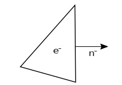
This study developed a method to approximate the covariance matrix associated with the simulation of water molecular diffusion inside the brain tissue. The computation implements the Discontinuous Galerkin method of the diffusion equation. A physically consistent numerical flux is applied to model the interaction between the axon walls and extracellular regions. This numerical flux yields an efficient GPU-CUDA implementation. We consider the two-dimensional case of high axon pack density, valid, for instance, in the brain's corpus callosum region.
Citation: Daniel Cervantes, Miguel angel Moreles, Joaquin Peña, Alonso Ramirez-Manzanares. A computational method for the covariance matrix associated with extracellular diffusivity on disordered models of cylindrical brain axons[J]. Mathematical Biosciences and Engineering, 2021, 18(5): 4961-4970. doi: 10.3934/mbe.2021252
This study developed a method to approximate the covariance matrix associated with the simulation of water molecular diffusion inside the brain tissue. The computation implements the Discontinuous Galerkin method of the diffusion equation. A physically consistent numerical flux is applied to model the interaction between the axon walls and extracellular regions. This numerical flux yields an efficient GPU-CUDA implementation. We consider the two-dimensional case of high axon pack density, valid, for instance, in the brain's corpus callosum region.

| [1] | D. Jones, Diffusion MRI theory methods and applications, Oxford University Press, (2011). |
| [2] |
M. G. Hall, D., C. Alexander, Convergence and parameter choice for monte-carlo simulations of diffusion MRI, IEEE Trans. Med Imaging, 28 (2009), 1354-1364. doi: 10.1109/TMI.2009.2015756

|
| [3] |
L. M. Burcaw, E. Fieremans, D. S. Novikov, Mesoscopic structure of neuronal tracts from time-dependent diffusion, NeuroImage, 114 (2015), 18-37. doi: 10.1016/j.neuroimage.2015.03.061
|
| [4] |
E. Fieremans, D. S. Novikov, J. H. Jensen, J. Helpern, Monte Carlo study of a two-compartment exchange model of diffusion, NMR Biomed., 23 (2010), 711-724. doi: 10.1002/nbm.1577
|
| [5] | F. Aboitiz, A. B. Scheibel, R. S. Fisher, E. Zaidel, Fiber composition of the human corpus callosum, Brain Res., 143 (1992), 143-153. |
| [6] |
E. Panagiotaki, T. Schneider, B. Siow, M. G. Hall, M. F. Lythgoe, D. C. Alexander, Compartment models of the diffusion mr signal in brain white matter: A taxonomy and comparison, NeuroImage, 59 (2012), 2241-2254. doi: 10.1016/j.neuroimage.2011.09.081
|
| [7] | W. S. Price, Pulsed-field gradient nuclear magnetic resonance as a tool for studying translational diffusion: Part 1, Basic theory, Concepts Magn. Reson., (1997), 299-336. |
| [8] | C. Pierpaoli, P. Jezzard, P. J. Basser, A. Barnett, G. Di Chiro, Diffusion tensor MR imaging of the human brain, Radiology, (1996), 637-648. |
| [9] | B. Cockburn, G. E. Karniadakis, C. W. Shu, Discontinuous Galerkin methods: Theory, computation and applications, volume 11, Springer Science & Business Media, (2012). |
| [10] | A. Szafer, J. Zhong, J. C. Gore, Theoretical model for water diffusion in tissues, Magn. Reson. Med., (1995), 697-712. |
| [11] |
T. B. Dyrby, L. V. Sogaard, M. G. Hall, M. Ptito, D. C. Alexander, Contrast and stability of the axon diameter index from microstructure imaging with diffusion MRI, Magn. Reson. Med., 70 (2013), 711-721. doi: 10.1002/mrm.24501
|


Figures(4)

Daniel Cervantes, Miguel angel Moreles, Joaquin Peña, Alonso Ramirez-Manzanares. A computational method for the covariance matrix associated with extracellular diffusivity on disordered models of cylindrical brain axons[J]. Mathematical Biosciences and Engineering, 2021, 18(5): 4961-4970. doi: 10.3934/mbe.2021252


 DownLoad: