Opinion paper Special Issues

Bone remodeling platforms: Understanding the need for multicellular lab-on-a-chip systems and predictive agent-based models

  • The purpose of this paper is to emphasize the need for more complex bone remodeling platforms that allow for investigations of intricate multicellular interactions that regulate this process. We discuss the efforts we have taken to develop lab-on-a-chip systems for bone remodeling and the motivation for pursuing more advanced multicellular models. Further, we discuss mathematical modeling opportunities that will allow experimental results to extend beyond the set laboratory conditions. We advocate for the development of an agent-based model comprised of multiple cellular automata of each bone cell type. In total, this work requires a combination of techniques from bone biology, microfluidics, cell mechanobiology, mechanics, and mathematical modeling. Thus, significant advancements within the field will require a collective contribution from a variety of research laboratories.

    Citation: Sharon L. Truesdell, Marnie M. Saunders. Bone remodeling platforms: Understanding the need for multicellular lab-on-a-chip systems and predictive agent-based models[J]. Mathematical Biosciences and Engineering, 2020, 17(2): 1233-1252. doi: 10.3934/mbe.2020063

    Related Papers:

    [1] Jason M. Graham, Bruce P. Ayati, Prem S. Ramakrishnan, James A. Martin . Towards a new spatial representation of bone remodeling. Mathematical Biosciences and Engineering, 2012, 9(2): 281-295. doi: 10.3934/mbe.2012.9.281
    [2] Xu Ji, Fan Bai, Jiang Jiang, Hongge Fu, Qingjie Sun, Weiyu Zhu . Numerical simulation and experimental study for ultrasonic vibration-assisted drilling of SiCp/AL6063. Mathematical Biosciences and Engineering, 2023, 20(2): 2651-2668. doi: 10.3934/mbe.2023124
    [3] Ariosto Silva, Alexander R. A. Anderson, Robert Gatenby . A multiscale model of the bone marrow and hematopoiesis. Mathematical Biosciences and Engineering, 2011, 8(2): 643-658. doi: 10.3934/mbe.2011.8.643
    [4] Wei Lin, Fengshuang Yang . Computational analysis of cutting parameters based on gradient Voronoi model of cancellous bone. Mathematical Biosciences and Engineering, 2022, 19(11): 11657-11674. doi: 10.3934/mbe.2022542
    [5] Tianran Yuan, Hongsheng Zhang, Hao Liu, Juan Du, Huiming Yu, Yimin Wang, Yabin Xu . Watertight 2-manifold 3D bone surface model reconstruction from CT images based on visual hyper-spherical mapping. Mathematical Biosciences and Engineering, 2021, 18(2): 1280-1313. doi: 10.3934/mbe.2021068
    [6] Ritu Agarwal, Pooja Airan, Mohammad Sajid . Numerical and graphical simulation of the non-linear fractional dynamical system of bone mineralization. Mathematical Biosciences and Engineering, 2024, 21(4): 5138-5163. doi: 10.3934/mbe.2024227
    [7] Yuzhong Zhao, Yihao Wang, Haolei Yuan, Weiwei Xie, Qiaoqiao Ding, Xiaoqun Zhang . A fully automated U-net based ROIs localization and bone age assessment method. Mathematical Biosciences and Engineering, 2025, 22(1): 138-151. doi: 10.3934/mbe.2025007
    [8] Jian Cao, Tao Liu, Ziyang Han, Bin Tu . Sulfate ions diffusion in concrete under coupled effect of compression load and dry-wet circulation. Mathematical Biosciences and Engineering, 2023, 20(6): 9965-9991. doi: 10.3934/mbe.2023437
    [9] Ana Isabel Muñoz, J. Ignacio Tello . On a mathematical model of bone marrow metastatic niche. Mathematical Biosciences and Engineering, 2017, 14(1): 289-304. doi: 10.3934/mbe.2017019
    [10] Mohamed Abbas, Ali Algahtani, Amir Kessentini, Hassen Loukil, Muneer Parayangat, Thafasal Ijyas, Abdul Wase Mohammed . Qualitative theoretical modeling to study the possibility of detecting multi-virus in blood flow using Nano-quartz crystal microbalance. Mathematical Biosciences and Engineering, 2020, 17(5): 4563-4577. doi: 10.3934/mbe.2020252
  • The purpose of this paper is to emphasize the need for more complex bone remodeling platforms that allow for investigations of intricate multicellular interactions that regulate this process. We discuss the efforts we have taken to develop lab-on-a-chip systems for bone remodeling and the motivation for pursuing more advanced multicellular models. Further, we discuss mathematical modeling opportunities that will allow experimental results to extend beyond the set laboratory conditions. We advocate for the development of an agent-based model comprised of multiple cellular automata of each bone cell type. In total, this work requires a combination of techniques from bone biology, microfluidics, cell mechanobiology, mechanics, and mathematical modeling. Thus, significant advancements within the field will require a collective contribution from a variety of research laboratories.


    LOC: lab-on-a-chip; BRLOC: bone remodeling lab-on-a-chip; CM: conditioned medium; dCM: double conditioned medium; PDMS: polydimethylsiloxane; BMU: basic multicellular unit; ABM: agent-based model; CA: cellular automaton; FEA: finite element analysis; PDE: partial differential equation

    In this article we deviate from a traditional article that discusses the state of the art of a field and instead focus on the efforts and challenges needed to advance an area of experimental, biomedical research. The focus here is on lab-on-a-chip (LOC) platforms for bone remodeling. We begin by briefly discussing the motivation and scope of the work and the opportunities that drive our interest in developing this field. We discuss the areas of research required for the development of systems (bone biology, microfluidics, cell mechanobiology and mechanics) utilized in the approach we have employed and discuss mathematical modeling opportunities. Our goal is to demonstrate the tremendous efforts that are required to advance the field, even modestly, and to encourage other interested researchers to lend their talents to this effort. Only through the combined efforts of multiple research laboratories will significant advancement be realized. However, in areas of research where relatively little has been done, there are ample opportunities to contribute.

    Bone is a highly dynamic structure that is continuously changing (adapting) throughout the course of one’s lifetime. There are three main types of bone cells responsible for this adaptation. Osteocytes sense external stimuli, osteoclasts resorb bone, and osteoblasts form new bone. This adaptive process is known as bone remodeling. Figure 1 shows a schematic diagram of the different phases that occur throughout this remodeling process: activation, resorption, reversal, formation, and mineralization. While remodeling may develop at random sites, remodeling may also be directed to sites of damage after osteocytes sense changes within their microenvironment [1]. Upon activation of remodeling, bone lining cells move away from the bone to create a raised canopy above the remodeling surface and osteoclast progenitor cells are recruited. These mononuclear cells fuse together and differentiate into multinucleated osteoclasts. During the resorption phase osteoclasts resorb old or damaged bone. Subsequently, there is a reversal phase, which links resorption to formation, during which mononuclear macrophage-like cells smooth the bone surface in preparation for matrix deposition. The formation phase is orchestrated by osteoblasts, which secrete osteoid. Some of the osteoblasts become trapped in the matrix and differentiate into osteocytes. Finally, remaining osteoblasts either apoptose or turn into bone lining cells as the osteoid mineralizes into new bone [2,3,4].

    Figure 1.  Phases of bone remodeling. Schematic diagram showing the different phases of bone remodeling: activation, resorption, reversal, formation, and mineralization. This figure was created using images from Servier Medical Art Commons Attribution 3.0 Unported License. (http://smart.servier.com). Servier Medical Art by Servier is licensed under a Creative Commons Attribution 3.0 Unported License.

    The field of mechanobiology recognizes that this ongoing adaptation is a response to the mechanical environment and the demands placed upon the tissue. Early work studying bone cell mechanobiology in the lab included efforts to elucidate means by which external forces result in a bone cell response. Mechanotransduction in bone largely focuses on the mechanisms and pathways by which physical stimuli (e.g. mechanical, electrical, magnetic) result in biochemical signals to which the bone cells respond with the net activity of creating bone, eliminating bone or maintaining the status quo (homeostasis). Early work in mechanotransduction involved subjecting cells to fluid shear forces in devices such as cone and plate viscometers and parallel plate flow chambers with the latter being particularly useful for bone. In the parallel plate flow chamber, a single cell type is seeded onto a glass slide that is inverted to form the lid of a parallel plate chamber. The plate has channels cut into it such that fluid enters the channels through tubing connected to ports. By utilizing some sort of a flow delivery system, shown here utilizing a commercial testing machine (Figure 2), fluid filled syringes deliver fluid to the cells. The machine is configured such that the plungers attached to the arm of the loading machine are cycled exposing the cells in the chamber to oscillatory flow. Modifications to viscosity and flow rate alter the shear stress for the given plate dimensions. Syringe pumps, which are cheaper and easier to operate are also an option for flow delivery. Following stimulation, cells can be either removed from the chamber and analyzed or incubated in fresh medium for a fixed period of time. After incubation, the medium can be collected for analysis and/or the cells can be used for molecular analysis.

    Figure 2.  (Top) Schematic of a commercial, pneumatic testing machine with setup to apply fluid shear stress to cells within a parallel plate flow chamber. An exploded view of the chamber shows that the cells are seeded on a glass slide inverted to form the lid of the chamber; a silastic gasket seals the slide and plate. A casing for long term incubation is omitted in the schematic. (Bottom) Photograph of a polycarbonate parallel plate flow chamber and casing. Utilizing a programmable sine wave, oscillatory flow is generated and mimics fluid gradient shifts that accompany loading and unloading of bone in walking.

    There are several limitations of conducting this type of mechanotransduction research for bone. The first is that a single cell type is utilized. While it is well understood by all bone researchers that the osteocytes, osteoclasts and osteoblasts interact, investigating the individual response of cell types to mechanical stimulation necessitates this type of an approach. Second, the cells are isolated in an artificial environment and subjected to an artificial stimulus that does not ideally model the in vivo environment. Third, the results of the experiments are often related to quantifying differential activity of bone markers or DNA/RNA activity and interpreted as being consistent or inconsistent with the formation of bone. As we continued to conduct this type of research in an engineering lab, we focused upon the models rather than their use in biologic research. Our driving goal has been to develop a model system or approach that would enable the multicellular interactions in bone to be represented on a single platform that ultimately enables the direct quantification of net bone formation and resorption.

    Simply stated our research program is aimed at developing in vitro bone remodeling platforms that adequately mimic biologic bone remodeling [5]. Bone is a dynamic organ that is constantly changing throughout the life of an individual. Bone’s innate remodeling ability allows it to optimize mass and strength (bone quality and quantity) to adjust to altered loading environments and functional needs. While this enables an athlete to adapt to increased demands, remodeling is also responsible for bone loss that occurs in extended bed rest, paralysis and spaceflight. Bone remodeling is an elegant and tightly regulated multicellular process that minimally involves osteocytes, osteoblasts and osteoclasts. While admittedly simplistic in its physiologic relevance, developing a multicellular model that incorporates these three bone cell types and enables quantification of bone remodeling was to us a worthwhile undertaking and a modest advancement to the field.

    The bone remodeling lab-on-a-chip (BRLOC) is shown in Figure 3. The cartoon rendering shows the initial design that incorporated three wells, one for each of the three bone cell types. The osteoblasts are seeded on polystyrene discs and the osteoclasts are seeded on bone wafers. The wells are in a triangular configuration separated by microchannels that allow them to be exposed to the fluid of adjacent compartments. The rationale for this design is to provide a platform that can systematically address the complexity of bone’s multicellular interactions. That is, cells interact indirectly via soluble signals as well as directly via direct contact (including cell-cell communication) and matrix interactions. As such the objective was to develop a platform that could be configured as necessary to incorporate the interactions of interest. For example, in Figure 3, the red osteocyte represents a population of osteocytes exposed to significant mechanical load; the direct effects of that activity can be quantified within the osteocyte. The indirect effects of that activity on osteoclast bone resorption and osteoblast bone formation can be independently quantified. In this configuration only the soluble interactions are being investigated.

    Figure 3.  Graphic representation of the bone remodeling lab-on-a-chip (BRLOC). The cells are seeded in wells on appropriate substrates for several weeks. The configuration of the BRLOC enables systematic investigation of isolated factors and contact-required factors. In this configuration, osteocytes are subjected to mechanical load and the soluble effects of the osteocyte response are independently introduced to the osteoclasts and osteoblasts and bone resorption and formation are quantified, respectively.

    From what we know about bone remodeling, bone resorption precedes bone formation. The configuration in Figure 4 enables osteocytes to be subjected to mechanical load and the osteoclasts to be subjected to the soluble signals from the osteocyte conditioned medium (CM). The osteoclasts will in turn condition this medium with soluble signals, creating essentially a double conditioned medium (dCM). Quantifying the effects of the osteoblasts exposed to the dCM more appropriately models the biologic sequence of events in remodeling. Resorption and formation are individually quantified on the bone and polystyrene discs, respectively. To build upon the complexity, the cells (osteoblasts and osteoclasts) can be co-cultured on the same disc and the quantification of bone remodeling can be completed in the context of a system that incorporates soluble activity and direct contact including intact cell-cell communication. Gap junction blockers, such as octanol and 18 alpha glycyrrhetinic acid [6,7], can be used to inhibit cell-cell communication to quantify the contribution to net remodeling. In this way, systematic analysis can be accomplished.

    Figure 4.  Graphic representation of the BRLOC configured to investigate bone remodeling signals in which bone resorption precedes bone formation.

    LOC technologies have been aimed at developing miniaturized devices and platforms that integrate various laboratory functions and analyses. Our work is an extension of this concept and was undertaken to mimic the biologic process of bone remodeling in vitro. A fidelic platform would adequately recapitulate the intricacy of this biologic process and would enable quantification of functional outcomes. Once vetted, the platform would enable critical discoveries in the role of multicellular interactions in growth/normal function and disease. The former could include elucidation of the effects of mechanical loading on development, while the latter could include investigation into diseases such as osteoporosis, osteonecrosis of the jaw/hip and bone metastasis. Practical advantages include low reagent consumption, precise control of fluids, compactness and high-throughput for commercial applications. And, as previously noted, unlike other mechanotransduction models, which indirectly quantify remodeling based upon marker activity, our platform enables direct quantification of bone cell activity (formation/resorption) while eliminating temporal and spatial limitations associated with in vivo bone remodeling.

    The main disadvantage of the LOC technology is that it is not fully developed; little work has been conducted in the bone field utilizing microfluidics or chip technologies [8,9]. However, the LOC technology has been successfully developed in a variety of fields and significant discoveries have been realized as a result. For example, while many are familiar with the use of LOC technologies for disease diagnosis such as HIV and malaria [10,11], the technology is having far more reaching applications including improving in vitro fertilization [12]. In the latter use, the LOC utilizes quantifiable fluid analysis to improve selection survival and replaces non-quantitative manual shape inspection methods. Furthermore, given the great strides this work has demonstrated, cutting edge research is also leading to the development of organs-on-a-chip, or organoids, that integrate microfluidic components on a single platform to recapitulate in vivo function [13]. This effort has led to organ models including lung [14], kidney [15,16], and blood vessels [17,18]. Augmentation of these systems theoretically is feasible, resulting in the human-on-a-chip model. However, bone is conspicuously absent from this work; an oversight which we hope to correct. A BRLOC platform that correctly recapitulates the in vivo function of bone offers tremendous opportunities for discovery and use in the bone field. Additionally, it has the potential as a component of a human-on-a-chip to lead to a sophisticated assay system for basic science and translational and clinical research aimed at disease eradication and drug discovery.

    Much of our approach to developing this technology has been previously published. Here, we describe briefly the volume of work that has been completed to develop the initial chip which has taken over 5 years to realize. We are working to share techniques, pitfalls, and loading device drawings to assist researchers interested in this work. We hope these efforts can expedite research progress.

    Given that our idea to develop a BRLOC that combines the multicellular interactions of osteocytes, osteoblasts and osteoclasts and enables quantification of functional activity is novel [19], initial work was extensive. We initially established culture conditions for osteocytes, osteoclast bone resorption, and osteoblast formation, which were then used as a baseline for determining culture conditions within the LOC environment (Figure 5). Additionally, development of the BRLOC required characterizing the chip material, fabricating loading platforms, conducting parametric stress/strain analyses of loading conditions, establishing all cellular assays and protocols, addressing long term sterility and cell viability in the LOC, and combining all these aspects in initial studies to establish proof-of-concept.

    Figure 5.  In establishing the BRLOC, standard conditions to enable osteoclast bone resorption and osteoblast bone formation in culture were established. These conditions served as baseline data to determine by trial and error, the necessary BRLOC culture conditions for the osteocytes, osteoclasts and osteoblasts. The top row is a graphic representation of wells with each cell type and the bottom row shows actual images of osteocyte morphology, osteoclast bone resorption on a bone wafer and osteoblast bone formation on polystyrene.

    A common substrate for LOC fabrication is polydimethylsiloxane (PDMS). A 10:1 ratio of elastomer base to curing agent (Sylgard 184, Dow Corning) was selected, which is consistent with much of the literature. Using this ratio, PDMS strips were fabricated and subjected to mechanical testing; structural and material property data were obtained and numerical modeling utilizing finite element analysis (FEA) was used to generate stress and strain profiles for various loading conditions as a function of PDMS material properties and dimensions [20]. A major goal of this work was to determine chip geometry (substrate thickness) that allowed for a range of sub to supraphysiologic levels of mechanical stimulation without exceeding the yield strength of the PDMS.

    Given our engineering background, much of our work focuses upon subjecting cells to mechanical stimuli. As such, we designed and fabricated a variety of loading platforms (metal machined and 3D printed) to reproducibly apply tension, pure axial tension and out-of-plane distention to osteocytes seeded on PDMS substrates [21,22,23,24,25]. Step profiles (thickness) were also investigated to subject cells cultured on a single substrate under identical conditions to a range of strains to correlate strain with cell viability [26]. Cell viability was determined using stains for cell activity (energy conversion).

    We had previously established osteocyte culture conditions in our lab utilizing the MLO-Y4 line [21] (a generous gift from Dr Lynda Bonewald). In the LOC, osteocytes contact the PDMS substrate via a thin layer of collagen type Ⅰ. It was necessary to complete characterization studies to verify that osteocyte morphology and function were not adversely affected by the use of the artificial substrate. This included growth and proliferation studies, biomarker activity and verification of cell-cell communication. Note, this work was then used as baseline data for comparison with osteocytes seeded within the LOC out to the necessary 72 h to optimize osteocyte LOC conditions [5].

    We had previously established osteoblast cultures in our lab utilizing several commercially-available cell lines. However our experience was not with cell systems that can form bone nodules in culture. We established the osteoblast cultures using the MC3T3-E1 preosteoblast line (ATCC) which successfully form bone nodules in standard culture, in our hands, at day 26 when seeded at 2,500 cells/cm2 and fed every 72 h. Note, this work was then used as baseline data for comparison with osteoblasts seeded within the LOC. Comparable osteoblast bone nodule formation in the LOC was observed at day 49 when seeded at 10,000 cells/cm2 and fed every 72 h [5].

    We worked to establish the osteoclast cultures in our lab utilizing the RAW 264.7 preosteoclast line (ATCC) which successfully resorb bone in standard culture, in our hands, at day 20 when seeded at 1,000 cells/well and fed every 48 h. Note, this work was then used as baseline data for comparison with osteoclasts seeded within the LOC. Comparable osteoclast bone resorption in the LOC was observed at day 30 when seeded at both 1,500 cells/well and 56,000 cells/well and fed every 72 h [5].

    Molds were fabricated to create single-, double-, and triple-well configurations separated by microchannels, for various types of analysis. For example, while early work correlating mechanical load and osteocyte activity was completed in a single-well model [27], the bone remodeling studies will minimally utilize a triple-well configuration. Leveling boxes were used to ensure a uniform chip thickness was created. The LOCs are self-contained, closed systems that allow for the long term culture of the bone cells. Significant effort was spent developing reproducible techniques to create chips to accommodate the extended culture period required for bone remodeling. Leakage and sterility were a major focus of these efforts.

    As previously noted, osteoblasts were cultured in LOC wells on polystyrene discs; osteoclasts were cultured in LOC wells on bone wafers. These substrates are standard culture substrates used in the literature. However, because the LOC is a sealed system, these substrates had to be attached to the wells of the LOC prior to closure. Thus, we had to develop procedures to seed cells on the substrates via access channels using a syringe pump. We optimized seeding densities for each cell type and flow rates for both seeding and feeding cells. Efforts were expended to reproducibly culture the cells and ensure they adhered to the substrates.

    The biologic community widely accepts the use of in vitro cell models in all areas of medical research. Initially our argument in support of the development of a BRLOC was that the multicellular interactions had to be more physiologically relevant than the experiments utilizing an isolated cell type. A naivety on our part was the failure to acknowledge the potential for error compounding in our multicellular models consisting of immortalized cell lines.

    Cellular models include primary cultures and immortalized systems. Primary cultures involve isolating cells from tissue that are then grown and split in culture to obtain a reasonable number of cells with which to complete an experiment. Each time an experiment is completed, new cultures of cells are required. As such, these models are time consuming and can involve the sacrifice of animals for this expressed purpose. Alternately some cells can be cultured from tissue harvested from waste during surgical procedures (e.g. osteoblasts from femoral head) or butchering (e.g. cartilage cells from cow hock joints). Immortalized lines comprise cells that have been manipulated to continue to grow in culture. While these lines can be conveniently grown and passaged for long periods of time in a lab, due to the manipulations that enable their continuous proliferation, these cell lines are not identical to the in vivo cells they are intended to model. For example, several osteoblastic bone cell lines have been developed from bone cancers, given that cancer cells continuously proliferate. As such, these cells have been referred to as ‘osteoblast-like’ or ‘osteoblastic’ and cannot identically duplicate osteoblast behavior; error is introduced. Researchers accept these limitations, but one must understand that in the case of bone, utilizing three cell lines, none of which are ideal, introduces significant error that may be exponentially compounded when trying to model their interactions in an artificial environment. Furthermore, as is the case with most biological processes, the outcome of bone remodeling is not solely based upon the interactions of these three cells but is also affected by factors such as additional cell types and a variety of environmental cues.

    Animal models will be necessary to establish fidelity. That is, BRLOC remodeling results compared to remodeling in an animal model under similar conditions will be required. Comparisons would need to be drawn under conditions of normal growth and development, as well as abnormal disease states and treatments. While we recognize that animal models are more reliable, their systemic complexity is inhibitive of mechanistic study, and tissue harvest enables only endpoint analyses. In vitro models that successfully recapitulate the multicellular bone interactions and remodeling would offer significant research opportunities in the bone field incorporating signaling pathways and mechanisms. Ultimately, a fidelic BRLOC could reduce the need for animal models in research and would be an ideal system for studying bone loss in environments such as microgravity.

    We are working to utilize the BRLOC and BRLOC protocols in research addressing mechanical loading and drug induced bone loss. An example of how we are working to address mechanical loading is shown in Figure 6. The experiment is aimed at subjecting osteocytes to mechanical damage and investigating their ability to coordinate a response by the osteoclasts and osteoblasts. In the top configuration, the red osteocyte (Ⅰ) represents a population of osteocytes that experience mechanical overload. The osteoclasts (Ⅱ) and osteoblasts (Ⅲ) are subjected to the CM from these osteocytes, and bone resorption and formation are quantified, respectively. In the bottom configuration, the red osteocyte (Ia) represents a population of osteocytes that experience mechanical overload. The osteocytes in Ib are exposed to the CM from the damaged osteocytes and are only indirectly exposed to the damage. The osteoclasts (Ⅱ) and osteoblasts (Ⅲ) are exposed to the dCM from the osteocytes indirectly exposed to damage and bone resorption and formation are quantified, respectively. By comparison of the results it is possible to determine whether damaged osteocytes or osteocytes indirectly exposed to damage are the more potent initiators of bone remodeling.

    Figure 6.  Graphic demonstrating the use of the BRLOC in bone remodeling experiments. In the top chip, the soluble signals from directly damaged osteocytes are introduced to osteoclasts and osteoblasts and the resulting bone resorption and formation is quantified. In the bottom chip, the osteoclast and osteoblast activity in response to soluble signals from osteocytes indirectly exposed to mechanical damage is quantified. The setup quantifies the potency of damaged osteocytes versus neighboring, intact osteocytes in eliciting a remodeling response.

    Improved in vitro model systems are essential to research aimed at addressing the multitude of bone-related issues facing society today. Models that successfully mimic the environment found in vivo and incorporate the bone multicellular interactions will critically impact the field, far outpacing research progress of in vitro systems consisting of a single cell type. Applications of this work include understanding metabolic bone diseases such as osteoporosis, unexplained anomalies such as osteonecrosis of the jaw, implant failure (particulate debris), fracture healing, functional bone regenerative strategies and bone loss in extended bed rest to microgravity. For example, introduction of implant debris into the LOC would enable the systematic study of particulate material, size and shape on bone cell activity. The role of acute versus chronic inflammation could then be addressed. Commercialization could lead to a product to quantify antiresorptive drug efficacy as well as any investigational new drug to quantify unintended side effects on bone activity.

    The platform can be used in basic research to better understand the bone remodeling process. As we learn more about bone, the complexity of this tissue is striking. We understand the basic function of the osteocyte, osteoclast and osteoblast but have relatively little understanding as to their interactions and are only beginning to acknowledge the intricacies. For example, little is known about the remodeling cues that signal the cessation of resorption and the initiation of formation. To complicate the matter, osteoclasts and osteoblasts are not spatially synchronized. We now appreciate that osteoclast signaling is likely not the primary contributor to osteoblast precursor recruitment, given that as much as 5 weeks can transpire between the osteoclast exit and the osteoblast appearance at a given remodeling site [28,29]. This further suggests the importance of the surroundings. In addition, the basic multicellular unit (BMU) for remodeling incorporates not only the osteoclasts and osteoblasts, but also includes cell types in the marrow space, such as adipocytes, macrophages, T-cells and hematopoietic precursors [29]. The BRLOC platform and approach can be expanded in complexity to better model the milieu and incorporate these additional cell types. However, this will require extensive experimental work.

    A significant limitation of the BRLOC is that results are specific to the conditions of the study and conclusions do not extend beyond these conditions. Thus, it is necessary to develop techniques to study isolated behaviors of multicellular systems that can be used on a global scale to accurately predict bone remodeling. To that end we advocate for the development of agent-based models (ABMs) constructed from cellular automata (CA), with experimental validation. The ABM approach does not rely on governing equations of state, but instead enables the model to imitate the experimental behavior such that observed data emerges, Figure 7. This is ideal for biological systems as they are easily augmented and can incorporate, for example, the effect of each relevant cytokine or hormone (environmental cues). These microscale systems can then be scaled-up to tissue level consequences while retaining the individual cellular characteristics. We will not discuss upscaling here.

    Figure 7.  Graphic demonstrating the difference between the classical partial differential equation (PDE) and agent-based model (ABM) approaches in modeling.

    In the same manner in which progress has been hampered by the lack of sophisticated biologic models, the field has been largely devoid of mathematical models. Historically, researchers have utilized FEA [30,31] and continuum-based approaches to study site-specific remodeling [32,33]. These models have been slowed by the need for a cumbersome, governing differential equation to define system state which does not readily accommodate system adaptation. Nor does it offer significant flexibility to represent key interactions without bulk assumptions or averaging when crossing length scales [34,35]. If mathematical models are to progress in the biological realm, approaches must be employed that can grow with system complexity and parallel biological discovery. We believe ABMs have this potential.

    In normal bone, remodeling serves to replace bone, either stochastically or in response to damage [1]. With respect to the latter, excessive stresses on bones induced by external forces cause microfractures. These microfractures are accepted to be a catalyst for a microscopic process in which cells interact to remove damaged cells/dead matrix and replace it with healthy cells/viable matrix [36,37,38]. Given that these processes occur at different time scales, mathematical modeling of bone remodeling would benefit from a multiscale approach. There exist a wide variety of mathematical models of bone remodeling [30,31,32,33,34,35], however the vast majority of such models are ordinary or partial differential equations (PDEs), in which cells and their behaviors/interactions are represented by averages. Such models have worked well in the past to give a general understanding of the bone remodeling process and to suggest new experiments. However, continuous population level models ignore a significant number of details necessary in order to create a more realistic, biomimetic mathematical model such as heterogeneity, or the acknowledgement that bone is comprised of geometrically complex cells of variable sizes with associated functions. For example, osteoclasts are multinucleated, with the degree of multinuclearity affecting activity and size; osteocytes possess spindly dendritic processes which allow them to link and provide a critical communication network with neighboring cells.

    An approach that would parallel the approach used in establishing the BRLOC would be to develop an ABM that is comprised of individual CA of the osteocytes, osteoclasts and osteoblasts. Each CA would effectively represent one well in the BRLOC which separates cells by type. We have utilized this approach to develop a simple CA of osteoblasts that was validated experimentally utilizing experimental culture data and bone formation quantification [39].

    CAs are discrete dynamic computational models that can simulate increasingly complex interactions and display self-organizing behavior [40]. A CA can be thought of as an n-dimensional array in space. A common example in the second dimension is a rectangular grid. Each element in the array can be in one of a countable set of states. The states of an element in the array can contain a wide variety of information about the properties of the element [41]. In this way, one can think of the state of an element in the array as a vector with numerical values for all the relevant characteristics that are included in the model. In a CA, time is split into finite intervals and at every time step, the state of each element in the array is updated. This update is stochastically based on its current state and the state of its spatial neighbors and optional, external inputs. There are several advantages of using modeling techniques such as the CA. For example, CA models are by design stochastic; biologic systems are stochastic [42]. Another advantage is that CA models are relatively easy to modify, reduce, augment, or grow in complexity, in comparison to PDEs. Initially, CAs can be constructed taking into consideration basic interactions and processes. More complex dynamics can then be incorporated into the model by minimally augmenting the code. This powerful flexibility makes CA-compiled ABMs an ideal modeling approach to utilize in biology since they can immediately reflect new knowledge and research discovery.

    Once the individual CAs are constructed, an ABM can be compiled from the CA to parallel the BRLOC configuration. The advantage of constructing this series of CA models is that each model is easier to construct, simpler to calibrate and is very flexible to new or changing data. Each CA can be thought of as a large grid or matrix. Each element in the grid represents a bone cell (or for larger cells, part of a cell). The properties of each cell changes depending on probabilities given by the neighboring cells and the CM. At the same time new properties of the cell may also modify the medium. At every time step, every cell is allowed to make a decision (resorb or form bone) with certain probability. The state of every cell, the amount of resorbed or formed bone and the state of the medium are updated with each time step. In our initial osteoblast CA we utilized a 25,000 by 25,000 grid with each time step representing a 24 h time period.

    At first, all the markers in the CM can be represented as a one dimensional vector that serves as the input for one of the CA models. As experimental data becomes available, the markers in the CM and their effect on the sheet of osteoclasts or osteoblasts can be represented as the dot product of a vector of markers (α) and a vector of weights (ω) that represents the effect(s) each marker has on osteoclast or osteoblast function: α1 α2 α3  αnω1 ω2 ω3  ωn. To calibrate the model, computer learning algorithms (Naïve Bayes and Higher Order Naïve Bayes method) could be used. A reasonable modeling strategy for a simple BRLOC configuration (Figure 4) is proposed in Table 1.

    Table 1.  Mathematical modeling strategy. The table outlines the construction of the individual cellular automata (CAs) to model the individual wells of a BRLOC. In the configuration, the effect of osteocyte damage signals on subsequent remodeling is quantified. The ABM is a compilation of the CAs. CA1 models osteocytes and the effects of mechanical loading by generating conditioned medium (CM). The CM from CA1 is then provided as input to CA2 which models osteoclasts and the probability of bone resorption. The output of CA2 is the resorbed substrate and the double conditioned medium (dCM). The dCM from CA2 is then provided as input to CA3 which models osteoblasts and the probability of bone formation. The output of CA3 is the substrate with bone deposits. The areas of the bone formation and resorption can be quantified and compared against experimental findings to validate the models.
    Simulation:
    Exposing osteocytes to mechanical load and quantifying the soluble effects on resorption and formation
    CA1 CA2 CA3 ABM 1 = CA1+CA2+CA3
    Setup/Program/Output:
    This setup would be the same for varying magnitudes of mechanical loading. Shown below are (top) FEA loading results and (bottom) the parallel loading utilized in the CA – modeled here as 10% strain.
    Setup:
    Sheet of osteocytes
    Program:
    Subject cells to load
    Osteocytes respond stochastically to load and release markers into the medium
    Output:
    CM
    Setup:
    Sheet of osteoclasts and CM from CA1
    Program:
    Based upon CM, each osteoclast will, with some probability, resorb bone and release markers to medium
    Output:
    Wafer of resorbed bone and dCM
    Setup:
    Sheet of osteoblasts and dCM from CA2
    Program:
    Based upon dCM, each osteoblast will, with some probability, form bone
    Output:
    Disc with formed bone

     | Show Table
    DownLoad: CSV

    The ABM mimics the BRLOC experiment run in the lab as it models mechanical load of the osteocytes and generation of CM that is applied to osteoclasts. Bone resorption is quantified and dCM is generated that contains soluble signals from the osteocytes and osteoclasts. The osteoblasts are subjected to the dCM and bone formation is quantified. Table 1 breaks down each CA-compiled ABM into its CA components and indicates how the output of a CA becomes input to the next component CA. The models are calibrated for each of the experiments based upon the in vitro findings. In order to assess how well the experimental CA models represent the physical experiment, a vector (Si for the simulations and Ii for experimental images) of relevant markers can be created for all experimental images and CA runs. For each image, Moran’s I and Mantel’s Test [43,44] can be obtained and stored in each image vector. Other entries of the vector can store information such as total amount of bone formed (or resorbed), number of clusters, activity area and activity intensity. An adequate metric could be defined to compare vectors from different images. A model can be considered a good simulation of the experiments if the following is true: d(Si, Ij)d(Ij,Ik) for some i, j, k. That is, good mathematical representation does not exceed greater variability with experimental results than the total range of experimental result variability.

    Research advances are slow to be realized. In the case of biomedical research, advances are realized only through the collective and concerted efforts of multidisciplinary researchers. Here we discuss efforts to develop a BRLOC platform utilizing technology and techniques from microfluidics, mechanics and bone biology. These efforts have helped to progress the fabrication of a chip and initial proof-of-concept. Opportunities to advance this research lie within predictive strategies that can accelerate and possibly replace experimental research by trial and error. We believe mathematical modeling offers one such strategy. As such, we have laid out a simple approach in which a CA has been used to create a model of osteoblast bone formation and we suggest the compilation of ABMs. As biomedical engineers and as non-mathematical experts, we have attempted to introduce the elegant complexity of bone remodeling and explain the far-reaching applications of elucidating its mechanisms. We hope to pique the interest of mathematical experts that may wish to apply their talents to developing mathematical models of bone remodeling.

    This work was supported by the National Science Foundation under Grant Nos. (CBET 1060990 and EBMS 1700299). Also, this material is based upon work supported by the National Science Foundation Graduate Research Fellowship Program under Grant No. (2018250692). Any opinions, findings, conclusions, or recommendations expressed in this material are those of the authors and do not necessarily reflect the views of the National Science Foundation.

    All authors declare no conflicts of interest in this paper.


    [1] B. Clarke, Normal bone anatomy and physiology, J. Am. Soc. Nephrol., 3 (2008), S131-S139.
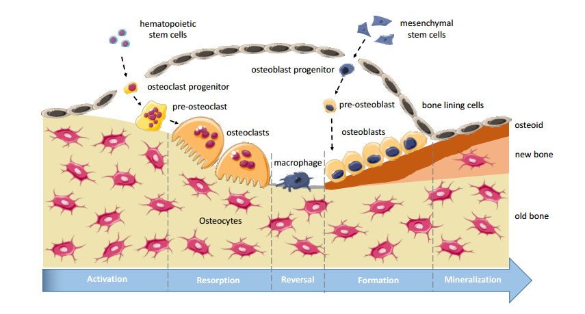
    [2] J. Kenkre and J. Bassett, The bone remodelling cycle, Ann. Clin. Biochem., 55 (2018), 308-327.
    [3] L. G. Raisz, Physiology and pathophysiology of bone remodeling, Clin. Chem., 45 (1999), 1353-1358.
    [4] D. J. Hadjidakis and I. I. Androulaskis, Bone remodeling, Ann. N. Y. Acad. Sci, 1092 (2006), 385-396.
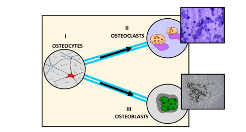
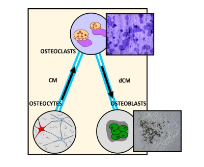
    [5] E. L. George, S. L. Truesdell, S. L. York, et al., Lab-on-a-chip platforms for quantification of multicellular interactions in bone remodeling, Exp. Cell Res., 365 (2018), 106-118.
    [6] W. Zhang, C. Green and N. S. Stott, Bone morphogenetic protein-2 modulation of chondrogenic differentiation in vitro involves gap junction-mediated intercellular communication, J. Cell Physiol., 193 (2002), 233-243.
    [7] J. Ilvesaro, K. Väänänen and J. Tuukkanen, Bone-resorbing osteoclasts contain gap-junctional connexin-43, J. Bone Miner. Res., 15 (2000), 919-926.
    [8] S. H. Park, W. Y. Sim, B. H. Min, et al., Chip-based comparison of the osteogenesis of human bone marrow- and adipose tissue-derived mesenchymal stem cells under mechanical stimulation, PLoS ONE, 7 (2012), e46689.
    [9] Y. Zhang, Z. Gazit, G. Pelled, et al., Patterning osteogenesis by inducible gene expression in microfluidic culture systems, Integr. Biol., 3 (2010), 39-47.
    [10] B. J. Taylor, A. Howell, K. A. Martin, et al., A lab-on-chip for malaria diagnosis and surveillance, Malar. J., 13 (2014), 179.
    [11] S. Wang, A. Ip, F. Xu, et al., Development of a microfluidic system for measuring HIV-1 viral load, Proc. SPIE, 7666 (2010), 76661H.
    [12] M. Wheeler and M. Rubessa, Integration of microfluidics and mammalian IVF, Mol. Hum. Reprod., 23 (2016), 248-256.
    [13] E. K. Sackmann, A. L. Fulton and D. J. Beebe, The present and future role of microfluidics in biomedical research, Nature, 507 (2014), 181-189.
    [14] D. Huh, B. D. Matthews, A. Mammoto, et al., Reconstituting organ-level lung functions on a chip, Science, 328 (2010), 1662-1668.
    [15] R. Baudoin, L. Griscom, M. Monge, et al., Development of a renal microchip for in vitro distal tubule models, Biotechnol. Prog., 23 (2007), 1245-1253.
    [16] K. J. Jang and K. Y. Suh, A multi-layer microfluidic device for efficient culture and analysis of renal tubular cells, Lab Chip, 10 (2010), 36-42.
    [17] L. L. Bischel, E. W. Young, B. R. Mader, et al., Tubeless microfluidic angiogenesis assay with three-dimensional endothelial-lined microvessels, Biomaterials, 34 (2013), 1471-1477.
    [18] M. Tsai, A. Kita, J. Leach, et al., In vitro modeling of the microvascular occlusion and thrombosis that occur in hematologic diseases using microfluidic technology, J. Clin. Invest., 122 (2012), 408-418.
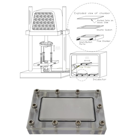
    [19] M. M. Saunders, Lab-on-a-chip (LOC) for biomimetic bone remodeling, (2018), US Pat. Appl., 1151.
    [20] K. S. Shah, S. L. York, P. Sethu, et al. Developing a microloading platform for applications in mechanotransduction research, Mechanics of Biological Systems and Materials, Volume 5, Conference Proceedings of the Society for Experimental Mechanics Series, New York: Springer International Publishing, (2013), pp. 197-205.
    [21] S. L. York, A. R. Arida, K. S. Shah, et al., Osteocyte characterization on polydimethylsiloxane substrates for microsystems applications, J. Biomim. Biomater. Tissue Eng., 16 (2012), 27-42.
    [22] S. L. York, J. D. King, A. S. Pietros, et al. Development of a microloading platform for in vitro mechanotransduction studies, Mechanics of Biological Systems and Materials, Volume 7, Conference Proceedings of the Society for Experimental Mechanics Series, New York: Springer International Publishing, (2015), pp. 53-59.
    [23] S. L. York, P. Sethu and M. M. Saunders, In vitro osteocytic microdamage and viability quantification using a microloading platform, Med. Eng. Phys., 38 (2016), 1115-1122.
    [24] J. D. King, S. L. York and M. M. Saunders, Design, fabrication and characterization of a pure uniaxial microloading system for biologic testing, Med. Eng. Phys., 38 (2016), 411-416.
    [25] S. L. Truesdell, E. L. George, C. E. Seno, et al., 3D printed loading device for inducing cellular mechanotransduction via matrix deformation, Exp. Mech., 59 (2019), 1223-1232.
    [26] J. D. King, D. Hayes, K. Shah, et al. Development of a multi-strain profile for cellular mechanotransduction testing, Mechanics of Biological Systems and Materials, Volume 7, Conference Proceedings of the Society for Experimental Mechanics Series, New York: Springer International Publishing, (2015), pp. 61-67.
    [27] S. L. York, P. Sethu and M. M. Saunders, Impact of gap junctional intercellular communication on MLO-Y4 sclerostin and soluble factor expression, Ann. Biomed. Eng., 44 (2016), 1170-1180.
    [28] J. M. Delaisse, The reversal phase of the bone-remodeling cycle: Cellular prerequisites for coupling resorption and formation, Bonekey Rep., 3 (2014), 561.
    [29] N. A. Sims and T. J. Martin, Coupling the activities of bone formation and resorption: A multitude of signals within the basic multicellular unit, Bonekey Rep., 3 (2014), 481.
    [30] R. Hambli, H. Katerchi, C. L. Benhamou, et al., Multiscale methodology for bone remodelling simulation using coupled finite element and neural network computation, Biomech. Model. Mechanobiol., 10 (2011), 133-145.
    [31] S. Ilic, K. Hackl and R. Gilbert, Application of the multiscale FEM to the modeling of cancellous bone, Biomech. Model. Mechanobiol., 9 (2010), 87-102.
    [32] M. D. Ryser, N. Nigam and S. V. Komarova, Mathematical modeling of spatio-temporal dynamics of a single bone multicellular unit, J. Bone Miner. Res., 24 (2009), 860-870.
    [33] S. Scheiner, P. Pivonka and C. Hellmich, Coupling systems biology with multiscale mechanics, for computer simulations of bone remodeling, Comput. Methods Appl. Mech. Eng., 254 (2013), 181-196.
    [34] V. Lemaire, F. L. Tobin, L. D. Greller, et al., Modeling the interactions between osteoblast and osteoclast activities in bone remodeling, J. Theor. Biol., 229 (2004), 293-309.
    [35] P. Pivonka and S. V. Komarova, Mathematical modeling in bone biology: from intracellular signaling to tissue mechanics, Bone, 47 (2010), 181-189.
    [36] S. A. Colopy, J. Benz-Dean, J. G. Barrett, et al., Response of the osteocyte syncytium adjacent to and distant from linear microcracks during adaptation to cyclic fatigue loading, Bone, 35 (2004), 881-891.
    [37] J. G. Hazenberg, M. Freeley, E. Foran, et al., Microdamage: A cell transducing mechanism based on ruptured osteocyte processes, J. Biomech., 39 (2006), 2096-2103.
    [38] O. Verborgt, G. J. Gibson and M. B. Schaffler, Loss of osteocyte integrity in association with microdamage and bone remodeling after fatigue in vivo, J. Bone Miner. Res., 15 (2000), 60-67.
    [39] G. K. Van Scoy, E. L. George, F. Opoku Asantewaa, et al., A cellular automata model of bone formation, Math. Biosci., 286 (2017), 58-64.
    [40] J. Eberhard, Y. Efendiev, R. Ewing, et al., Coupled cellular models for biofilm growth and hydrodynamic flow in a pipe, Int. J. Multiscale Compt. Eng., 3 (2005), 499-516. doi: 10.1615/IntJMultCompEng.v3.i4.70
    [41] D. G. Mallet and L. G. De Pillis, A cellular automata model of tumor-immune system interactions, J. Theor. Biol., 239 (2006), 334-350.
    [42] A. Prieto-Langarica, H. Kojouharov, B. Chen-Charpentier, et al., A cellular automata model of infection control on medical implants, Appl. Appl. Math., 6 (2011), 1-10.
    [43] R. Bivand, E. Pebesma and V. Gómez Rubio, Applied spatial data analysis with R, 2013.
    [44] P. A. Moran, Notes on continuous stochastic phenomena, Biometrika, 37 (1950), 17-23.
  • This article has been cited by:

    1. Laila A. Damiati, Safa A. Damiati, Samar Damiati, 2022, 9780128244692, 423, 10.1016/B978-0-12-824469-2.00025-7
    2. Yang Zhang, Taozhao Yu, Jingyi Ding, Zida Li, Bone-on-a-chip platforms and integrated biosensors: Towards advanced in vitro bone models with real-time biosensing, 2023, 219, 09565663, 114798, 10.1016/j.bios.2022.114798
    3. Colleen McCarthy, Gulden Camci-Unal, Low Intensity Pulsed Ultrasound for Bone Tissue Engineering, 2021, 12, 2072-666X, 1488, 10.3390/mi12121488
    4. Wen-Ying Lin, Kameshwara Kumar Dharini, Cheng-Huan Peng, Chung-Yen Lin, Kuang-Ting Yeh, Wen-Chih Lee, Ming-Der Lin, Zebrafish models for glucocorticoid-induced osteoporosis, 2022, 34, 1016-3190, 373, 10.4103/tcmj.tcmj_80_22
    5. M.A.M. Vis, F. Zhao, E.S.R. Bodelier, C.M. Bood, J. Bulsink, M. van Doeselaar, H. Eslami Amirabadi, K. Ito, S. Hofmann, Osteogenesis and osteoclastogenesis on a chip: Engineering a self-assembling 3D coculture, 2023, 173, 87563282, 116812, 10.1016/j.bone.2023.116812
    6. Aaqil Rifai, D. Kavindi Weerasinghe, Gebreselassie Addisu Tilaye, David Nisbet, Jason M. Hodge, Julie A. Pasco, Lana J. Williams, Rasika M. Samarasinghe, Richard J. Williams, Biofabrication of functional bone tissue: defining tissue-engineered scaffolds from nature, 2023, 11, 2296-4185, 10.3389/fbioe.2023.1185841
    7. Ya-Ming Wang, Jiang-Tao Shen, Chitosan-based promising scaffolds for the construction of tailored nanosystems against osteoporosis: Current status and future prospects, 2024, 22, 2280-8000, 10.1177/22808000241266487
    8. Yamei Deng, Yonglu Chen, Qian He, Xu Wang, Yong Liao, Jue Liu, Zhaoran Liu, Jianwei Huang, Ting Song, Bone age assessment from articular surface and epiphysis using deep neural networks, 2023, 20, 1551-0018, 13133, 10.3934/mbe.2023585
    9. Yamei Deng, Ting Song, Xu Wang, Yonglu Chen, Jianwei Huang, Region fine-grained attention network for accurate bone age assessment, 2024, 21, 1551-0018, 1857, 10.3934/mbe.2024081
    10. Ali Azadi, Mahdis Teimoori, Arash Khojasteh, 2025, Chapter 308-1, 978-3-031-36962-9, 1, 10.1007/978-3-031-36962-9_308-1
  • Reader Comments
  • © 2020 the Author(s), licensee AIMS Press. This is an open access article distributed under the terms of the Creative Commons Attribution License (http://creativecommons.org/licenses/by/4.0)
通讯作者: 陈斌, bchen63@163.com
  • 1. 

    沈阳化工大学材料科学与工程学院 沈阳 110142

  1. 本站搜索
  2. 百度学术搜索
  3. 万方数据库搜索
  4. CNKI搜索

Metrics

Article views(7005) PDF downloads(1320) Cited by(10)

Figures and Tables

Figures(7)  /  Tables(1)

Other Articles By Authors

/

DownLoad:  Full-Size Img  PowerPoint
Return
Return

Catalog