Citation: Ana Isabel Muñoz, J. Ignacio Tello. On a mathematical model of bone marrow metastatic niche[J]. Mathematical Biosciences and Engineering, 2017, 14(1): 289-304. doi: 10.3934/mbe.2017019
[1] | Ariosto Silva, Alexander R. A. Anderson, Robert Gatenby . A multiscale model of the bone marrow and hematopoiesis. Mathematical Biosciences and Engineering, 2011, 8(2): 643-658. doi: 10.3934/mbe.2011.8.643 |
[2] | Jason M. Graham, Bruce P. Ayati, Prem S. Ramakrishnan, James A. Martin . Towards a new spatial representation of bone remodeling. Mathematical Biosciences and Engineering, 2012, 9(2): 281-295. doi: 10.3934/mbe.2012.9.281 |
[3] | Katrine O. Bangsgaard, Morten Andersen, Vibe Skov, Lasse Kjær, Hans C. Hasselbalch, Johnny T. Ottesen . Dynamics of competing heterogeneous clones in blood cancers explains multiple observations - a mathematical modeling approach. Mathematical Biosciences and Engineering, 2020, 17(6): 7645-7670. doi: 10.3934/mbe.2020389 |
[4] | Evans K. Afenya . Using Mathematical Modeling as a Resource in Clinical Trials. Mathematical Biosciences and Engineering, 2005, 2(3): 421-436. doi: 10.3934/mbe.2005.2.421 |
[5] | Urszula Ledzewicz, Heinz Schättler . Controlling a model for bone marrow dynamics in cancer chemotherapy. Mathematical Biosciences and Engineering, 2004, 1(1): 95-110. doi: 10.3934/mbe.2004.1.95 |
[6] | Thanh Nam Nguyen, Jean Clairambault, Thierry Jaffredo, Benoît Perthame, Delphine Salort . Adaptive dynamics of hematopoietic stem cells and their supporting stroma: a model and mathematical analysis. Mathematical Biosciences and Engineering, 2019, 16(5): 4818-4845. doi: 10.3934/mbe.2019243 |
[7] | Eugene Kashdan, Svetlana Bunimovich-Mendrazitsky . Hybrid discrete-continuous model of invasive bladder cancer. Mathematical Biosciences and Engineering, 2013, 10(3): 729-742. doi: 10.3934/mbe.2013.10.729 |
[8] | Fabien Crauste . Global Asymptotic Stability and Hopf Bifurcation for a Blood Cell Production Model. Mathematical Biosciences and Engineering, 2006, 3(2): 325-346. doi: 10.3934/mbe.2006.3.325 |
[9] | Zamra Sajid, Morten Andersen, Johnny T. Ottesen . Mathematical analysis of the Cancitis model and the role of inflammation in blood cancer progression. Mathematical Biosciences and Engineering, 2019, 16(6): 8268-8289. doi: 10.3934/mbe.2019418 |
[10] | Avner Friedman, Harsh Vardhan Jain . A partial differential equation model of metastasized prostatic cancer. Mathematical Biosciences and Engineering, 2013, 10(3): 591-608. doi: 10.3934/mbe.2013.10.591 |
Metastasis, (meta, "next", and stasis "placement") is the process by which a primary tumor spreads to a secondary distance location.
This process occurs in anatomical sites providing the necessary environment of vascularization, oxygen and food allowing to camouflage its action for triggering the fast growing of cancer.
The process presents several stages: Invasion to the surrounding extracellular matrix, intravasation, dissemination thorough the circulation system, extravasation in different organs, settlement into latency in a pre-metastatic niche, reactivation and generation of a new tumor. Only a small percentage of metastatic cancer cells survive to the circulation system, and those that get out of the circulatory system rapidly die if they can not find the appropriate micro environment.
Metastasis is the final stage of cancer and the major cause of death in patients with cancer. For that reason, many anticancer therapies focus on Metastasis inhibition. Thus anti-metastasis treatments have different targets depending on the stage of the process they act. Angiogenesis, Cancer Stem Cells and metastatic niche, the role played by the inmune system (promoting or inhibiting metastasis) are some of the interesting issues in the area at extracellular level.
The motivation of this work relies on creating a mathematical model to describe the tumor cells movement towards a metastasis location into the bone marrow and the influence of the inhibition of chemoattractant receptors in metastatic tumor cells due to the drugs action. The mathematical model shows the importance of the balance between the chemotactic and random movements to reach the pre-metastatic niche.
Prostate cancer skeletal involvement is a complicated process, in which bone provides a favorable medium for tumor growth, resulting in alterations in bone remodeling and development of cancer-associated bone lesions. Prostate cancer and the bone microenvironment communicate and interact with each other through the progression of skeletal metastasis.
The pre-metastatic niche is a microenvironment in a specific location which facilitates the invasion and survival of metastatic tumor cells and may host a secondary tumor once the metastatic cancer cells start to proliferate (see [12]). The pre-metastatic niche microenvironment is formed by different types of cells: endothelial cells, mesenchymal progenitor cells, cancer associated fibroblasts (CAFs), myeloid cells and osteoclast; chemical signals: CXCL-12 and TGF-
Three different sources of metastatic niche have been already reported (see [12]):
1.-Native stem cell niches that metastatic cells may occupy in the host tissues;
2.-Niche functions provided by stromal cells not belonging to stem cell niches;
3.-Stem cell niche components that the cancer cells themselves may produce.
There exist at least two different types of pre-metastatic niches in the bone marrow, the osteoblast or endosteal niche and the vascular niche.
Osteoblast and osteoclast populations are the main agents involved in bone remodeling. In particular, osteoblasts mineralize the bone, while osteoclasts are responsible for bone resorption. There exists a dynamic equilibrium between osteoblasts and osteoclasts to maintain the bone tissue in normal adult vertebrates. This dynamical equilibrium existing in a healthy bone is perturbed by the chemical factors secreted by tumor cells.
The osteoblast niche is composed by osteoblast cells lining the endosteal surface which secretes a large variety of cytokines, growth factors and other signaling molecules that regulate the differentiation, the self-renewal and proliferation of hematopoietic stem cells (HSCs). The hematopoietic stem cells are a blood cell type which are host at the osteoblast niche in normal vertebrates (see [3]). The differentiation of HSCs produces myeloid cells, as macrophages, neutrophils, dendritic cells, etc, and lymphoid lineages as T-cells and B-cells among others.
Among the signaling molecules, CXCL-12, also known as stroma derivated factor 1 (SDF-1), is relatively highly expressed in the bone marrow (BM) (see [6]) and its receptor, CXCR-4, is expressed in HSCs and progenitors cells (see [9]). The retention in the BM of HSCs is regulated by the balance between CXCL-12 and Sphingosine-1-phosphate (S1P) produced by mature red blood cells in the circulatory system, which recruits HSCs to the circulation.
CXCL-12 is mainly produced by osteoblast cells, but it is also secreted by mesenchymal cells, endothelial precursor cells, cancer associated fibroblasts (CAFs) among others (see [13] and [16] for more details). The osteoblasts are activated by parathyroid hormone (PTH) and PTH-related protein (PTHrP), through the PTH/PTHrP receptor (PPR) (see [2] and [3]).
Prostate cancer cells produce other pro-osteoblastic factors that enhance bone mineralization as wingless int (Wnt), transforming growth factor-
It is known that CXCR-4 is overexpresed in metastatic solid tumor in prostate and breast cancer. The path CXCL-12 / CXCR-4 is used to localize the osteoblast niche (see [17]) and drives the cancer cells to the niche following the chemical gradients of CXCL-12 (chemotaxis).
Recently, several therapeutic strategies have been investigated to target the path CXCL-12/CXCR-4 to inhibit chemotactic movement (see for instance [7] and [11]). In [11], the authors present a novel therapeutic strategy and study the blockade of the path CXCL-12/CXCR-4 by AMD11070 in melanomas, comparing its effects with the inhibitor AMD3100. In this work, we consider the action of the inhibitor by reducing the chemosensitivity of the tumor cells, two different types of simulations are presented:
● when inhibitor is not present, and the tumor cell movement is composed of the combination of chemotaxis and random walk
● and when the action of chemotaxis is strongly reduced by the drug.
In the first case, the computational model shows how the tumor cell arrives at the niche while in the second case, the random movement is dominant and the cell doesn't arrive at the niche, at least during the computational time. The simulations show the existence of a threshold value in the rate chemotaxis / random coefficients:
In the following section, we introduce a mathematical model to describe the tumor cells movement towards a metastasis location into the BM. The description of the numerical scheme of resolution and some numerical simulations are performed in sections 3 and 4 respectively. The simulations illustrate the qualitative behavior of the solutions, which reproduce the migration of tumor cells from blood vessels to the osteoblastic niche in BM following the chemical gradients of CXCL-12.
In this section we present a mathematical model to describe tumor cells migration from blood vessels in BM to the osteoblast niche. The domain
We consider two different types of cells, osteoblasts, whose density shall be denoted by ''
We consider a finite number of MCCs,
(x′i,y′i)=χ(w)∇w(t,xi(t),yi(t))+D(cos(2πθij(t)),sin(2πθij(t))) | (1) |
where
Experiments show that cell movement is also influenced by the existing fibers in the extracellular matrix. On this occasion, to simplify the system we assume a homogeneous distribution of fibers in space and time and no influence in the cell movement.
Since the time from extravasation to the arrival of the pre-metastatic niche is small, we do not consider mitosis. Cancer cell death occurs if the cancer cell doesn
We denote by
N∑i=112√πϵie−14ϵi[(x−xi(t))2+(y−yi(t))2]. |
Notice that
limϵi→01√2πϵie−14ϵi[(x−xi(t))2+(y−yi(t))2]=δ(xi,yi) |
where
ut−DuΔu+u(μu+b1+u)=N∑i=11√2πϵie−14ϵi[(x−xi(t))2+(y−yi(t))2], | (2) |
which models the evolution of the concentration of tumor factors
u(0,x,y)=u0(x,y),(x,y)∈ˉΩ |
and the following boundary conditions
−Du∂u∂n=−Fuatx=1,−Du∂u∂n=0otherwise, |
where
Bone remodeling is regulated by osteoblasts located at the endosteal surface in dynamic equilibrium with osteoclasts, which are responsible of bone reabsorption. The process involves several substances and different types of progenitors cells in a basic multicellular unit (BMU). TGF-
b′=α2cbg22−β2b,α2=4 day−1,β2=0.02 day−1,g22≥0. |
As before mentioned, we assume that osteoblast are located only at the boundary
bt=g(b,u)b, |
where
bt=αb(μb+μbuu1+u−b). | (3) |
where
f(u)=μbuu1+u. |
Notice that we do not consider diffusion and transport of osteoblasts, see also [8]. The equation (3) is completed with an initial condition:
b(0,y)=b0(y),y∈[0,1]. |
CXCL-12 is secreted by osteoblasts, acting as a chemoattractant for the metastatic cancer cells which present CXCR-4 receptors at the surface. We denote by
Hence, we assume that the evolution and distribution of
wt−DwΔw+μww=0. | (4) |
Diffusion of
−Dw∂w∂n=−b, at x=0and−Dw∂w∂n=0,otherwise. |
The problem is completed with a given initial datum,
w(0,x,y)=w0(x,y),(x,y)∈ˉΩ. |
In this section, we shall describe the numerical scheme of resolution of the system (1)-(4).
Due to the parabolic feature of the problem, we use a time marching scheme. Then, the solutions are computed with a decoupling iterative method. For this, we proceed to solve sequentially (1)-(4), in the following way: first, we solve the problem for the variable
The numerical resolution of the corresponding decoupled systems at each step in time will be carried out by means of well known methods. To be precise, the resulting problems for the variables
Next, we shall briefly describe the overall process of resolution.
Let
um(x,y)=u(tm,x,y),wm(x,y)=w(tm,x,y),bm(y)=b(tm,y) |
and(xi,m,yi,m)=(xi(tm),yi(tm)). |
Notice that
For each
● Problem for
Given the location of the cells at the
∫Ω(1+Δtμu)umϕdxdy+ΔtDu∫Ω∇um∇ϕdxdy=∫Ωum−1ϕdxdy−Δt∫Ωμuum−1bm−11+um−1dxdy+ΔtN∑i=1∫Ω12√πϵie−14ϵi[(x−xi(t))2+(y−yi(t))2]ϕdxdy+Δt∫Γ1Fuϕdy,∀ϕ∈H1(Ω), |
where
● Problem for the osteoblast density:
Given
bm=bm−1+Δtαbm−1(μb+μbuumum+1−bm−1). |
● Problem for the concentration of CXCL-12 secreted by the osteoblasts:
Given
∫Ω(1+Δtμw)wmζdxdy+ΔtDw∫Ω∇um∇ζdxdy= |
∫Ωwm−1ζdxdy+Δt∫Γ0bmζdy,∀ζ∈H1(Ω), |
where
● And finally, the location for the tumor cells is computed with the updated values of
(xi,m,yi,m)≈(xi,m−1,yi,m−1)+Δtχ(wm)∇wm(xi,m−1,yi,m−1)+ |
ΔtD(cos(2πθi(tm−1)),sin(2πθi(tm−1))), |
in the sense that the positions of the cells are restricted to grids points, and
Pi,m−1=(xi,m−1,yi,m−1)+Δtχ(wm)∇wm(xi,m−1,yi,m−1)+ |
ΔtD(cos(2πθi(tm−1)),sin(2πθi(tm−1))) |
could not coincide with the coordinate of a grid point. To cope with this fact, we define
In this section we present some numerical results and their biological interpretation in order to illustrate the dynamical behavior of the different variables.
As we mentioned in previous sections, we consider a square domain
We consider two scenarios in order to illustrate the effect of a therapeutic strategy consisting in targeting the path CXCL-12-CXCR-4 to inhibit chemotactic movement of the metastatic tumor cell. In the first scenario, we assume that no treatment has been applied and hence, the tumor cell movement is directed by a combination of chemotaxis and random walk. In this case the values of the parameters which we considered are:
χ0=10,D=1,μu=1,α=1,μb=0.5,Fu=10−5,ϵi=10−4,μbu=100,μw=100 and Du=Dw=5⋅103. | (5) |
In the second scenario, a drug is used to inhibit the chemoattractant action. In this case, the parameter values which were considered are:
χ0=0.01,D=1,μu=1,α=1,μb=0.5,Fu=10−5,ϵi=10−4,μbu=100,μw=100 and Du=Dw=5⋅103. | (6) |
In both scenarios, we assume that a cell, coming form the blood vessels, arrives in the domain and that is located initially at the point
b0(y)=10e−100(x−0.4)2. |
The roles of the different parameters are the expected taking into account their positions in the equations.
● We observe the existence of a threshold value in the rate chemotaxis / random coefficients,
● Increasing the diffusion parameter
● As expected, we have obtained that the value of
● The role of the rest of the parameters are the expected.
In Figures 2-3, we illustrate the paths followed by the cells in both scenarios. We can observe that in the first case, the cells arrive at the niche, as there is no inhibition of the chemoattractant receptors, however in the second case (
In Figures 4-5, we present the results obtained for the tumor factors
12√πϵie−14ϵi[(x−xi(t))2+(y−yi(t))2] |
in the profile of
Finally, in Figures 6-7, we can see the results obtained for the CXCL-12 concentration, which resembles the behavior of
We have presented a mathematical model to describe the tumor cells movement towards a metastasis location into the bone marrow and the influence of the inhibition of the chemoattractant receptors in the metastatic tumor cell due to the drugs action. Recently, several therapeutic strategies have been investigated to target the path CXCL-12 / CXCR-4 to inhibit chemotactic movement (see for instance [7] and [11]). In the model, we consider the evolution of signaling molecules CXCL-12 secreted by osteoblasts (bone cells responsible of the mineralization of the bone) and PTHrP (secreted by tumor cells) which activates osteoblast growth. The model consists of a coupled system of second order PDEs describing the evolution of CXCL-12 and PTHrP, an ODE of logistic type to model the osteoblasts density and an extra equation for each cancer cell. We simulate the system to illustrate the qualitative behavior of the solutions and the numerical method of resolution is presented in detail. In particular, we consider the following two scenarios to illustrate the effect of a therapeutic strategy which target the path CXCL-12/CXCR-4 to inhibit chemotactic movement of the tumor cell: The first scenario reflects the case where the inhibitor is not present, and the tumor cell movement is composed of the combination of chemotaxis and random walk. In the second scenario, the action of chemotaxis is strongly reduced by the drug used to inhibit the chemoattractant receptors. The simulations show the existence of a threshold value in the rate chemotaxis / random coefficients:
The first author would like to thank MEC of Spain for supporting Projects TEC2012-39095-C03-02 and MTM2014-57158-R, and the second author for supporting Project MTM2013-42907-P.
[1] | [ R. A. Adams, Sobolev Spaces Academic Press, New York-London, 1975. |
[2] | [ A. A. Bryden,S. Islam,A. J. Freemont,J. H. Shanks,N. J. George,N. W. Clarke, Parathyroid hormone-related peptide: Expression in prostate cancer bone metastases, Prostate Cancer Prostatic Dis, 5 (2002): 59-62. |
[3] | [ L. M. Calvi,G. B. Adams,K. W. Weibrecht,J. M. Weber,D. P. Olson,M. C. Knight,R. P. Martin,E. Schipani,P. Divieti,F. R. Bringhurst,L. A. Milner,H. M. Kronenberg,D. T. Scadden, Osteoblastic cells regulate the haematopoietic stem cell niche, Nature, 425 (2003): 841-846. |
[4] | [ S. L. Chang, S. P. Cavnar, S. Takayama, G. D. Luker and J. J. Linderman, Cell, isoform, and environment factors shape gradients and modulate chemotaxis PLoS One 10 (2015), e0123450. |
[5] | [ N. L. Coggins,D. Trakimas,S. L. Chang,A. Ehrlich,P. Ray,K. E. Luker,J. J. Linderman,G. D. Luker, CXCR7 controls competition for recruitment of β-arrestin 2 in cells expressing both CXCR4 and CXCR7, PLoS One, 9 (2014): 841-846. |
[6] | [ K. Golan,O. Kollet,T. Lapidot, Dynamic cross talk between S1P and CXCL12 regulates hematopoietic stem cells migration, development and bone remodeling, Pharmaceuticals, 6 (2013): 1145-1169. |
[7] | [ G. Innamorati,M. T. Valenti,F. Giovinazzo,L. Dalle Carbonare,M. Parenti,C. Bassi, Molecular approaches to target gpcrs in cancer therapy, Pharmaceuticals, 4 (2011): 567-589. |
[8] | [ S. V. Komarova,R. J. Smith,S. J. Dixon,S. M. Sims,L. M. Wahlb, Mathematical model predicts a critical role for osteoclast autocrine regulation in the control of bone remodeling, Bone, 33 (2003): 206-215. |
[9] | [ A. J. Lilly, W. E. Johnson and C. M. Bunce, The haematopoietic stem cell niche: New insights into the mechanisms regulating haematopoietic stem cell behaviour Stem Cells International, 2011 (2011), ID 274564. |
[10] | [ A. I. Muñoz, Numerical resolution of a model of tumor growth, Mathematical Medicine and Biology, 33 (2016): 1-29. |
[11] | [ G. O'Boyle, I. Swidenbank, H. Marshall, C. E. Barker, J. Armstrong, S. A. White, S. P. Fricker, R. Plummer, M. Wright and P. E. Lovat, Inhibition of CXCR4/CXCL12 chemotaxis in melanoma by AMD11070, Br J Cancer., 108 (2013), 1634--1640, http://www.ncbi.nlm.nih.gov/pmc/articles/PMC3668477/ |
[12] | [ T. Oskarsson,E. Batlle,J. Massague, Metastatic stem cells: Sources, niches, and vital pathways, Cell Stem Cell, 14 (2014): 306-321. |
[13] | [ A. A. Rose,P. M. Siegel, Emerging therapeutic targets in breast cancer bone metastasis, Future Oncol., 6 (2010): 55-74. |
[14] | [ M. D. Ryser,N. Nigam,S. V. Komarova, Mathematical modeling of spatio-temporal dynamics of a single bone multicellular unit, J. of Bone and Mineral Research, 24 (2009): 860-870. |
[15] | [ J. Sceneay,M. J. Smyth,A. Möller, The pre-metastatic niche: Finding common ground, Cancer Metastasis Rev., 32 (2013): 449-464. |
[16] | [ Y. X. Sun,J. Wang,C. E. Shelburne,D. E. Lopatin,A. M. Chinnaiyan,M. A. Rubin,K. J. Pienta,R. S. Taichman, Expression of CXCR4 and CXCL12 (SDF-1) in human prostate cancers (PCa) in vivo, Journal of Cellular Biochemistry, 89 (2003): 462-473. |
[17] | [ R. S. Taichman,C. Cooper,E. T. Keller,K. J. Pienta,N. S. Taichman,L. K. McCauley, Use of the stromal cell-derived factor-1/CXCR4 pathway in prostate cancer metastasis to bone, Cancer Research, 62 (2002): 1832-1837. |
[18] | [ J. I. Tello, On a mathematical model of tumor growth based on cancer stem cells, Math. Biosc. Eng., 10 (2013): 263-278. |
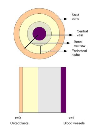
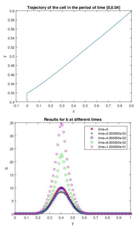
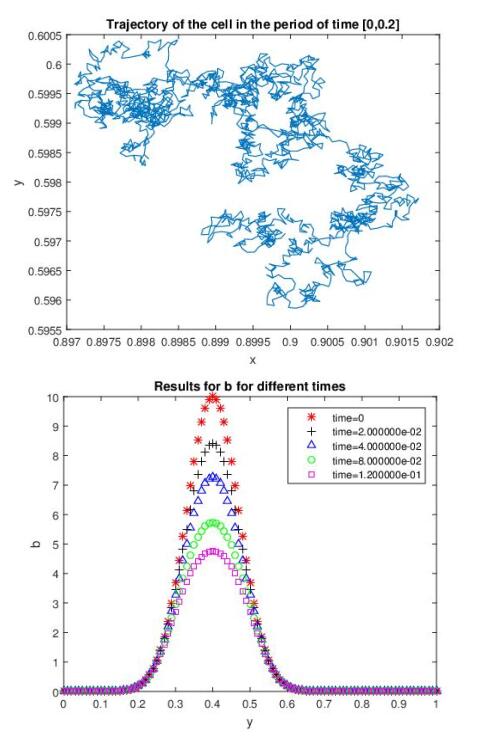
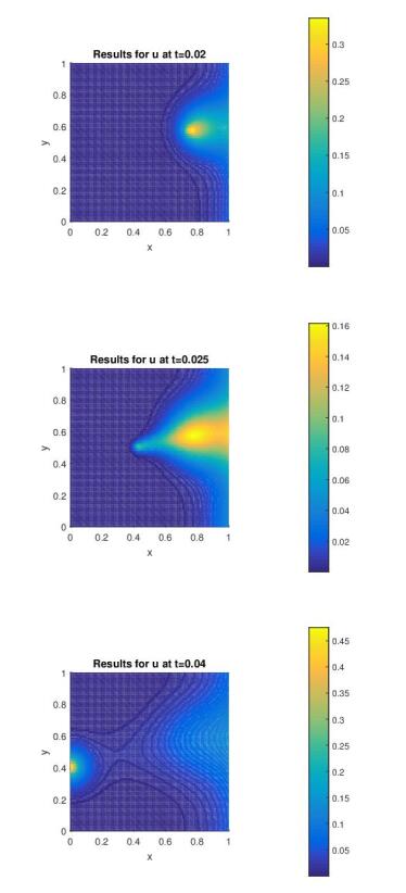
1. | Tiina E. Kähkönen, Jenni Bernoulli, Jussi M. Halleen, Mari I. Suominen, Novel and Conventional Preclinical Models to Investigate Bone Metastasis, 2019, 5, 2198-6428, 48, 10.1007/s40610-019-0114-5 | |
2. | Alexander Jinnah, Benjamin Zacks, Chukwuweike Gwam, Bethany Kerr, Emerging and Established Models of Bone Metastasis, 2018, 10, 2072-6694, 176, 10.3390/cancers10060176 | |
3. | Victoria L. Thai, Katherine H. Griffin, Steven W. Thorpe, R. Lor Randall, J. Kent Leach, Tissue engineered platforms for studying primary and metastatic neoplasm behavior in bone, 2021, 115, 00219290, 110189, 10.1016/j.jbiomech.2020.110189 | |
4. | Naoya Hatanaka, Mitsuru Futakuchi, Takashi Suzuki, 2021, Chapter 16, 978-981-16-4865-6, 235, 10.1007/978-981-16-4866-3_16 | |
5. | Silvia Jerez, Emilene Pliego, Francisco J. Solis, Anna K. Miller, Antigen receptor therapy in bone metastasis via optimal control for different human life stages, 2021, 83, 0303-6812, 10.1007/s00285-021-01673-4 |