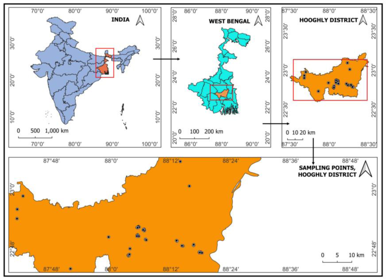
Aquaculture is one of the major economic activities in India, providing livelihoods and nutritional security to millions of people. In recent times, fish diseases have come to the limelight resulting in significant economic losses. We aimed to identify pathogenicity and virulence profiling of thirty-six pathogenic bacterial strains isolated from diseased Labeo rohita in the district of Hooghly, West Bengal, India. The bacterial strains were characterized through a comprehensive approach involving the examination of morphological features, biochemical properties, amplification, and sequencing of the 16S rRNA, species-specific genes, and virulence genes. Considering the prevalence frequency, virulence potential, and statistical significance Aeromonas hydrophila and Pseudomonas aeruginosa were selected for a survival assay followed by the examination of histopathological features to elucidate their effects. The identified bacterial isolates were arranged based on their predominance frequency, i.e., Aeromonas hydrophila (25%), Aeromonas veronii (22%), Pseudomonas aeruginosa (22%), Enterococcus faecalis (14%), Klebsiella pneumoniae (6%), Staphylococcus aureus (6%) and Escherichia coli (5%). Sixteen virulence-associated genes related to pathogenicity were amplified across the thirty-six isolates; aer, alt, lip and hlyA for A. hydrophila; exoS, lasB, toxA, oprL and phzM for P. aeruginosa; entB, fimH and uge in K. pneumoniae; aer in A. veronii; hlyA in E. coli; hlb in S. aureus and gelE for E. faecalis. The log-probit analysis revealed that A. hydrophila was notably more pathogenic than P. aeruginosa, as indicated by its lower lethal dose of 1.5×10⁴ CFU/mL. Additionally, histological examination revealed notable pathological changes, including tissue degeneration, inflammatory cell infiltration and vacuolation observed in the liver, kidney, gill and intestine of the challenged fish. We highlighted several potent aquatic microbial pathogens in order to manage and prevent such aquacultural maladies.
Citation: Abhijit Pakhira, Prasenjit Paria, Biswanath Malakar, Manoharmayum Shaya Devi, Vikash Kumar, Basanta Kumar Das, Asim Kumar Samanta, Santanu Chakrabarti, Bijay Kumar Behera. Identification and virulence gene characterization of pathogenic bacteria from diseased Labeo rohita (Hamilton, 1822): Insight into aquatic animal health management in Indian aquaculture[J]. AIMS Molecular Science, 2024, 11(3): 277-302. doi: 10.3934/molsci.2024017
Aquaculture is one of the major economic activities in India, providing livelihoods and nutritional security to millions of people. In recent times, fish diseases have come to the limelight resulting in significant economic losses. We aimed to identify pathogenicity and virulence profiling of thirty-six pathogenic bacterial strains isolated from diseased Labeo rohita in the district of Hooghly, West Bengal, India. The bacterial strains were characterized through a comprehensive approach involving the examination of morphological features, biochemical properties, amplification, and sequencing of the 16S rRNA, species-specific genes, and virulence genes. Considering the prevalence frequency, virulence potential, and statistical significance Aeromonas hydrophila and Pseudomonas aeruginosa were selected for a survival assay followed by the examination of histopathological features to elucidate their effects. The identified bacterial isolates were arranged based on their predominance frequency, i.e., Aeromonas hydrophila (25%), Aeromonas veronii (22%), Pseudomonas aeruginosa (22%), Enterococcus faecalis (14%), Klebsiella pneumoniae (6%), Staphylococcus aureus (6%) and Escherichia coli (5%). Sixteen virulence-associated genes related to pathogenicity were amplified across the thirty-six isolates; aer, alt, lip and hlyA for A. hydrophila; exoS, lasB, toxA, oprL and phzM for P. aeruginosa; entB, fimH and uge in K. pneumoniae; aer in A. veronii; hlyA in E. coli; hlb in S. aureus and gelE for E. faecalis. The log-probit analysis revealed that A. hydrophila was notably more pathogenic than P. aeruginosa, as indicated by its lower lethal dose of 1.5×10⁴ CFU/mL. Additionally, histological examination revealed notable pathological changes, including tissue degeneration, inflammatory cell infiltration and vacuolation observed in the liver, kidney, gill and intestine of the challenged fish. We highlighted several potent aquatic microbial pathogens in order to manage and prevent such aquacultural maladies.

| [1] | Béné C, Barange M, Subasinghe R, et al. (2015) Feeding 9 billion by 2050–Putting fish back on the menu. Food Sec 7: 261-274. https://doi.org/10.1007/s12571-015-0427-z |
| [2] | Hall SJ, Delaporte A, Phillips MJ, et al. (2011) Blue frontiers: Managing the environmental costs of aquaculture. Penang, Malaysia: The WorldFish Center. |
| [3] | Department of Fisheries, Ministry of Fisheries, Animal Husbandry & Dairying, Government of IndiaHandbook of fisheries statistics 2023 (2023). |
| [4] | Mishra SS, Rakesh D, Dhiman M, et al. (2017) Present status of fish disease management in freshwater aquaculture in India: state-of-the-art-review. J Aquac Fisheries 1: 003. http://doi.org/10.24966/AAF-5523/100003 |
| [5] | Lai XH, Wang SY, Edebro H, et al. (2003) Francisella strains express hemolysins of distinct characteristics. FEMS Microbiol Lett 224: 91-95. https://doi.org/10.1016/S0378-1097(03)00431-2 |
| [6] | Sun J, Zhang X, Gao X, et al. (2016) Characterization of virulence properties of Aeromonas veronii isolated from diseased Gibel Carp (Carassius gibelio). Int J Mol Sci 17: 496. https://doi.org/10.3390/ijms17040496 |
| [7] | Sambrook J, Russel DW (2001) Molecular cloning: A laboratory manual, third edition. CSH Laboratory Press, Cold Spring Harbor . |
| [8] | Behera BK, Paria P, Das A, et al. (2021) Molecular identification and pathogenicity study of virulent Citrobacter freundii associated with mortality of farmed Labeo rohita (Hamilton 1822), in India. Aquaculture 547: 737437. https://doi.org/10.1016/j.aquaculture.2021.737437 |
| [9] | Saitou N, Nei M (1987) The neighbor-joining method: A new method for reconstructing phylogenetic trees. Mol Biol Evol 4: 406-425. https://doi.org/10.1093/oxfordjournals.molbev.a040454 |
| [10] | Kumar S, Stecher G, Li M, et al. (2018) MEGA X: Molecular evolutionary genetics analysis across computing platforms. Mol Biol Evol 35: 1547-1549. https://doi.org/10.1093/molbev/msy096 |
| [11] | Tamura K, Nei M, Kumar S (2004) Prospects for inferring very large phylogenies by using the neighbor-joining method. Proc Natl Acad Sci 101: 11030-11035. https://doi.org/10.1073/pnas.0404206101 |
| [12] | Felsenstein J (1985) Confidence limits on phylogenies: An approach using the bootstrap. Evolution 39: 783-791. https://doi.org/10.2307/2408678 |
| [13] | Martino ME, Fasolato L, Montemurro F, et al. (2011) Determination of microbial diversity of Aeromonas strains on the basis of multilocus sequence typing, phenotype, and presence of putative virulence genes. Appl Environ Microbiol 77: 4986-5000. https://doi.org/10.1128/aem.00708-11 |
| [14] | Persson S, Al-Shuweli S, Yapici S, et al. (2015) Identification of clinical Aeromonas species by rpoB and gyrB sequencing and development of a multiplex PCR method for detection of Aeromonas hydrophila, A. caviae, A. veronii, and A. media. J. Clin Microbiol 53: 653-656. https://doi.org/10.1128/jcm.01963-14 |
| [15] | Wirth T, Falush D, Lan R, et al. (2006) Sex and virulence in Escherichia coli: An evolutionary perspective. Mol Microbiol 60: 1136-1151. https://doi.org/10.1111/j.1365-2958.2006.05172.x |
| [16] | Guo C, Yang X, Wu Y, et al. (2015) MLST-based inference of genetic diversity and population structure of clinical Klebsiella pneumoniae, China. Sci Rep 5: 7612. https://doi.org/10.1038/srep07612 |
| [17] | Enright MC, Day NPJ, Davies CE, et al. (2000) Multilocus sequence typing for characterization of methicillin-resistant and methicillin-susceptible clones of Staphylococcus aureus. J Clin Microbiol 38: 1008-1015. https://doi.org/10.1128/jcm.38.3.1008-1015.2000 |
| [18] | Curran B, Jonas D, Grundmann H, et al. (2004) Development of a multilocus sequence typing scheme for the opportunistic pathogen Pseudomonas aeruginosa. J Clin Microbiol 42: 5644-5649. https://doi.org/10.1128/jcm.42.12.5644-5649.2004 |
| [19] | Ruiz-Garbajosa P, Bonten MJM, Robinson DA, et al. (2006) Multilocus sequence typing scheme for Enterococcus faecalis reveals hospital-adapted genetic complexes in a background of high rates of recombination. J Clin Microbiol 44: 2220-2228. https://doi.org/10.1128/jcm.02596-05 |
| [20] | Kong RYC, Lee SKY, Law SHW, et al. (2002) Rapid detection of six types of bacterial pathogens in marine waters by multiplex PCR. Water Res 36: 2802-2812. https://doi.org/10.1016/s0043-1354(01)00503-6 |
| [21] | Yang ZS, Fang H (2003) Human and animal pathogenic bacteriology. Shijiazhuan, China: Hebei Science and Technology Press 1550-1610. |
| [22] | Nawaz M, Khan SA, Khan AA, et al. (2010) Detection and characterization of virulence genes and integrons in Aeromonas veronii isolated from catfish. Food Microbiol 27: 327-331. https://doi.org/10.1016/j.fm.2009.11.007 |
| [23] | Sen K, Rodgers M (2004) Distribution of six virulence factors in Aeromonas species isolated from US drinking water utilities: A PCR identification. J. Appl Microbiol 97: 1077-1086. https://doi.org/10.1111/j.1365-2672.2004.02398.x |
| [24] | Wong CYF, Heuzenroeder MW, Flower RLP (1998) Inactivation of two haemolytic toxin genes in Aeromonas hydrophila attenuates virulence in a suckling mouse model. Microbiology 144: 291-298. https://doi.org/10.1099/00221287-144-2-291 |
| [25] | Kerényi M, Allison HE, Bátai I, et al. (2005) Occurrence of hlyA and sheA genes in extraintestinal Escherichia coli strains. J Clin Microbiol 43: 2965-2968. https://doi.org/10.1128/jcm.43.6.2965-2968.2005 |
| [26] | Chapman TA, Wu XY, Barchia I, et al. (2006) Comparison of virulence gene profiles of Escherichia coli strains isolated from healthy and diarrheic swine. Appl Environ Microbiol 72: 4782-4795. https://doi.org/10.1128/aem.02885-05 |
| [27] | Paton AW, Paton JC (1998) Detection and characterization of Shiga toxigenic Escherichia coli by using multiplex PCR assays for stx 1, stx 2, eaeA, enterohemorrhagic E. coli hlyA, rfb O111, and rfb O157. J Clin Microbiol 36: 598-602. https://doi.org/10.1128/jcm.36.2.598-602.1998 |
| [28] | Russo TA, Olson R, MacDonald U, et al. (2014) Aerobactin mediates virulence and accounts for increased siderophore production under iron-limiting conditions by hypervirulent (hypermucoviscous) Klebsiella pneumoniae. Infect Immun 82: 2356-2367. https://doi.org/10.1128/iai.01667-13 |
| [29] | Alcántar-Curiel MD, Blackburn D, Saldaña Z, et al. (2013) Multi-functional analysis of Klebsiella pneumoniae fimbrial types in adherence and biofilm formation. Virulence 4: 129-138. https://doi.org/10.4161/viru.22974 |
| [30] | Zhang S, Yang G, Ye Q, et al. (2018) Phenotypic and genotypic characterization of Klebsiella pneumoniae isolated from retail foods in China. Front Microbiol 9: 289. https://doi.org/10.3389/fmicb.2018.00289 |
| [31] | Fang CT, Chuang YP, Shun CT, et al. (2004) A novel virulence gene in Klebsiella pneumoniae strains causing primary liver abscess and septic metastatic complications. J Exp Med 199: 697-705. https://doi.org/10.1084/jem.20030857 |
| [32] | Nadasy KA, Domiati-Saad R, Tribble MA (2007) Invasive Klebsiella pneumoniae syndrome in North America. Clin Infect Dis 45: e25-e28. https://doi.org/10.1086/519424 |
| [33] | Jarraud S, Mougel C, Thioulouse J, et al. (2002) Relationships between Staphylococcus aureus genetic background, virulence factors, agr groups (alleles), and human disease. Infect Immun 70: 631-641. https://doi.org/10.1128/iai.70.2.631-641.2002 |
| [34] | Li X, Huang T, Xu K, et al. (2019) Molecular characteristics and virulence gene profiles of Staphylococcus aureus isolates in Hainan, China. BMC Infect Dis 19: 873. https://doi.org/10.1186/s12879-019-4547-5 |
| [35] | Mehrotra M, Wang G, Johnson WM (2000) Multiplex PCR for detection of genes for Staphylococcus aureus enterotoxins, exfoliative toxins, toxic shock syndrome toxin 1, and methicillin resistance. J Clin Microbiol 38: 1032-1035. https://doi.org/10.1128/jcm.38.3.1032-1035.2000 |
| [36] | Fazeli N, Momtaz H (2014) Virulence gene profiles of multidrug-resistant Pseudomonas aeruginosa isolated from Iranian hospital infections. Iran Red Crescent Med J 16: e15722. https://doi.org/10.5812/ircmj.15722 |
| [37] | Xu J, Moore JE, Murphy PG, et al. (2004) Early detection of Pseudomonas aeruginosa – comparison of conventional versus molecular (PCR) detection directly from adult patients with cystic fibrosis (CF). Ann Clin Microbiol Antimicrob 3: 21. https://doi.org/10.1186/1476-0711-3-21 |
| [38] | Matar GM, Ramlawi F, Hijazi N, et al. (2002) Transcription levels of Pseudomonas aeruginosa exotoxin A gene and severity of symptoms in patients with otitis externa. Curr Microbiol 45: 350-354. https://doi.org/10.1007/s00284-002-3703-z |
| [39] | Finnan S, Morrissey JP, O'gara F, et al. (2004) Genome diversity of Pseudomonas aeruginosa isolates from cystic fibrosis patients and the hospital environment. J Clin Microbiol 42: 5783-5792. https://doi.org/10.1128/jcm.42.12.5783-5792.2004 |
| [40] | Vankerckhoven V, Van Autgaerden T, Vael C, et al. (2004) Development of a multiplex PCR for the detection of asa1, gelE, cylA, esp, and hyl genes in enterococci and survey for virulence determinants among European hospital isolates of Enterococcus faecium. J Clin Microbiol 42: 4473-4479. https://doi.org/10.1128/jcm.42.10.4473-4479.2004 |
| [41] | Creti R, Imperi M, Bertuccini L, et al. (2004) Survey for virulence determinants among Enterococcus faecalis isolated from different sources. J Clin Microbiol 53: 13-20. https://doi.org/10.1099/jmm.0.05353-0 |
| [42] | Maheshwari DG, Shaikh NK (2016) An overview on-toxicity testing method. Int J Pharm Technol 8: 3834-3849. |
| [43] | Luna LG (1968) Manual of histologic staining methods of the Armed Forces Institute of Pathology. New York: Blakiston Division, McGraw-Hill. |
| [44] | Sherwani RAK, Shakeel H, Awan WB, et al. (2021) Analysis of COVID-19 data using neutrosophic Kruskal Wallis H test. BMC Med Res Methodol 21: 215. https://doi.org/10.1186/s12874-021-01410-x |
| [45] | Snieszko SF (1972) Nutritional fish diseases. Fish Nutr : 403-437. |
| [46] | Martin-Carnahan A, Joseph SW (2005) Family I. Aeromonadaceae. Bergey's manual of systematic bacteriology 2: 556-587. https://doi.org/10.1007/0-387-28022-7_12 |
| [47] | Beaz-Hidalgo R, Figueras MJ (2013) Aeromonas spp. whole genomes and virulence factors implicated in fish disease. J Fish Dis 36: 371-388. https://doi.org/10.1111/jfd.12025 |
| [48] | Samayanpaulraj V, Sivaramapillai M, Palani SN, et al. (2020) Identification and characterization of virulent Aeromonas hydrophila Ah17 from infected Channa striata in river Cauvery and in vitro evaluation of shrimp chitosan. Food Sci Nutr 8: 1272-1283. https://doi.org/10.1002/fsn3.1416 |
| [49] | Das A, Acharya S, Behera BK, et al. (2018) Isolation, identification and characterization of Klebsiella pneumoniae from infected farmed Indian Major Carp Labeo rohita (Hamilton 1822) in West Bengal, India. Aquaculture 482: 111-116. https://doi.org/10.1016/j.aquaculture.2017.08.037 |
| [50] | Visnuvinayagam S, Joseph TC, Murugadas V, et al. (2015) Status on methicillin resistant and multiple drug resistant Staphylococcus aureus in fishes of Cochin and Mumbai coast, India. J Environ Biol 36: 571-575. |
| [51] | Vaiyapuri M, Joseph TC, Rao BM, et al. (2019) Methicillin-resistant Staphylococcus aureus in seafood: Prevalence, laboratory detection, clonal nature, and control in seafood chain. J Food Sci 84: 3341-3351. https://doi.org/10.1111/1750-3841.14915 |
| [52] | Eissa NME, El-Ghiet EA, Shaheen AA, et al. (2010) Characterization of Pseudomonas species isolated from tilapia “Oreochromis niloticus” in Qaroun and Wadi-El-Rayan lakes, Egypt. Global Vet 5: 116-121. http://dx.doi.org/10.13140/2.1.5002.4961 |
| [53] | Paria P, Behera BK, Mohapatra PKD, et al. (2021) Virulence factor genes and comparative pathogenicity study of tdh, trh and tlh positive Vibrio parahaemolyticus strains isolated from Whiteleg shrimp, Litopenaeus vannamei (Boone, 1931) in India. Infect Genet Evol 95: 105083. https://doi.org/10.1016/j.meegid.2021.105083 |
| [54] | Zhang XJ, Qin GM, Bing XW, et al. (2011) Phenotypic and molecular characterization of Photobacterium damselae, a pathogen of the cultured tongue sole Cynoglossus semilaevis in China. N Z J Mar Freshwater Res 45: 1-13. https://doi.org/10.1080/00288330.2010.531745 |
| [55] | Cole JR, Wang Q, Cardenas E, et al. (2009) The Ribosomal Database Project: Improved alignments and new tools for rRNA analysis. Nucleic Acids Res 37: D141-D145. https://doi.org/10.1093/nar/gkn879 |
| [56] | Wang G, Clark CG, Liu C, et al. (2003) Detection and characterization of the hemolysin genes in Aeromonas hydrophila and Aeromonas sobria by multiplex PCR. J Clin Microbiol 41: 1048-1054. https://doi.org/10.1128/jcm.41.3.1048-1054.2003 |
| [57] | Devi MS, Paria P, Kumar V, et al. (2022) Molecular identification and pathogenicity study of virulent Vibrio cholerae non O1/O139 serotype associated with mortality of farmed Labeo rohita (Hamilton, 1822), in India. Aquaculture 547: 737529. https://doi.org/10.1016/j.aquaculture.2021.737529 |
| [58] | Wang X, Wang Q, Xiao J, et al. (2010) Hemolysin EthA in Edwardsiella tarda is essential for fish invasion in vivo and in vitro and regulated by two-component system EsrA–EsrB and nucleoid protein HhaEt. Fish Shellfish Immun 29: 1082-1091. https://doi.org/10.1016/j.fsi.2010.08.025 |
| [59] | Preena PG, Dharmaratnam A, Swaminathan TR (2022) A peek into mass mortality caused by antimicrobial resistant Edwardsiella tarda in goldfish, Carassius auratus in Kerala. Biologia 77: 1161-1171. https://doi.org/10.1007/s11756-022-01007-9 |
| [60] | Locke JB, Colvin KM, Varki N, et al. (2007) Streptococcus iniae β-hemolysin streptolysin S is a virulence factor in fish infection. Dis Aquat Organ 76: 17-26. https://doi.org/10.3354/dao076017 |
| [61] | Batt CA (2016) Virulence. Reference module in food science . Amsterdam, The Netherlands: Elsevier. https://doi.org/10.1016/B978-0-08-100596-5.03453-3 |
| [62] | Remya PA, Shanthi M, Sekar U (2019) Characterisation of virulence genes associated with pathogenicity in Klebsiella pneumoniae. Indian J Med Microbiol 37: 210-218. https://doi.org/10.4103/ijmm.ijmm_19_157 |
| [63] | Sivaraman GK, Rajan V, Vijayan A, et al. (2021) Antibiotic resistance profiles and molecular characteristics of extended-spectrum beta-lactamase (ESBL)-producing Escherichia coli and Klebsiella pneumoniae isolated from shrimp aquaculture farms in Kerala, India. Front Microbiol 12: 622891. https://doi.org/10.3389/fmicb.2021.622891 |
| [64] | Regué M, Hita B, Piqué N, et al. (2004) A gene, uge, is essential for Klebsiella pneumoniae virulence. Infect Immun 72: 54-61. https://doi.org/10.1128/iai.72.1.54-61.2004 |
| [65] | Abrami L, Fivaz M, Glauser PE, et al. (2003) Sensitivity of polarized epithelial cells to the pore-forming toxin aerolysin. Infect Immun 71: 739-746. https://doi.org/10.1128/IAI.71.2.739-746.2003 |
| [66] | Nhinh DT, Le DV, Van KV, et al. (2021) Prevalence, virulence gene distribution and alarming the multidrug resistance of Aeromonas hydrophila associated with disease outbreaks in freshwater aquaculture. Antibiotics 10: 532. https://doi.org/10.3390/antibiotics10050532 |
| [67] | Muduli C, Tripathi G, Paniprasad K, et al. (2021) Virulence potential of Aeromonas hydrophila isolated from apparently healthy freshwater food fish. Biologia 76: 1005-1015. https://doi.org/10.2478/s11756-020-00639-z |
| [68] | Chopra AK, Xu XJ, Ribardo D, et al. (2000) The cytotoxic enterotoxin of Aeromonas hydrophila induces proinflammatory cytokine production and activates arachidonic acid metabolism in macrophages. Infect Immun 68: 2808-2818. https://doi.org/10.1128/iai.68.5.2808-2818.2000 |
| [69] | Ribardo DA, Kuhl KR, Boldogh I, et al. (2002) Early cell signaling by the cytotoxic enterotoxin of Aeromonas hydrophila in macrophages. Microb Pathog 32: 149-163. https://doi.org/10.1006/mpat.2001.0490 |
| [70] | Hayati HR, Hassan MD, Ong BL, et al. (2015) Virulence genes detection of Aeromonas hydrophila originated from diseased freshwater fishes. Adv Environ Biol 9: 22-26. |
| [71] | Tomás JM (2012) The main Aeromonas pathogenic factors. ISRN Microbiol 2012: 256261. https://doi.org/10.5402/2012/256261 |
| [72] | Ristow LC, Welch RA (2016) Hemolysin of uropathogenic Escherichia coli: A cloak or a dagger?. BBA-Biomembranes 1858: 538-545. https://doi.org/10.1016/j.bbamem.2015.08.015 |
| [73] | Chuang YC, Chiou SF, Su JH, et al. (1997) Molecular analysis and expression of the extracellular lipase of Aeromonas hydrophila MCC-2. Microbiology 143: 803-812. https://doi.org/10.1099/00221287-143-3-803 |
| [74] | Lee KK, Ellis AE (1990) Glycerophospholipid: Cholesterol acyltransferase complexed with lipopolysaccharide (LPS) is a major lethal exotoxin and cytolysin of Aeromonas salmonicida: LPS stabilizes and enhances toxicity of the enzyme. J Bacteriol 172: 5382-5393. https://doi.org/10.1128/jb.172.9.5382-5393.1990 |
| [75] | Pemberton JM, Kidd SP, Schmidt R (1997) Secreted enzymes of Aeromonas. FEMS Microbiol Lett 152: 1-10. https://doi.org/10.1111/j.1574-6968.1997.tb10401.x |
| [76] | Nawaz M, Khan SA, Khan AA, et al. (2010) Detection and characterization of virulence genes and integrons in Aeromonas veronii isolated from catfish. Food Microbiol 27: 327-331. https://doi.org/10.1016/j.fm.2009.11.007 |
| [77] | Chen F, Sun J, Han Z, et al. (2019) Isolation, identification and characteristics of Aeromonas veronii from diseased crucian carp (Carassius auratus gibelio). Front Microbiol 10: 2742. https://doi.org/10.3389/fmicb.2019.02742 |
| [78] | Kong C, Neoh HM, Nathan S (2016) Targeting Staphylococcus aureus toxins: A potential form of anti-virulence therapy. Toxins 8: 72. https://doi.org/10.3390/toxins8030072 |
| [79] | Alagarsamy S, Thampuran N, Joseph TC (2010) Virulence genes, serobiotypes and antibiotic resistance profile of Escherichia coli strains isolated from aquaculture and other sources. Aquac Res 41: 1003-1014. https://doi.org/10.1111/j.1365-2109.2009.02384.x |
| [80] | Bauer ME, Welch RA (1996) Characterization of an RTX toxin from enterohemorrhagic Escherichia coli O157: H7. Infect Immun 64: 167-175. https://doi.org/10.1128/iai.64.1.167-175.1996 |
| [81] | Gentschev I, Dietrich G, Goebel W (2002) The E. coli α-hemolysin secretion system and its use in vaccine development. Trends Microbiol 10: 39-45. https://doi.org/10.1016/S0966-842X(01)02259-4 |
| [82] | Welch RA, Dellinger EP, Minshew B, et al. (1981) Haemolysin contributes to virulence of extra-intestinal E. coli infections. Nature 294: 665-667. https://doi.org/10.1038/294665a0 |
| [83] | Benz R (2020) RTX-toxins. Toxins 12: 359. https://doi.org/10.3390/toxins12060359 |
| [84] | Alonso B, Fernández-Barat L, Di Domenico EG, et al. (2020) Characterization of the virulence of Pseudomonas aeruginosa strains causing ventilator-associated pneumonia. BMC Infect Dis 20: 909. https://doi.org/10.1186/s12879-020-05534-1 |
| [85] | Algammal AM, Mabrok M, Sivaramasamy E, et al. (2020) Emerging MDR-Pseudomonas aeruginosa in fish commonly harbor oprL and toxA virulence genes and blaTEM, blaCTX-M, and tetA antibiotic-resistance genes. Sci Rep 10: 15961. https://doi.org/10.1038/s41598-020-72264-4 |
| [86] | Vareechon C, Zmina SE, Karmakar M, et al. (2017) Pseudomonas aeruginosa effector ExoS inhibits ROS production in human neutrophils. Cell Host Microbe 21: 611-618. https://doi.org/10.1016/j.chom.2017.04.001 |
| [87] | Kaminski A, Gupta KH, Goldufsky JW, et al. (2018) Pseudomonas aeruginosa ExoS induces intrinsic apoptosis in target host cells in a manner that is dependent on its GAP domain activity. Sci Rep 8: 14047. https://doi.org/10.1038/s41598-018-32491-2 |
| [88] | Rust L, Pesci EC, Iglewski BH (1996) Analysis of the Pseudomonas aeruginosa elastase (lasB) regulatory region. J Bacteriol 178: 1134-1140. https://doi.org/10.1128/jb.178.4.1134-1140.1996 |
| [89] | Todar K Bacterial Protein Toxins In: Todar's Online textbook of bacteriology (2012). Available from: http://textbookofbacteriology.net/ |
| [90] | Nowroozi J, Sepahi AA, Rashnonejad A (2012) Pyocyanine biosynthetic genes in clinical and environmental isolates of Pseudomonas aeruginosa and detection of pyocyanine's antimicrobial effects with or without colloidal silver nanoparticles. Cell J 14: 7-18. |
| [91] | Lim A, De Vos D, Brauns M, et al. (1997) Molecular and immunological characterization of OprL, the 18 KDa outer-membrane peptidoglycan-associated lipoprotein (PAL) of Pseudomonas aeruginosa. Microbiology 143: 1709-1716. https://doi.org/10.1099/00221287-143-5-1709 |
| [92] | Remans K, Vercammen K, Bodilis J, et al. (2010) Genome-wide analysis and literature-based survey of lipoproteins in Pseudomonas aeruginosa. Microbiology 156: 2597-2607. https://doi.org/10.1099/mic.0.040659-0 |
| [93] | Bradbury RS, Roddam LF, Merritt A, et al. (2010) Virulence gene distribution in clinical, nosocomial and environmental isolates of Pseudomonas aeruginosa. J Med Microbiol 59: 881-890. https://doi.org/10.1099/jmm.0.018283-0 |
| [94] | Cezairliyan B, Vinayavekhin N, Grenfell-Lee D, et al. (2013) Identification of Pseudomonas aeruginosa phenazines that kill Caenorhabditis elegans. PLoS Pathog 9: e1003101. https://doi.org/10.1371/journal.ppat.1003101 |
| [95] | Aljebory IS (2018) PCR detection of some virulence genes of Pseudomonas aeruginosa in Kirkuk city, Iraq. J Pharm Sci Res 10: 1068-1071. |
| [96] | Zheng JX, Wu Y, Lin ZW, et al. (2017) Characteristics of and virulence factors associated with biofilm formation in clinical Enterococcus faecalis isolates in China. Front Microbiol 8: 2338. https://doi.org/10.3389/fmicb.2017.02338 |
| [97] | Anderson AC, Jonas D, Huber I, et al. (2016) Enterococcus faecalis from food, clinical specimens, and oral sites: prevalence of virulence factors in association with biofilm formation. Front Microbiol 6: 1534. https://doi.org/10.3389/fmicb.2015.01534 |
| [98] | Kayaoglu G, Ørstavik D (2004) Virulence factors of Enterococcus faecalis: Relationship to endodontic disease. Crit Rev Oral Biol Med 15: 308-320. https://doi.org/10.1177/154411130401500506 |
| [99] | Park SY, Kim KM, Lee JH, et al. (2007) Extracellular gelatinase of Enterococcus faecalis destroys a defense system in insect hemolymph and human serum. Infect Immun 75: 1861-1869. https://doi.org/10.1128/iai.01473-06 |
| [100] | Thomas VC, Thurlow LR, Boyle D, et al. (2008) Regulation of autolysis-dependent extracellular DNA release by Enterococcus faecalis extracellular proteases influences biofilm development. J Bacteriol 190: 5690-5698. https://doi.org/10.1128/jb.00314-08 |
| [101] | Mohamed JA, Murray BE (2005) Lack of correlation of gelatinase production and biofilm formation in a large collection of Enterococcus faecalis isolates. J Clin Microbiol 43: 5405-5407. https://doi.org/10.1128/JCM.43.10.5405-5407.2005 |
| [102] | Behera BK, Paria P, Das A, et al. (2017) Molecular characterization and pathogenicity of a virulent Acinetobacter baumannii associated with mortality of farmed Indian Major Carp Labeo rohita (Hamilton 1822). Aquaculture 471: 157-162. https://doi.org/10.1016/j.aquaculture.2017.01.018 |
| [103] | Abdelhamed H, Ibrahim I, Baumgartner W, et al. (2017) Characterization of histopathological and ultrastructural changes in channel catfish experimentally infected with virulent Aeromonas hydrophila. Front Microbiol 8: 1519. https://doi.org/10.3389/fmicb.2017.01519 |
 molsci-11-03-017-s001.pdf molsci-11-03-017-s001.pdf |
 |


Figures(7) / Tables(4)

Abhijit Pakhira, Prasenjit Paria, Biswanath Malakar, Manoharmayum Shaya Devi, Vikash Kumar, Basanta Kumar Das, Asim Kumar Samanta, Santanu Chakrabarti, Bijay Kumar Behera. Identification and virulence gene characterization of pathogenic bacteria from diseased Labeo rohita (Hamilton, 1822): Insight into aquatic animal health management in Indian aquaculture[J]. AIMS Molecular Science, 2024, 11(3): 277-302. doi: 10.3934/molsci.2024017


 DownLoad: