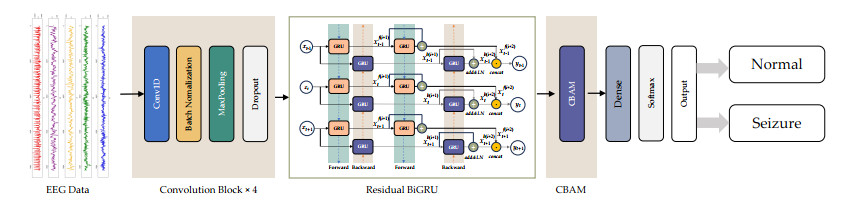
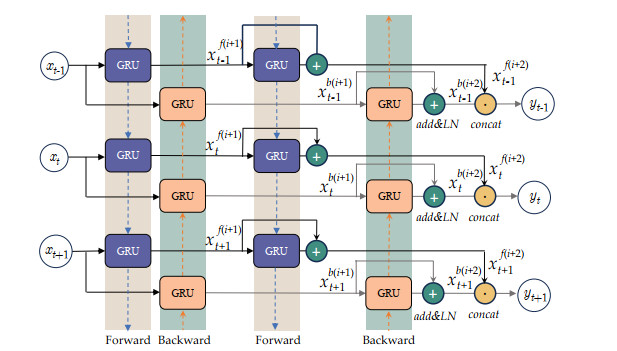
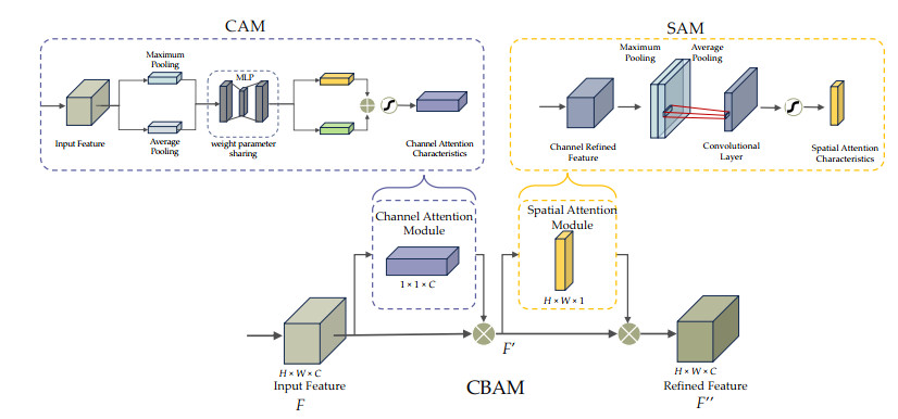
Epileptic seizures, a prevalent neurological condition, necessitate precise and prompt identification for optimal care. Nevertheless, the intricate characteristics of electroencephalography (EEG) signals, noise, and the want for real-time analysis require enhancement in the creation of dependable detection approaches. Despite advances in machine learning and deep learning, capturing the intricate spatial and temporal patterns in EEG data remains challenging. This study introduced a novel deep learning framework combining a convolutional neural network (CNN), bidirectional gated recurrent unit (BiGRU), and convolutional block attention module (CBAM). The CNN extracts spatial features, the BiGRU captures long-term temporal dependencies, and the CBAM emphasizes critical spatial and temporal regions, creating a hybrid architecture optimized for EEG pattern recognition. Evaluation of a public EEG dataset revealed superior performance compared to existing methods. The model achieved 99.00% accuracy in binary classification, 96.20% in three-class tasks, 92.00% in four-class scenarios, and 89.00% in five-class classification. High sensitivity (89.00–99.00%) and specificity (89.63–99.00%) across all tasks highlighted the model's robust ability to identify diverse EEG patterns. This approach supports healthcare professionals in diagnosing epileptic seizures accurately and promptly, improving patient outcomes and quality of life.
Citation: Sakorn Mekruksavanich, Wikanda Phaphan, Anuchit Jitpattanakul. Epileptic seizure detection in EEG signals via an enhanced hybrid CNN with an integrated attention mechanism[J]. Mathematical Biosciences and Engineering, 2025, 22(1): 73-105. doi: 10.3934/mbe.2025004
Epileptic seizures, a prevalent neurological condition, necessitate precise and prompt identification for optimal care. Nevertheless, the intricate characteristics of electroencephalography (EEG) signals, noise, and the want for real-time analysis require enhancement in the creation of dependable detection approaches. Despite advances in machine learning and deep learning, capturing the intricate spatial and temporal patterns in EEG data remains challenging. This study introduced a novel deep learning framework combining a convolutional neural network (CNN), bidirectional gated recurrent unit (BiGRU), and convolutional block attention module (CBAM). The CNN extracts spatial features, the BiGRU captures long-term temporal dependencies, and the CBAM emphasizes critical spatial and temporal regions, creating a hybrid architecture optimized for EEG pattern recognition. Evaluation of a public EEG dataset revealed superior performance compared to existing methods. The model achieved 99.00% accuracy in binary classification, 96.20% in three-class tasks, 92.00% in four-class scenarios, and 89.00% in five-class classification. High sensitivity (89.00–99.00%) and specificity (89.63–99.00%) across all tasks highlighted the model's robust ability to identify diverse EEG patterns. This approach supports healthcare professionals in diagnosing epileptic seizures accurately and promptly, improving patient outcomes and quality of life.

| [1] |
A. Guekht, M. Brodie, M. Secco, S. Li, N. Volkers, S. Wiebe, The road to a world health organization global action plan on epilepsy and other neurological disorders, Epilepsia, 62 (2021), 1057–1063. https://doi.org/10.1111/epi.16856 doi: 10.1111/epi.16856

|
| [2] |
I. E. Scheffer, S. Berkovic, G. Capovilla, M. B. Connolly, J. French, L. Guilhoto, et al., Ilae classification of the epilepsies: Position paper of the ilae commission for classification and terminology, Epilepsia, 58 (2017), 512–521. https://doi.org/10.1111/epi.13709 doi: 10.1111/epi.13709
|
| [3] |
K. M. Fiest, K. M. Sauro, S. Wiebe, S. B. Patten, C. S. Kwon, J. Dykeman, et al., Prevalence and incidence of epilepsy, Neurology, 88 (2017), 296–303. https://doi.org/10.1212/WNL.000000000000350 doi: 10.1212/WNL.000000000000350
|
| [4] |
M. K. Alharthi, K. M. Moria, D. M. Alghazzawi, H. O. Tayeb, Epileptic disorder detection of seizures using eeg signals, Sensors, 22 (2022), 6592. https://doi.org/10.3390/s22176592 doi: 10.3390/s22176592
|
| [5] |
Y. Tang, Q. Wu, H. Mao, L. Guo, Epileptic seizure detection based on path signature and Bi-LSTM network with attention mechanism, IEEE Trans. Neural Syst. Rehabil. Eng., 32 (2024), 304–313. https://doi.org/10.1109/TNSRE.2024.3350074 doi: 10.1109/TNSRE.2024.3350074
|
| [6] |
J. Jing, H. Sun, J. A. Kim, A. Herlopian, I. Karakis, M. Ng, et al., Development ofexpert-level automated detection of epileptiform discharges during electroencephalogram interpretation, JAMA Neurol., 77 (2020), 103–108. https://doi.org/10.1001/jamaneurol.2019.3485 doi: 10.1001/jamaneurol.2019.3485
|
| [7] |
Y. Roy, H. Banville, I. Albuquerque, A. Gramfort, T. H. Falk, J. Faubert, Deep learning-based electroencephalography analysis: A systematic review, J. Neural Eng., 16 (2019), 051001. https://doi.org/10.1088/1741-2552/ab260c doi: 10.1088/1741-2552/ab260c
|
| [8] |
A. Shoeibi, M. Khodatars, N. Ghassemi, M. Jafari, P. Moridian, R. Alizadehsani, et al., Epileptic seizures detection using deep learning techniques: A review, Int. J. Environ. Res. Public Health, 18 (2021), 5780. https://doi.org/10.3390/ijerph18115780 doi: 10.3390/ijerph18115780
|
| [9] |
M. Kaseris, I. Kostavelis, S. Malassiotis, A comprehensive survey on deep learning methods in human activity recognition, Mach. Learn. Knowl. Extr., 6 (2024), 842–876. https://doi.org/10.3390/make6020040 doi: 10.3390/make6020040
|
| [10] |
A. Shoeibi, N. Ghassemi, R. Alizadehsani, M. Rouhani, H. Hosseini-Nejad, A. Khosravi, et al., A comprehensive comparison of handcrafted features and convolutional autoencoders for epileptic seizures detection in eeg signals, Expert Syst. Appl., 163 (2021), 113788. https://doi.org/10.1016/j.eswa.2020.113788 doi: 10.1016/j.eswa.2020.113788
|
| [11] |
G. Xu, T. Ren, Y. Chen, W. Che, A one-dimensional cnn-lstm model for epileptic seizure recognition using eeg signal analysis, Front. Neurosci., 14 (2020), 1–9. https://doi.org/10.3389/fnins.2020.578126 doi: 10.3389/fnins.2020.578126
|
| [12] | K. He, X. Zhang, S. Ren, J. Sun, Deep residual learning for image recognition, in 2016 IEEE Conference on Computer Vision and Pattern Recognition (CVPR), (2016), 770–778. https://doi.org/10.1109/CVPR.2016.90 |
| [13] | A. Vaswani, N. Shazeer, N. Parmar, J. Uszkoreit, L. Jones, A. N. Gomez, et al., Attention is all you need, in 31st International Conference on Neural Information Processing Systems (NIPS'17), (2017), 6000–6010. |
| [14] | S. Woo, J. Park, J. Y. Lee, I. S. Kweon, Cbam: Convolutional block attention module, in European Conference on Computer Vision (ECCV) (eds. V. Ferrari, M. Hebert, C. Sminchisescu and Y. Weiss), Springer International Publishing, Cham, (2018), 3–19. |
| [15] |
A. Ju, Z. Wang, Convolutional block attention module based on visual mechanism for robot image edge detection, EAI Endorsed Trans. Scalable Inf. Syst., 9 (2021), 1–9. https://doi.org/10.4108/eai.19-11-2021.172214 doi: 10.4108/eai.19-11-2021.172214
|
| [16] |
S. J. M. Smith, EEG in the diagnosis, classification, and management of patients with epilepsy, J. Neurol. Neurosurg. Psychiatry, 76 (2005), ii2–ii7. https://doi.org/10.1136/jnnp.2005.069245 doi: 10.1136/jnnp.2005.069245
|
| [17] |
A. T. Tzallas, M. G. Tsipouras, D. I. Fotiadis, Epileptic seizure detection in eegs using time–frequency analysis, IEEE Trans. Inf. Technol. Biomed., 13 (2009), 703–710. https://doi.org/10.1109/TITB.2009.2017939 doi: 10.1109/TITB.2009.2017939
|
| [18] |
U. R. Acharya, S. V. Sree, G. Swapna, R. J. Martis, J. S. Suri, Automated eeg analysis of epilepsy: A review, Knowl.-Based Syst., 45 (2013), 147–165. https://doi.org/10.1016/j.knosys.2013.02.014 doi: 10.1016/j.knosys.2013.02.014
|
| [19] |
T. N. Alotaiby, S. A. Alshebeili, T. Alshawi, I. Ahmad, F. E. A. El-Samie, Eeg seizure detection and prediction algorithms: A survey, EURASIP J. Adv. Signal Process., 2014 (2014), 183. https://doi.org/10.1186/1687-6180-2014-183 doi: 10.1186/1687-6180-2014-183
|
| [20] |
S. Ramgopal, S. Thome-Souza, M. Jackson, N. E. Kadish, I. Fernández, J. Klehm, et al., Seizure detection, seizure prediction, and closed-loop warning systems in epilepsy, Epilepsy Behav., 37 (2014), 291–307. https://doi.org/10.1016/j.yebeh.2014.06.023 doi: 10.1016/j.yebeh.2014.06.023
|
| [21] |
B. Maimaiti, H. Meng, Y. Lv, J. Qiu, Z. Zhu, Y. Xie, et al., An overview of eeg-based machine learning methods in seizure prediction and opportunities for neurologists in this field, Neuroscience, 481 (2022), 197–218. https://doi.org/10.1016/j.neuroscience.2021.11.017 doi: 10.1016/j.neuroscience.2021.11.017
|
| [22] |
A. Shoeb, A. Kharbouch, J. Soegaard, S. Schachter, J. Guttag, A machine-learning algorithm for detecting seizure termination in scalp eeg, Epilepsy Behav., 22 (2011), S36–S43. https://doi.org/10.1016/j.yebeh.2011.08.040 doi: 10.1016/j.yebeh.2011.08.040
|
| [23] |
A. K. Tiwari, R. B. Pachori, V. Kanhangad, B. K. Panigrahi, Automated diagnosis of epilepsy using key-point-based local binary pattern of eeg signals, IEEE J. Biomed. Health Inf., 21 (2017), 888–896. https://doi.org/10.1109/JBHI.2016.2589971 doi: 10.1109/JBHI.2016.2589971
|
| [24] |
H. Al-Hadeethi, S. Abdulla, M. Diykh, R. C. Deo, J. H. Green, Adaptive boost ls-svm classification approach for time-series signal classification in epileptic seizure diagnosis applications, Expert Syst. Appl., 161 (2020), 113676. https://doi.org/10.1016/j.eswa.2020.113676 doi: 10.1016/j.eswa.2020.113676
|
| [25] |
J. Vicnesh, Y. Hagiwara, Accurate detection of seizure using nonlinear parameters extracted from eeg signals, J. Mech. Med. Biol., 19 (2019), 1940004. https://doi.org/10.1142/S0219519419400049 doi: 10.1142/S0219519419400049
|
| [26] |
R. Rosas-Romero, E. Guevara, K. Peng, D. K. Nguyen, F. Lesage, P. Pouliot, et al., Prediction of epileptic seizures with convolutional neural networks and functional near-infrared spectroscopy signals, Comput. Biol. Med., 111 (2019), 103355. https://doi.org/10.1016/j.compbiomed.2019.103355 doi: 10.1016/j.compbiomed.2019.103355
|
| [27] |
Y. Zhang, Y. Guo, P. Yang, W. Chen, B. Lo, Epilepsy seizure prediction on eeg using common spatial pattern and convolutional neural network, IEEE J. Biomed. Health Inf., 24 (2020), 465–474. https://doi.org/10.1109/JBHI.2019.2933046 doi: 10.1109/JBHI.2019.2933046
|
| [28] | X. Ma, S. Qiu, Y. Zhang, X. Lian, H. He, Predicting epileptic seizures from intracranial eeg using lstm-based multi-task learning, in Pattern Recognition and Computer Vision (eds. J. H. Lai, C. L. Liu, X. Chen, J. Zhou, T. Tan, N. Zheng and H. Zha), Springer International Publishing, Cham, (2018), 157–167. https://doi.org/10.1007/978-3-030-03335-4_14 |
| [29] |
H. Daoud, M. A. Bayoumi, Efficient epileptic seizure prediction based on deep learning, IEEE Trans. Biomed. Circuits Syst., 13 (2019), 804–813. https://doi.org/10.1109/TBCAS.2019.2929053 doi: 10.1109/TBCAS.2019.2929053
|
| [30] |
G. C. Jana, R. Sharma, A. Agrawal, A 1D-CNN-spectrogram based approach for seizure detection from eeg signal, Procedia Comput. Sci., 167 (2020), 403–412. https://doi.org/10.1016/j.procs.2020.03.248 doi: 10.1016/j.procs.2020.03.248
|
| [31] |
X. Hu, S. Yuan, F. Xu, Y. Leng, K. Yuan, Q. Yuan, Scalp eeg classification using deep Bi-LSTM network for seizure detection, Comput. Biol. Med., 124 (2020), 103919. https://doi.org/10.1016/j.compbiomed.2020.103919 doi: 10.1016/j.compbiomed.2020.103919
|
| [32] |
K. Tsiouris, V. Pezoulas, M. Zervakis, S. Konitsiotis, D. Koutsouris, D. Fotiadis, A long short-term memory deep learning network for the prediction of epileptic seizures using eeg signals, Comput. Biol. Med., 99 (2018), 24–37. https://doi.org/10.1016/j.compbiomed.2018.05.019 doi: 10.1016/j.compbiomed.2018.05.019
|
| [33] |
J. Wang, S. Cheng, J. Tian, Y. Gao, A 2D CNN-LSTM hybrid algorithm using time series segments of eeg data for motor imagery classification, Biomed. Signal Process. Control., 83 (2023), 104627. https://doi.org/10.1016/j.bspc.2023.104627 doi: 10.1016/j.bspc.2023.104627
|
| [34] |
A. M. Roy, Adaptive transfer learning-based multiscale feature fused deep convolutional neural network for eeg mi multiclassification in brain–computer interface, Eng. Appl. Artif. Intell., 116 (2022), 105347. https://doi.org/10.1016/j.engappai.2022.105347 doi: 10.1016/j.engappai.2022.105347
|
| [35] |
R. G. Andrzejak, K. Lehnertz, F. Mormann, C. Rieke, P. David, C. E. Elger, Indications of nonlinear deterministic and finite-dimensional structures in time series of brain electrical activity: Dependence on recording region and brain state, Phys. Rev. E, 64 (2001), 061907. https://doi.org/10.1103/PhysRevE.64.061907 doi: 10.1103/PhysRevE.64.061907
|
| [36] |
S. Hochreiter, The vanishing gradient problem during learning recurrent neural nets and problem solutions, Int. J. Uncertain. Fuzziness Knowl. Based Syst., 6 (1998), 107–116. https://doi.org/10.1142/S0218488598000094 doi: 10.1142/S0218488598000094
|
| [37] | K. Cho, B. van Merriënboer, D. Bahdanau, Y. Bengio, On the properties of neural machine translation: Encoder–decoder approaches, in Eighth Workshop on Syntax, Semantics and Structure in Statistical Translation (SSST-8), Association for Computational Linguistics, Doha, Qatar, (2014), 103–111. https://doi.org/10.3115/v1/W14-4012 |
| [38] | J. Chung, C. Gulcehre, K. Cho, Y. Bengio, Empirical evaluation of gated recurrent neural networks on sequence modeling, in NIPS 2014 Deep Learning and Representation Learning Workshop, (2014), 1–9. https://doi.org/10.48550/arXiv.1412.3555 |
| [39] |
S. Agac, O. D. Incel, On the use of a convolutional block attention module in deep learning-based human activity recognition with motion sensors, Diagnostics, 13 (2023), 1861. https://doi.org/10.3390/diagnostics13111861 doi: 10.3390/diagnostics13111861
|
| [40] |
P. Nagabushanam, S. T. George, S. Radha, Eeg signal classification using LSTM and improved neural network algorithms, Soft Comput., 24 (2020), 9981–10003. https://doi.org/10.1007/s00500-019-04515-0 doi: 10.1007/s00500-019-04515-0
|
| [41] |
S. Mallick, V. Baths, Novel deep learning framework for detection of epileptic seizures using eeg signals, Front. Comput. Neurosci., 18 (2024), 1–17. https://doi.org/10.3389/fncom.2024.1340251 doi: 10.3389/fncom.2024.1340251
|
| [42] |
A. L. Goldberger, L. A. N. Amaral, L. Glass, J. M. Hausdorff, P. C. Ivanov, R. G. Mark, et al., Physiobank, physiotoolkit, and physionet: Components of a new research resource for complex physiologic signals, Circulation, 101 (2000), e215–e220. https://doi.org/10.1161/01.CIR.101.23.e215 doi: 10.1161/01.CIR.101.23.e215
|
| [43] |
Y. Song, C. Fan, X. Mao, Optimization of epilepsy detection method based on dynamic eeg channel screening, Neural Networks, 172 (2024), 106119. https://doi.org/10.1016/j.neunet.2024.106119 doi: 10.1016/j.neunet.2024.106119
|
| [44] |
H. I. Fawaz, G. Forestier, J. Weber, L. Idoumghar, P. A. Muller, Deep learning for time series classification: A review, Data Min. Knowl. Discovery, 33 (2019), 917–963. https://doi.org/10.1007/s10618-019-00619-1 doi: 10.1007/s10618-019-00619-1
|
| [45] |
K. Akyol, Stacking ensemble based deep neural networks modeling for effective epileptic seizure detection, Expert Syst. Appl., 148 (2020), 113239. https://doi.org/10.1016/j.eswa.2020.113239 doi: 10.1016/j.eswa.2020.113239
|
| [46] |
D. K. Thara, B. G. PremaSudha, F. Xiong, Epileptic seizure detection and prediction using stacked bidirectional long short term memory, Pattern Recognit. Lett., 128 (2019), 529–535. https://doi.org/10.1016/j.patrec.2019.10.034 doi: 10.1016/j.patrec.2019.10.034
|
| [47] | K. Shekokar, S. Dour, G. Ahmad, Epileptic seizure classification using lstm, in 2021 8th International Conference on Signal Processing and Integrated Networks (SPIN), (2021), 591–594. https://doi.org/10.1109/SPIN52536.2021.9566118 |
| [48] |
A. Zahra, N. Kanwal, N. ur Rehman, S. Ehsan, K. D. McDonald-Maier, Seizure detection from eeg signals using multivariate empirical mode decomposition, Comput. Biol. Med., 88 (2017), 132–141. https://doi.org/10.1016/j.compbiomed.2017.07.010 doi: 10.1016/j.compbiomed.2017.07.010
|
| [49] |
S. Montaha, S. Azam, A. K. M. R. H. Rafid, P. Ghosh, M. Z. Hasan, M. Jonkman, et al., Breastnet18: A high accuracy fine-tuned vgg16 model evaluated using ablation study for diagnosing breast cancer from enhanced mammography images, Biology, 10 (2021), 1347. https://doi.org/10.3390/biology10121347 doi: 10.3390/biology10121347
|
| [50] | C. de Vente, L. H. Boulogne, K. V. Venkadesh, C. Sital, N. Lessmann, C. Jacobs, et al., Improving automated covid-19 grading with convolutional neural networks in computed tomography scans: An ablation study, preprint, arXiv: 2009.09725. |
| [51] | R. Meyes, M. Lu, C. W. de Puiseau, T. Meisen, Ablation studies in artificial neural networks, preprint, arXiv: 1901.08644. |
Figures(4) / Tables(15)

Sakorn Mekruksavanich, Wikanda Phaphan, Anuchit Jitpattanakul. Epileptic seizure detection in EEG signals via an enhanced hybrid CNN with an integrated attention mechanism[J]. Mathematical Biosciences and Engineering, 2025, 22(1): 73-105. doi: 10.3934/mbe.2025004


 DownLoad: