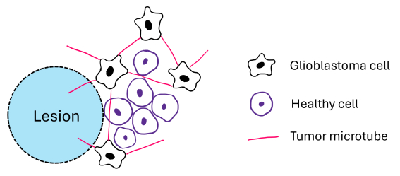
Recently, glioblastoma tumors were shown to form tumor microtubes, which are thin, long protrusions that help the tumor grow and spread. Follow-up experiments were conducted on mice in order to test what impact the tumor microtubes have on tumor regrowth after the partial removal of a tumor region. The surgery was performed in isolation and along with growth-inhibiting treatments such as a tumor microtube-inhibiting treatment and an anti-inflammatory treatment. Here, we have proposed a partial differential equation model applicable to describe the microtube-driven regrowth of the cancer in the lesion. We found that the model is able to replicate the main trends seen in the experiments such as fast regrowth, larger cancer density in the lesion, and further spread into healthy tissue. The model indicates that the dominant mechanisms of re-growth are growth-inducing wound-healing mechanisms and the proliferative advantage from the tumor microtubes. In addition, tumor microtubes provide orientational guidance from the untreated tissue into the lesion.
Citation: Alexandra Shyntar, Thomas Hillen. Mathematical modeling of microtube-driven regrowth of gliomas after local resection[J]. Mathematical Biosciences and Engineering, 2025, 22(1): 52-72. doi: 10.3934/mbe.2025003
Recently, glioblastoma tumors were shown to form tumor microtubes, which are thin, long protrusions that help the tumor grow and spread. Follow-up experiments were conducted on mice in order to test what impact the tumor microtubes have on tumor regrowth after the partial removal of a tumor region. The surgery was performed in isolation and along with growth-inhibiting treatments such as a tumor microtube-inhibiting treatment and an anti-inflammatory treatment. Here, we have proposed a partial differential equation model applicable to describe the microtube-driven regrowth of the cancer in the lesion. We found that the model is able to replicate the main trends seen in the experiments such as fast regrowth, larger cancer density in the lesion, and further spread into healthy tissue. The model indicates that the dominant mechanisms of re-growth are growth-inducing wound-healing mechanisms and the proliferative advantage from the tumor microtubes. In addition, tumor microtubes provide orientational guidance from the untreated tissue into the lesion.

| [1] |
S. H. Torp, O. Solheim, A. J. Skjulsvik, The who 2021 classification of central nervous system tumours: a practical update on what neurosurgeons need to know—a minireview, Acta Neurochir., 164 (2022), 2453–2464. https://doi.org/10.1007/s00701-022-05301-y doi: 10.1007/s00701-022-05301-y

|
| [2] |
M. Osswald, E. Jung, F. Sahm, G. Solecki, V. Venkataramani, J. Blaes, et al., Brain tumour cells interconnect to a functional and resistant network, Nature, 528 (2015), 93–98. https://doi.org/10.1038/nature16071 doi: 10.1038/nature16071
|
| [3] |
S. Weil, M. Osswald, G. Solecki, J. Grosch, E. Jung, D. Lemke, et al., Tumor microtubes convey resistance to surgical lesions and chemotherapy in gliomas, Neuro-Oncology, 19 (2017), 1316–1326. https://doi.org/10.1093/neuonc/nox070 doi: 10.1093/neuonc/nox070
|
| [4] |
D. Hausmann, D. C. Hoffmann, V. Venkataramani, E. Jung, S. Horschitz, S. K. Tetzlaff, et al., Autonomous rhythmic activity in glioma networks drives brain tumour growth, Nature, 613 (2023), 179–186. https://doi.org/10.1038/s41586-022-05520-4 doi: 10.1038/s41586-022-05520-4
|
| [5] |
K. Goslin, D. J. Schreyer, J. P. Skene, G. Banker, Development of neuronal polarity: Gap-43 distinguishes axonal from dendritic growth cones, Nature, 336 (1988), 672–674. https://doi.org/10.1038/336672a0 doi: 10.1038/336672a0
|
| [6] |
O. Garraud, W. N. Hozzein, G. Badr, Wound healing: time to look for intelligent, 'natural'immunological approaches?, BMC Immunol., 18 (2017), 1–8. https://doi.org/10.1186/s12865-017-0207-y doi: 10.1186/s12865-017-0207-y
|
| [7] | D. MacKay, A. L. Miller, Nutritional support for wound healing, Altern. Med. Rev., 8 (2003), 359. |
| [8] |
D. Hanahan, R. A. Weinberg, Hallmarks of cancer: the next generation, Cell, 144 (2011), 646–674. https://doi.org/10.1016/j.cell.2011.02.013 doi: 10.1016/j.cell.2011.02.013
|
| [9] |
D. Hanahan, Hallmarks of cancer: new dimensions, Cancer Dis., 12 (2022), 31–46. https://doi.org/10.1158/2159-8290.CD-21-1059 doi: 10.1158/2159-8290.CD-21-1059
|
| [10] |
J. Juliano, O. Gil, A. Hawkins-Daarud, S. Noticewala, R. C. Rockne, J. Gallaher, et al., Comparative dynamics of microglial and glioma cell motility at the infiltrative margin of brain tumours, J. R. Soc. Interface, 15 (2018), 20170582. https://doi.org/10.1098/rsif.2017.0582 doi: 10.1098/rsif.2017.0582
|
| [11] |
D. Hambardzumyan, D. H. Gutmann, H. Kettenmann, The role of microglia and macrophages in glioma maintenance and progression, Nature Neurosci., 19 (2016), 20–27. https://doi.org/10.1038/nn.4185 doi: 10.1038/nn.4185
|
| [12] |
K. Sadowski, A. Jażdżewska, J. Kozłowski, A. Zacny, T. Lorenc, et al., Revolutionizing glioblastoma treatment: A comprehensive overview of modern therapeutic approaches, Int. J. Mol. Sci., 25 (2024), 5774, https://doi.org/10.3390/ijms25115774 doi: 10.3390/ijms25115774
|
| [13] | C. McKinnon, M. Nandhabalan, S. A. Murray, P. Plaha, Glioblastoma: clinical presentation, diagnosis, and management, BMJ, 374 (2021). https://doi.org/10.1136/bmj.n1560 |
| [14] |
X. Gao, J. T. McDonald, L. Hlatky, H. Enderling, Acute and fractionated irradiation differentially modulate glioma stem cell division kinetics, Cancer Res., 73 (2013), 1481–1490. https://doi.org/10.1158/0008-5472.CAN-12-3429 doi: 10.1158/0008-5472.CAN-12-3429
|
| [15] | M. Lê, H. Delingette, J. Kalpathy-Cramer, E. R. Gerstner, T. Batchelor, J. Unkelbach, et al., Bayesian personalization of brain tumor growth model, in Medical Image Computing and Computer-Assisted Intervention–MICCAI 2015: 18th International Conference, Springer, (2015), 424–432. https://doi.org/10.1007/978-3-319-24571-3_51 |
| [16] |
K. R. Swanson, E. C. Alvord Jr, J. D. Murray, A quantitative model for differential motility of gliomas in grey and white matter, Cell Proliferation, 33 (2000), 317–329. https://doi.org/10.1046/j.1365-2184.2000.00177.x doi: 10.1046/j.1365-2184.2000.00177.x
|
| [17] |
J. Jacobs, R. C. Rockne, A. J. Hawkins-Daarud, P. R. Jackson, S. K. Johnston, P. Kinahan, et al., Improved model prediction of glioma growth utilizing tissue-specific boundary effects, Math. Biosci., 312 (2019), 59–66. https://doi.org/10.1016/j.mbs.2019.04.004 doi: 10.1016/j.mbs.2019.04.004
|
| [18] |
P. G. Gritsenko, O. Ilina, P. Friedl, Interstitial guidance of cancer invasion, J. Pathol., 226 (2012), 185–199. https://doi.org/10.1002/path.3031 doi: 10.1002/path.3031
|
| [19] |
M. C. Colombo, C. Giverso, E. Faggiano, C. Boffano, F. Acerbi, P. Ciarletta, Towards the personalized treatment of glioblastoma: integrating patient-specific clinical data in a continuous mechanical model, PLoS One, 10 (2015), e0132887. https://doi.org/10.1371/journal.pone.0143032 doi: 10.1371/journal.pone.0143032
|
| [20] |
A. Gholami, A. Mang, G. Biros, An inverse problem formulation for parameter estimation of a reaction–diffusion model of low grade gliomas, J. Math. Biol., 72 (2016), 409–433. https://doi.org/10.1007/s00285-015-0888-x doi: 10.1007/s00285-015-0888-x
|
| [21] |
K. Painter, T. Hillen, Mathematical modelling of glioma growth: the use of diffusion tensor imaging (DTI) data to predict the anisotropic pathways of cancer invasion, J. Theor. Biol., 323 (2013), 25–39. https://doi.org/10.1016/j.jtbi.2013.01.014 doi: 10.1016/j.jtbi.2013.01.014
|
| [22] |
M. Conte, C. Surulescu, Mathematical modeling of glioma invasion: acid-and vasculature mediated go-or-grow dichotomy and the influence of tissue anisotropy, Appl. Math. Comput., 407 (2021), 126305. https://doi.org/10.1016/j.amc.2021.126305 doi: 10.1016/j.amc.2021.126305
|
| [23] |
A. Swan, T. Hillen, J. C. Bowman, A. D. Murtha, A patient-specific anisotropic diffusion model for brain tumour spread, Bull. Math. Biol., 80 (2018), 1259–1291. https://doi.org/10.1007/s11538-017-0271-8 doi: 10.1007/s11538-017-0271-8
|
| [24] |
T. Hillen, N. Loy, K. J. Painter, R. Thiessen, Modelling microtube driven invasion of glioma, J. Math. Biol., 88 (2024). https://doi.org/10.1007/s00285-023-02025-0 doi: 10.1007/s00285-023-02025-0
|
| [25] |
I. Bica, T. Hillen, K. J. Painter, Aggregation of biological particles under radial directional guidance, J. Theor. Biol., 427 (2017), 77–89. https://doi.org/10.1016/j.jtbi.2017.05.039 doi: 10.1016/j.jtbi.2017.05.039
|
| [26] |
T. Hillen, K. J. Painter, A. C. Swan, A. D. Murtha, Moments of von Mises and Fisher distributions and applications, Math. Biosci. Eng., 14 (2017), 673–694. https://doi.org/10.3934/mbe.2017038 doi: 10.3934/mbe.2017038
|
| [27] | T. Hillen, K. J. Painter, Transport and anisotropic diffusion models for movement in oriented habitats, in Dispersal, individual movement and spatial ecology: A mathematical perspective, Springer, (2013), 177–222. https://doi.org/10.1007/978-3-642-35497-7_7 |
| [28] |
T. Hillen, $M^5$, mesoscopic and macroscopic models for mesenchymal motion, J. Math. Biol., 53 (2006), 585–616. https://doi.org/10.1007/s00285-006-0017-y doi: 10.1007/s00285-006-0017-y
|
| [29] |
H. Othmer, S. Dunbar, W. Alt, Models of dispersal in biological systems, J. Math. Biol., 26 (1988), 263–298. https://doi.org/10.1007/BF00277392 doi: 10.1007/BF00277392
|
| [30] | J. Murray, Mathematical Biology. Ⅰ: An Introduction, 3rd edition, Springer-Verlag, New York, 2002. |
| [31] |
D. G. Iworima, R. K. Baker, C. Ellis, C. Sherwood, L. Zhan, A. Rezania, et al., Metabolic switching, growth kinetics and cell yields in the scalable manufacture of stem cell-derived insulin-producing cells, Stem Cell Res. Therapy, 15 (2024), 1. https://doi.org/10.1186/s13287-023-03574-3 doi: 10.1186/s13287-023-03574-3
|
| [32] |
D. Giulian, J. Chen, J. Ingeman, J. George, M. Noponen, The role of mononuclear phagocytes in wound healing after traumatic injury to adult mammalian brain, J. Neurosci., 9 (1989), 4416–4429. https://doi.org/10.1523/JNEUROSCI.09-12-04416.1989 doi: 10.1523/JNEUROSCI.09-12-04416.1989
|
| [33] |
T. Hillen, K. Painter, M. Winkler, Anisotropic diffusion in oriented environments can lead to singularity formation, Eur. J. Appl. Math., 24 (2013), 371–413. https://doi.org/10.1017/S0956792512000447 doi: 10.1017/S0956792512000447
|
| [34] | H. Wang, Mathematical modeling I-preliminary, Bookboon, 2012. |
| [35] |
P. W. Tsao, T. Suzuki, R. Totsuka, T. Murata, T. Takagi, et al., The effect of dexamethasone on the expression of activated nf-$\kappa$b in adjuvant arthritis, Clin. Immunol. Immunopathol., 83 (1997), 173–178. https://doi.org/10.1006/clin.1997.4333 doi: 10.1006/clin.1997.4333
|
| [36] |
K. D. Bosscher, W. V. Berghe, L. Vermeulen, S. Plaisance, E. Boone, G. Haegeman, Glucocorticoids repress nf-$\kappa$b-driven genes by disturbing the interaction of p65 with the basal transcription machinery, irrespective of coactivator levels in the cell, PNAS, 97 (2000), 3919–3924. https://doi.org/10.1073/pnas.97.8.391 doi: 10.1073/pnas.97.8.391
|
| [37] |
K. Yakimchuk, Mathematical modeling of immune modulation by glucocorticoids, Biosystems, 187 (2020), 104066. https://doi.org/10.1016/j.biosystems.2019.104066 doi: 10.1016/j.biosystems.2019.104066
|
| [38] |
A. Wandler, B. Huang, Loss of glucocorticoid receptor expression mediates in vivo dexamethasone resistance in t-cell acute lymphoblastic leukemia, Leukemia, 34 (2020), 2025–2037. https://doi.org/10.1038/s41375-020-0748-6 doi: 10.1038/s41375-020-0748-6
|
| [39] |
A. S. Rosenberg, From mechanism to resistance – changes in the use of dexamethasone in the treatment of multiple myeloma, Leuk. Lymphoma, 64 (2023), 283–291. https://doi.org/10.1080/10428194.2022.2136950 doi: 10.1080/10428194.2022.2136950
|
| [40] |
J. Gong, M. M. Dos Santos, C. Finlay, T. Hillen, Are more complicated tumour control probability models better?, Math. Med. Biol. J. IMA, 30 (2013), 1–19. https://doi.org/10.1093/imammb/dqr023 doi: 10.1093/imammb/dqr023
|
| [41] |
K. Owens, I. Bozic, Modeling car t-cell therapy with patient preconditioning, Bull. Math. Biol., 83 (2021), 42. https://doi.org/10.1007/s11538-021-00869-5 doi: 10.1007/s11538-021-00869-5
|


Figures(9) / Tables(2)

Alexandra Shyntar, Thomas Hillen. Mathematical modeling of microtube-driven regrowth of gliomas after local resection[J]. Mathematical Biosciences and Engineering, 2025, 22(1): 52-72. doi: 10.3934/mbe.2025003


 DownLoad: