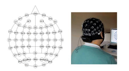
Most studies on drug addiction degree are made based on statistical scales, addicts' account, and subjective judgement of rehabilitation doctors. No objective, quantified evaluation has been made. This paper uses devises the synchronous bimodal signal collection and experimentation paradigm with electroencephalogram (EEG) and forehead high-density near-infrared spectroscopy (NIRS) device. The drug addicts are classified into mild, moderate and severe groups with reference to the suggestions of researchers and medical experts. Data of 45 drug addicts (mild: 15; moderate: 15; and severe: 15) is collected, and then used to design an addiction degree testing algorithm based on decision fusion. The algorithm is used to classify mild, moderate and severe addiction. This paper pioneers to use two types of Convolutional Neural Network (CNN) to abstract the EEG and NIR data of drug addicts, and introduces batch normalization to CNN, thus accelerating training process, reducing parameter sensitivity, and enhancing system robustness. The characteristics output by two CNNs are transformed into dimensions. Two new characteristics are assigned with a weight of 50% each. The data is used for decision fusion. In the networks, 27 subjects are used as training sets, 9 as validation sets, and 9 as testing sets. The 3-class accuracy remains to be 63.15%, preliminarily justifying this method as an effective approach to measure drug addiction degree. And the method is ready to use, objective, and offers results in real time.
Citation: Xuelin Gu, Banghua Yang, Shouwei Gao, Lin Feng Yan, Ding Xu, Wen Wang. Application of bi-modal signal in the classification and recognition of drug addiction degree based on machine learning[J]. Mathematical Biosciences and Engineering, 2021, 18(5): 6926-6940. doi: 10.3934/mbe.2021344
Most studies on drug addiction degree are made based on statistical scales, addicts' account, and subjective judgement of rehabilitation doctors. No objective, quantified evaluation has been made. This paper uses devises the synchronous bimodal signal collection and experimentation paradigm with electroencephalogram (EEG) and forehead high-density near-infrared spectroscopy (NIRS) device. The drug addicts are classified into mild, moderate and severe groups with reference to the suggestions of researchers and medical experts. Data of 45 drug addicts (mild: 15; moderate: 15; and severe: 15) is collected, and then used to design an addiction degree testing algorithm based on decision fusion. The algorithm is used to classify mild, moderate and severe addiction. This paper pioneers to use two types of Convolutional Neural Network (CNN) to abstract the EEG and NIR data of drug addicts, and introduces batch normalization to CNN, thus accelerating training process, reducing parameter sensitivity, and enhancing system robustness. The characteristics output by two CNNs are transformed into dimensions. Two new characteristics are assigned with a weight of 50% each. The data is used for decision fusion. In the networks, 27 subjects are used as training sets, 9 as validation sets, and 9 as testing sets. The 3-class accuracy remains to be 63.15%, preliminarily justifying this method as an effective approach to measure drug addiction degree. And the method is ready to use, objective, and offers results in real time.

| [1] |
X. B. Chen, Y. L. Ma, X. Y. Liu, W. Z. Kong, X. G. Xi, Analysis of corticomuscular connectivity during walking using vine copula, Math. Biosci. Eng., 18 (2021), 4341-4357. doi: 10.3934/mbe.2021218

|
| [2] |
A. S. Huhn, R. K. Brooner, M. M. Sweeney, S. W. Yip, H. Ayaz, K. E. Dunn, Increased neural activity in the right dorsolateral prefrontal cortex during a risky decision-making task is associated with cocaine use in methadone-maintained patients, Drug. Alcohol. Depend., 205 (2019), 107650. doi: 10.1016/j.drugalcdep.2019.107650
|
| [3] |
R. Madanu, F. Rahman, M. F. Abbod, S. Z. Fan, J. S. Shieh, Depth of anesthesia prediction via EEG signals using convolutional neural network and ensemble empirical mode decomposition, Math. Biosci. Eng., 18 (2021), 5047-5068. doi: 10.3934/mbe.2021257
|
| [4] | M. Ferrari, V. Quaresima, A brief review on the history of human functional near infrared spectroscopy (fNIRS) development and fields of application, Neuroimage, 63 (2021), 921-935. |
| [5] | A. M. Kroczek, F. B. Haeussinger, A. J. Fallgatter, A. Batra, A. C. Ehlis, Prefrontal functional connectivity measured with near-infrared spectroscopy during smoking cue exposure, Addict. Biol., 22 (2015), 513-522. |
| [6] | T. Liu, X. C. Liu, L. Yi, C. Z. Zhu, P. S. Markey, M. Pelowski, Assessing autism at its social and developmental roots, A review of Autism Spectrum Disorder studies using functional near-infrared spectroscopy, Neuroimage, 185 (2017), 955-967. |
| [7] |
L. Huang, S. Y. Guo, Y. Wang, S. Wang, Q. B. Chu, L. Li, et al., Attention based residual network for medicinal fungi near infrared spectroscopy analysis, Math. Biosci. Eng., 16 (2019), 3003-3017. doi: 10.3934/mbe.2019149
|
| [8] |
F. Al-Shargie, T. B. Tang, M. Kiguchi, Assessment of mental stress effects on prefrontal cortical activities using canonical correlation analysis: an fNIRS-EEG study, Biomed. Opt. Express., 8 (2017), 2583-2598. doi: 10.1364/BOE.8.002583
|
| [9] |
B. Abibullaev, J. An, S. H. Lee, J. I. Moon, Design and evaluation of action observation and motor imagery based BCIs using near-infrared spectroscopy, Measurement, 98 (2017), 250-261. doi: 10.1016/j.measurement.2016.12.001
|
| [10] |
A. Kassab, J. L. Lan, J. Tremblay, P. Vannasing, M. Dehbozorgi, P. Pouliot, et al., Multichannel wearable fNIRS-EEG system for long-term clinical monitoring, Hum. Brain. Mapp., 39 (2018), 7-23. doi: 10.1002/hbm.23849
|
| [11] | A. Lühmann, J. Addesa, S. Chandra, A. Das, M. Hayashibe, A. Dutta, Neural interfacing non-invasive brain stimulation with NIRS-EEG joint imaging for closed-loop control of neuroenergetics in ischemic stroke, IEEE Embc., (2017), 349-353. |
| [12] |
T. Zhang, L. Guo, K. M. Li, C. F. Jing, Y. Yin, D. J. Zhu, et al, Predicting functional cortical ROIs via DTI-derived fiber shape models, Cerebral. Cortex., 22 (2012), 854-864. doi: 10.1093/cercor/bhr152
|
| [13] |
Y. F. Xia, H. L. Zhang, L. Xu, Z. F. Gao, H. Y. Zhang, H. F. Liu, et al., An automatic cardiac arrhythmia classification system with wearable electrocardiogram, IEEE. Access., 6 (2018), 16529-16538. doi: 10.1109/ACCESS.2018.2807700
|
| [14] |
F. Wallois, M. Mahmoudzadeh, A. Patil, R. Grebe, Usefulness of simultaneous EEG-NIRS recording in language studies, Brain. Lang., 121 (2012), 110-23. doi: 10.1016/j.bandl.2011.03.010
|
| [15] |
M. Balconi, E. Grippa, M. E. Vanutelli, What hemodynamic (fNIRS), electrophysiological (EEG) and autonomic integrated measures can tell us about emotional processing, Brain. Cogn., 95 (2015), 67-76. doi: 10.1016/j.bandc.2015.02.001
|
| [16] | S. Fazli, J. Mehnert, J. Steinbrink, G. Curio, A. Villringer, K. R. Müller, et al., Enhanced preformance by a hybrid NIRS-EEG brain computer interface, Neuroimage, 59 (2012), 591-529. |
| [17] |
Y. Tomita, F. B. Vialatte, G. Dreyfus, Y. Mitsukura, H. Bakardjian, A. Cichocki, Bimodal BCI using simultaneously NIRS and EEG, IEEE. Trans. Biomed. Eng., 61 (2014), 1274-1284. doi: 10.1109/TBME.2014.2300492
|
| [18] |
P. Pouliot, T. P. Y. Tran, V. Birca, P. Vannasing, J. Tremblay, M. Lassonde, et al., Hemodynamic changes during posterior epilepsies: an EEG-fNIRS study, Epilepsy. Res., 108 (2014), 883-890. doi: 10.1016/j.eplepsyres.2014.03.007
|
| [19] | U. Jindal, M. Sood, A. Dutta, S. R. Chowdhury, Development of point of care testing device for neurovascular coupling from simultaneous recording of EEG and NIRS during anodal transcranial direct current stimulation, IEEE. J. Transl. Eng. He., 3 (2015), 1-12. |
| [20] | M. J. Khan, M. J. Y. Hong, K. S. Hong, Decoding of four movement directions using hybrid NIRS-EEG brain-computer interface. Front. Hum. Neurosci., 8 (2014), 412-432. |
| [21] | F. Putze, S. Hesslinger, C. Y. Tse, Y. Y. Huang, C. Herff, C. T. Guan, et al., Hybrid fNIRS-EEG based classification of auditory and visual perception processes, Neuroimage, 8 (2014), 19-29. |
| [22] |
C. Zich, S. Debener, A. K. Thoene, L. C. Chen, C. Kranczioch, Simultaneous EEG-fNIRS reveals how age and feedback affect motor imagery signatures, Neurobiol. Aging., 49 (2017), 183-197. doi: 10.1016/j.neurobiolaging.2016.10.011
|
| [23] | J. Safaie, R. Grebe, H. A. Moghaddam, F. Wallois, Toward a fully integrated wireless wearable EEG-NIRS bimodal acquisition system, J. Neural. Eng., 10 (2013), 637-637. |
| [24] |
M. Sawan, M. T. Salam, J. L. Lan, A. Kassab, S. Gelinas, P. Vannasing, et al., Wireless recording systems: from noninvasive EEG-NIRS to invasive EEG devices, IEEE. T. Biomed. Circ. S., 7 (2013), 186-195. doi: 10.1109/TBCAS.2013.2255595
|
| [25] | R. K. Almajidy, Y. Boudria, U. G. Hofmann, W. Besio, K, Mankodiya, A multimodal 2D brain computer interface, IEEE Embc., (2015), 1067-1070. |
| [26] |
A. P. Buccino, H. O. Keles, A. Omurtag, Hybrid EEG-fNIRS asynchronous brain-computer interface for multiple motor tasks, PLoS One, 11 (2016), e0146610. doi: 10.1371/journal.pone.0146610
|
| [27] |
J. Shin, D. W. Kim, K. R. Muller, H. J. Hwang, Improvement of information transfer rates using a hybrid EEG-NIRS brain-computer interface with a short trial length: offline and pseudo-online analyses, Sensors, 18 (2018), 1827. doi: 10.3390/s18061827
|
| [28] | D. Guo, G. M. Liu, Deep learning-based bimodal emotion recognition from facial expression and body posture, in Nanjing University of Posts and Telecommunications, 2019. |
| [29] |
J. M. Kim, J. K. Choi, M. Choi, M. Ji, G. Hwang, S. B. Ko, Assessment of cerebral autoregulation using continuous-wave near-infrared spectroscopy during squat-stand maneuvers in subjects with symptoms of orthostatic intolerance, Sci. Rep., 8 (2018), 13257. doi: 10.1038/s41598-018-31685-y
|
| [30] |
J. Shin, J. Kwon, J. Choi, C. H. Im, Performance enhancement of a brain-computer interface using high-density multi-distance NIRS, Sci. Rep., 7 (2017), 16545. doi: 10.1038/s41598-017-16639-0
|
| [31] |
H. F. Song, W. W. Yang, S. S. Dai, L. Du, Y. C. Sun, Using dual-channel CNN to classify hyperspectral image based on spatial-spectral information, Math. Biosci. Eng., 17 (2020), 3450-3477. doi: 10.3934/mbe.2020195
|
| [32] | C. L. Liu, P. Cui, T. Huang, Identification of cell cycle-regulated genes by convolutional neural network, Comb. Chem. High. Throughput. Screen., 20 (2017), 603-611. |
| [33] |
S. Raghu, N. Sriraam, Y. Temel, S. V. Rao, P. L. Kubben, EEG based multi-class seizure type classification using convolutional neural network and transfer learning, Neural. Netw., 124 (2020), 202-212. doi: 10.1016/j.neunet.2020.01.017
|
| [34] |
J. Y. Li, Q. Z. Xu, M. X. Wu, T. Huang, Y. D. Wang, Pan-cancer cassification based on self-normalizing neural networks and feature selection, Front. Bioeng. Biotechnol., 8 (2020), 766. doi: 10.3389/fbioe.2020.00766
|
| [35] | B. A. Mohammed, M. S. Al-Ani, An efficient approach to diagnose brain tumors through deep CNN, Math. Biosci. Eng., 18 (2020), 851-867. |
| [36] |
L. Chen, X. Y, Pan, Y. H. Zhang, M. Liu, T. Huang, Y. D. Cai, Classification of widely and rarely expressed genes with recurrent neural network, Comput. Struct. Biotechnol. J., 17 (2019), 49-60. doi: 10.1016/j.csbj.2018.12.002
|
| [37] |
P. Croce, F. Zappasodi, L. Marzetti, A. Merla, V. Pizzella, A. M. Chiarelli, Deep convolutional neural networks for feature-less automatic classification of independent components in multi-channel electrophysiological brain recordings, IEEE. Trans. Biomed. Eng., 66 (2019), 2372-2380. doi: 10.1109/TBME.2018.2889512
|
| [38] | X. Y. Pan, L. Chen, I. Liu, Z. B. Niu, T. Huang, Y. D. Cai, Identifying protein subcellular locations with embeddings-based node2loc, IEEE. ACM. T. Comput. Bi., 2021. |
| [39] | A. Das, D. Guhathakurta, R. Sengupta, A. Dutta, EEG-NIRS joint-image based assessment of neurovascular coupling in stroke, Int. J. Stroke., 11 (2016), 271-272. |
| [40] | S. F. Xing, Y. X. Zhang, H. L. Wang, J. Y. Tu, Study on the criteria of fine needle aspiration cytology for the diagnosis and classification of malignant lymphomas, Chin. J. Pathol., 16 (1987), 174. |
| [41] |
J. Shin, A. von Luhmann, D. W. Kim, J. Mehnert, H. J. Hwang, K. R. Muller, Data descriptor: simultaneous acquisition of EEG and NIRS during cognitive tasks for an open access dataset, Sci. Data, 5 (2018), 180003. doi: 10.1038/sdata.2018.3
|


Figures(11) / Tables(2)

Xuelin Gu, Banghua Yang, Shouwei Gao, Lin Feng Yan, Ding Xu, Wen Wang. Application of bi-modal signal in the classification and recognition of drug addiction degree based on machine learning[J]. Mathematical Biosciences and Engineering, 2021, 18(5): 6926-6940. doi: 10.3934/mbe.2021344


 DownLoad: