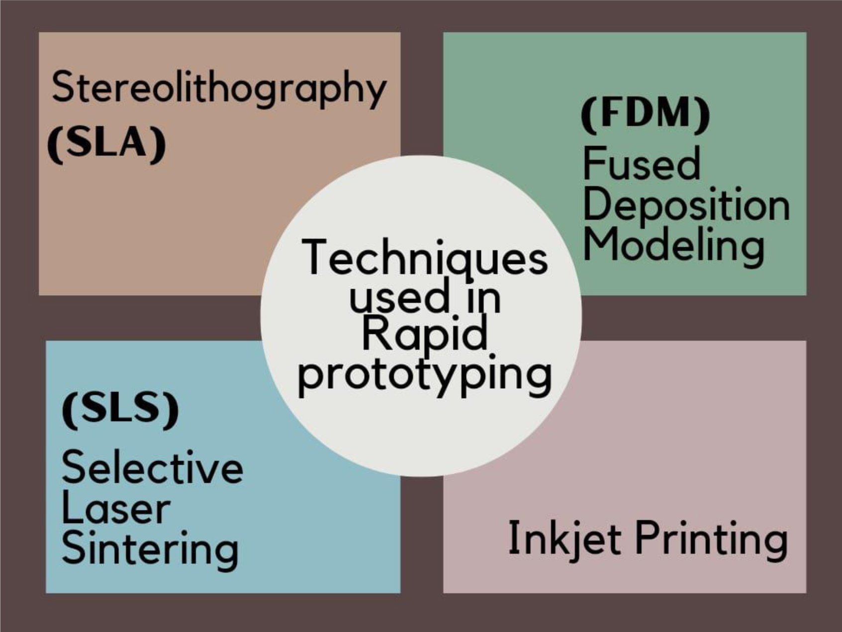
The term “rapid prototyping” (RP) refers to a variety of methods for creating “physical models based on computer-aided design and computer-aided manufacturing”. With the aid of RP technology, practically any variation of the surface and interior anatomical structure may be replicated in a medical model that is constructed layer by layer. To create the physical model, layer-by-layer construction is carried out using a variety of processes, including stereolithography, selective laser sintering, inkjet printing, and fused deposit modeling. Data for RP is received from magnetic resonance imaging and computed tomography scans, which are then turned into digital images and then into standard triangulation language files. The use of this computerized programming in orthodontics incorporates “diagnosis and treatment planning”, the creation of removable “orthodontic appliances”, “impression trays” for indirect bonding, “3D printed occlusal splints and aligners”, prototype models used in various orthognathic surgeries, and the production of a distractor for distraction osteogenesis. It increases a crucial understanding at the time of preoperative treatment planning and raises the effectiveness of the therapy, yet, clinical judgment is still essential. Applications of RP for an orthodontist vary, and if we utilize it creatively, the future appears more hopeful. This article briefly reviews key advancements, challenges, and prospects in the integration of rapid prototyping and 3D printing, shaping a promising future for orthodontics.
Citation: Simran Rajesh Katyari, Prateeksha Lakhe, Amit Reche. Rapid prototyping: A future in orthodontic[J]. AIMS Bioengineering, 2024, 11(1): 66-84. doi: 10.3934/bioeng.2024005
The term “rapid prototyping” (RP) refers to a variety of methods for creating “physical models based on computer-aided design and computer-aided manufacturing”. With the aid of RP technology, practically any variation of the surface and interior anatomical structure may be replicated in a medical model that is constructed layer by layer. To create the physical model, layer-by-layer construction is carried out using a variety of processes, including stereolithography, selective laser sintering, inkjet printing, and fused deposit modeling. Data for RP is received from magnetic resonance imaging and computed tomography scans, which are then turned into digital images and then into standard triangulation language files. The use of this computerized programming in orthodontics incorporates “diagnosis and treatment planning”, the creation of removable “orthodontic appliances”, “impression trays” for indirect bonding, “3D printed occlusal splints and aligners”, prototype models used in various orthognathic surgeries, and the production of a distractor for distraction osteogenesis. It increases a crucial understanding at the time of preoperative treatment planning and raises the effectiveness of the therapy, yet, clinical judgment is still essential. Applications of RP for an orthodontist vary, and if we utilize it creatively, the future appears more hopeful. This article briefly reviews key advancements, challenges, and prospects in the integration of rapid prototyping and 3D printing, shaping a promising future for orthodontics.

| [1] | Biglino G, Schievano S, Taylor MA (2011) The use of rapid prototyping in clinical applications. Advance Applications of Rapid Prototyping Technology in Modern Engineering . United Kingdom: Intech Open 21-40. http://dx.doi.org/10.5772/24128 |
| [2] |
Choi JY, Choi JH, Kim NK, et al. (2002) Analysis of errors in medical rapid prototyping models. Int J Oral Max Surg 31: 23-32. https://doi.org/10.1054/ijom.2000.0135 
|
| [3] | Bozkurt Y, Karayel E (2021) 3D printing technology; methods, biomedical applications, future opportunities and trends. J Mater Res 14: 1430-1450. https://doi.org/10.1016/j.jmrt.2021.07.050 |
| [4] |
Li H, Fan W, Zhu X (2020) Three-dimensional printing: the potential technology widely used in medical fields. J Biomed Mater Res 108: 2217-2229. https://doi.org/10.1002/jbm.a.36979
|
| [5] |
Tareq MS, Rahman T, Hossain M, et al. (2021) Additive manufacturing and the COVID-19 challenges: an in-depth study. J Manuf Syst 60: 787-798. https://doi.org/10.1016/j.jmsy.2020.12.021
|
| [6] |
Khalid MY, Arif ZU, Noroozi R, et al. (2023) 3D/4D printing of cellulose nanocrystals-based biomaterials: additives for sustainable applications. Int J Biol Macromol 251: 126287. https://doi.org/10.1016/j.ijbiomac.2023.126287
|
| [7] |
Seoane-Viaño I, Januskaite P, Alvarez-Lorenzo C, et al. (2021) Semi-solid extrusion 3D printing in drug delivery and biomedicine: personalised solutions for healthcare challenges. J Control Release 332: 367-389. https://doi.org/10.1016/j.jconrel.2021.02.027
|
| [8] |
Agarwal T, Tan SA, Onesto V, et al. (2021) Engineered herbal scaffolds for tissue repair and regeneration: recent trends and technologies. Adv Biomed Eng 2: 100015. https://doi.org/10.1016/j.bea.2021.100015
|
| [9] |
Żyłka E, Irzmańska E, Saramak J, et al. (2023) Functional 3D-printed polymeric materials with metallic reinforcement for use in cut-resistant gloves. Materials 17: 90. https://doi.org/10.3390/ma17010090
|
| [10] | Ravichandran D, Xu W, Kakarla M, et al. (2021) Multiphase direct ink writing (MDIW) for multilayered polymer/nanoparticle composites. Addit Manuf 47: 102322. https://doi.org/10.1016/j.addma.2021.102322 |
| [11] |
Shah SA, Sohail M, Khan S, et al. (2019) Biopolymer-based biomaterials for accelerated diabetic wound healing: a critical review. Int J Biolog Macromol 139: 975-993. https://doi.org/10.1016/j.ijbiomac.2019.08.007
|
| [12] |
Abdal-hay A, Raveendran NT, Fournier B, et al. (2020) Fabrication of biocompatible and bioabsorbable polycaprolactone/ magnesium hydroxide 3D printed scaffolds: degradation and in vitro osteoblasts interactions. Compos B Eng 197: 108158. https://doi.org/10.3389/fbioe.2023.1272348
|
| [13] |
Tahouni Y, Cheng T, Lajewski S, et al. (2023) Codesign of biobased cellulose-filled filaments and mesostructures for 4D printing humidity responsive smart structures. 3D Print Addit Manuf 10: 1-14. https://doi.org/10.1089/3dp.2022.0061
|
| [14] |
Khoo ZX, Teoh JEM, Liu Y, et al. (2015) 3D printing of smart materials: a review on recent progresses in 4D printing. Virtual Phys Prototyp 10: 103-122. https://doi.org/10.3390/mi11090796
|
| [15] |
Khalid MY, Arif ZU, Noroozi R, et al. (2022) 4D printing of shape memory polymer composites: a review on fabrication techniques, applications, and future perspectives. J Manuf Process 81: 759-797. http://dx.doi.org/10.1016/j.jmapro.2022.07.035
|
| [16] |
Cui X, Ruan Q, Zhuo X, et al. (2023) Photothermal nanomaterials: a powerful light-to-heat converter. Chem Rev 123: 6891-6952. https://doi.org/10.1021/acs.chemrev.3c00159
|
| [17] |
Tariq A, Arif ZU, Khalid MY, et al. (2023) Recent advances in the additive manufacturing of stimuli-responsive soft polymers. Adv Eng Mater 25: 2301074. http://dx.doi.org/10.1002/adem.202301074
|
| [18] |
Kermavnar T, Shannon A, O'Sullivan LW (2021) The application of additive manufacturing/3D printing in ergonomic aspects of product design: a systematic review. Appl Ergon 97: 103528. https://doi.org/10.1016/j.apergo.2021.103528
|
| [19] | Royer P (1958) Disorders of tubular phosphate mechanism. Monatsschr Kinderh 106: 167-168. https://doi.org/10.7861/clinmedicine.12-5-476 |
| [20] |
Liu Q, Leu MC, Schmitt SM (2006) Rapid prototyping in dentistry: technology and application. Int J Adv Manuf Technol 29: 317-335. http://dx.doi.org/10.1007/s00170-005-2523-2
|
| [21] |
Madhav VNV, Daule R (2013) Rapid prototyping and its application in dentistry. J Dent Allied Sci 2: 57. http://dx.doi.org/10.4103/2277-4696.159285
|
| [22] |
Negi S, Dhiman S, Kumar Sharma R (2014) Basics and applications of rapid prototyping medical models. Rapid Prototyp J 20: 256-267 . http://dx.doi.org/10.1108/RPJ-07-2012-0065
|
| [23] |
Chan DCN, Frazier KB, Tse LA, et al. (2004) Application of rapid prototyping to operative dentistry curriculum. J Dent Educ 68: 64-70. http://dx.doi.org/10.1002/j.0022-0337.2004.68.1.tb03737.x
|
| [24] |
Bidra AS, Taylor TD, Agar JR (2013) Computer-aided technology for fabricating complete dentures: systematic review of historical background, current status, and future perspectives. J Prosthet Dent 109: 361-366. https://doi.org/10.1016/s0022-3913(13)60318-2
|
| [25] |
Chocholata P, Kulda V, Babuska V (2019) Fabrication of scaffolds for bone-tissue regeneration. Mater 12: 568. https://doi.org/10.3390/ma12040568
|
| [26] |
Zhang B, Song J (2018) 3D-Printed biomaterials for guided tissue regeneration. Small Methods 2: 1700306. http://dx.doi.org/10.1002/smtd.201700306
|
| [27] |
Di Marzio N, Eglin D, Serra T, et al. (2020) Bio-fabrication: convergence of 3D bioprinting and nano-biomaterials in tissue engineering and regenerative medicine. Front Bioeng Biotechnol 8: 326. https://doi.org/10.3389/fbioe.2020.00326
|
| [28] | Midha S, Dalela M, Sybil D, et al. (2019) Advances in three-dimensional bioprinting of bone: progress and challenges. J Tissue Eng Regen Med 13: 925-945. https://doi.org/10.1002/term.2847 |
| [29] |
Mohammed A, Elshaer A, Sareh P, et al. (2020) Additive manufacturing technologies for drug delivery applications. Int J Pharm 580: 119245. https://doi.org/10.1016/j.ijpharm.2020.119245
|
| [30] |
Ghilan A, Chiriac AP, Nita LE, et al. (2020) Trends in 3D printing processes for biomedical field: opportunities and challenges. J Polym Environ 28: 1345-1367. https://doi.org/10.1007/s10924-020-01722-x
|
| [31] |
Daly R, Harrington TS, Martin GD, et al. (2015) Inkjet printing for pharmaceutics – a review of research and manufacturing. Int J Pharm 494: 554-567. https://doi.org/10.1016/j.ijpharm.2015.03.017
|
| [32] |
Khorsandi D, Fahimipour A, Abasian P, et al. (2021) 3D and 4D printing in dentistry and maxillofacial surgery: printing techniques, materials, and applications. Acta Biomater 122: 26-49. https://doi.org/10.1016/j.actbio.2020.12.044
|
| [33] |
Zheng Z, Patel M, Patel R (2022) Hyaluronic acid-based materials for bone regeneration: a review. React Funct Polym 171: 105151. http://dx.doi.org/10.1016/j.reactfunctpolym.2021.105151
|
| [34] |
Javaid M, Haleem A (2020) Significant advancements of 4D printing in the field of orthopaedics. J Clin Orthop Trauma 11: S485-S490. https://doi.org/10.1016/j.jcot.2020.04.021
|
| [35] |
Rajabi N, Rezaei A, Kharaziha M, et al. (2021) Recent advances on bioprinted gelatin methacrylate-based hydrogels for tissue repair. Tissue Eng Part A 27: 679-702. https://doi.org/10.1089/ten.tea.2020.0350
|
| [36] |
Rokaya D, Kongkiatkamon S, Heboyan A, et al. (2022) 3D-printed biomaterials in biomedical application. Functional Biomaterials: Drug Delivery and Biomedical Applications . Singapore: Springer Singapore 319-339. http://dx.doi.org/10.1007/978-981-16-7152-4_12
|
| [37] |
Lui YS, Sow WT, Tan LP, et al. (2019) 4D printing and stimuli-responsive materials in biomedical aspects. Acta Biomater 92: 19-36. https://doi.org/10.1016/j.actbio.2019.05.005
|
| [38] |
Osouli-Bostanabad K, Masalehdan T, Kapsa RMI, et al. (2022) Traction of 3D and 4D printing in the healthcare industry: from drug delivery and analysis to regenerative medicine. ACS Biomater Sci Eng 8: 2764-2797. https://doi.org/10.1021/acsbiomaterials.2c00094
|
| [39] |
Zhao D, Pang B, Zhu Y, et al. (2022) A stiffness-switchable, biomimetic smart material enabled by supramolecular reconfiguration. Adv Mater 34: 2107857. https://doi.org/10.1002/adma.202107857
|
| [40] |
Su JW, Tao X, Deng H, et al. (2018) 4D printing of a self-morphing polymer driven by a swellable guest medium. Soft Matter 14: 765-772. https://doi.org/10.3390/polym11111864
|
| [41] | Haleem A, Javaid M, Singh RP, et al. (2021) Significant roles of 4D printing using smart materials in the field of manufacturing. Adv Ind Eng Polym Res 4: 301-311. http://dx.doi.org/10.1016/j.aiepr.2021.05.001 |
| [42] |
Palmara G, Frascella F, Roppolo I, et al. (2021) Functional 3D printing: approaches and bioapplications. Biosens Bioelectron 175: 112849. https://doi.org/10.1016/j.bios.2020.112849
|
| [43] |
Heidarian P, Kaynak A, Paulino M, et al. (2021) Dynamic nanocellulose hydrogels: recent advancements and future outlook. Carbohydr Polym 270: 118357. https://doi.org/10.1016/j.carbpol.2021.118357
|
| [44] |
Heidarian P, Kouzani AZ, Kaynak A, et al. (2020) Dynamic plant-derived polysaccharide-based hydrogels. Carbohydr Polym 231: 115743. https://doi.org/10.1016/j.carbpol.2019.115743
|
| [45] |
Bom S, Ribeiro R, Ribeiro HM, et al. (2022) On the progress of hydrogel-based 3D printing: correlating rheological properties with printing behaviour. Int J Pharm 615: 121506. https://doi.org/10.1016/j.ijpharm.2022.121506
|
| [46] |
Wang Y, Li X (2021) 4D-printed bi-material composite laminate for manufacturing reversible shape-change structures. Compos B Eng 219: 108918. http://dx.doi.org/10.1016/j.compositesb.2021.108918
|
| [47] |
Zhao T, Yu R, Li X, et al. (2018) 4D printing of shape memory polyurethane via stereolithography. Eur Polym J 101: 120-126. http://dx.doi.org/10.1016/j.eurpolymj.2018.02.021
|
| [48] |
Lada ZG, Soto Beobide A, Mathioudakis GN, et al. (2021) Fe(II) spin crossover/polymer hybrid materials: investigation of the sco behavior via temperature-dependent Raman spectroscopy, physicochemical characterization and migration release study. Mol 26: 201.
|
| [49] |
Maaz Arif M, Khan SM, Gull N, et al. (2021) Polymer-based biomaterials for chronic wound management: promises and challenges. Int J Pharm 598: 120270. https://doi.org/10.1016/j.ijpharm.2021.120270
|
| [50] |
Pingale P, Dawre S, Dhapte-Pawar V, et al. (2023) Advances in 4D printing: from stimulation to simulation. Drug Deliv Transl Res 13: 164-188. https://doi.org/10.1007/s13346-022-01200-y
|
| [51] |
Kaczmarek-Szczepańska B, Polkowska I, Małek M, et al. (2023) The characterization of collagen-based scaffolds modified with phenolic acids for tissue engineering application. Sci Rep 13: 9966. https://doi.org/10.1038/s41598-023-37161-6
|
| [52] | Antezana PE, Municoy S, Álvarez-Echazú MI, et al. (2022) The 3D bioprinted scaffolds for wound healing. Pharm 14: 464. https://doi.org/10.3390/pharmaceutics14020464 |
| [53] |
Rokaya D, Singh AK, Sanohkan S, et al. (2022) Advanced polymers for craniomaxillofacial reconstruction. Specialty Polymers . Leiden: CRC Press 397-409. http://dx.doi.org/10.1201/9781003278269-26
|
| [54] |
Cuperus AMR, Harms MC, Rangel FA, et al. (2012) Dental models made with an intraoral scanner: a validation study. Am J Orthod Dentofac Orthop 142: 308-313. https://doi.org/10.1016/j.ajodo.2012.03.031
|
| [55] |
Jiao T, Lian Q, Zhao T, et al. (2021) Preparation, mechanical and biological properties of inkjet printed alginate/gelatin hydrogel. J Bionic Eng 18: 574-583. http://dx.doi.org/10.1007/s42235-021-0036-9
|
| [56] | Kokich V, Spear F, Mathews D (1996) An interdisciplinary approach to implant therapy. interview by Phillip Bonner. Dent Today 15: 62, 64-69. http://dx.doi.org/10.1038/bdj.2006.106 |
| [57] |
Kazemian M, Zarch SHH, Banihashemi E, et al. (2015) Frequency of impacted teeth in patients referred to a radiology center and the radiology department of Mashhad School of Dentistry. Bangladesh J Med Sci 14: 165-168. http://dx.doi.org/10.3329/bjms.v14i2.17965
|
| [58] |
Pirinen S, Arte S, Apajalahti S (1996) Palatal displacement of canine is genetic and related to congenital absence of teeth. J Dent Res 75: 1742-1746. https://doi.org/10.1177/00220345960750100601
|
| [59] |
Camilleri S (2005) Maxillary canine anomalies and tooth agenesis. Eur J Orthod 27: 450-456. https://doi.org/10.1093/ejo/cji040
|
| [60] |
Thilander B, Jakobsson SO (1968) Local factors in impaction of maxillary canines. Acta Odontol Scand 26: 145-168. https://doi.org/10.3109/00016356809004587
|
| [61] | Rayne J (1969) The unerupted maxillary canine. Dent Pract Dent Rec 19: 194-204. https://doi.org/10.26650/eor.20190055 |
| [62] |
Faber J, Berto PM, Quaresma M (2006) Rapid prototyping as a tool for diagnosis and treatment planning for maxillary canine impaction. Am J Orthod Dentofac Orthop 129: 583-589. https://doi.org/10.1016/j.ajodo.2005.12.015
|
| [63] |
Pessa JE (2001) The potential role of stereolithography in the study of facial aging. Am J Orthod Dentofac Orthop 119: 117-120. https://doi.org/10.1067/mod.2001.110984
|
| [64] |
Djeu G, Shelton C, Maganzini A (2005) Outcome assessment of invisalign and traditional orthodontic treatment compared with the American Board of Orthodontics objective grading system. Am J Orthod Dentofac Orthop 128: 292-298. https://doi.org/10.1016/j.ajodo.2005.06.002
|
| [65] |
Lauren M, McIntyre F (2008) A new computer-assisted method for design and fabrication of occlusal splints. Am J Orthod Dentofac Orthop 133: S130-S135. https://doi.org/10.1016/j.ajodo.2007.11.018
|
| [66] |
Nasef AA, El-Beialy AR, Mostafa YA (2014) Virtual techniques for designing and fabricating a retainer. Am J Orthod Dentofac Orthop 146: 394-398. https://doi.org/10.1016/j.ajodo.2014.01.025
|
| [67] |
Al Mortadi N, Eggbeer D, Lewis J, et al. (2012) CAD/CAM/AM applications in the manufacture of dental appliances. Am J Orthod Dentofac Orthop 142: 727-733. https://doi.org/10.1016/j.ajodo.2012.04.023
|
| [68] |
Tevlin R, Seo EY, Marecic O, et al. (2017) Pharmacological rescue of diabetic skeletal stem cell niches. Sci Transl Med 9: eaag2809. https://doi.org/10.1126/scitranslmed.aag2809
|
| [69] | Yu Y, Yu T, Wang X, et al. (2022) Functional hydrogels and their applications in craniomaxillofacial bone regeneration. Pharm 15: 150. https://doi.org/10.3390/pharmaceutics15010150 |
| [70] |
Lin Y, Zhang S, Chen X, et al. (2006) A novel method in the design and fabrication of dental splints based on 3D simulation and rapid prototyping technology. Int J Adv Manuf Technol 28: 919-922. http://dx.doi.org/10.1007/s00170-004-2197-1
|
| [71] |
Gateno J, Xia J, Teichgraeber JF, et al. (2003) The precision of computer-generated surgical splints. J Oral Max Surg 61: 814-817. https://doi.org/10.1016/s0278-2391(03)00240-4
|
| [72] |
Prpic V, Spehar F, Stajdohar D, et al. (2023) Mechanical properties of 3D-printed occlusal splint materials. Dent J 11: 199. https://doi.org/10.3390/dj11080199
|
| [73] |
Gibreel M, Perea-Lowery L, Vallittu PK, et al. (2022) Two-body wear and surface hardness of occlusal splint materials. Dent Mater J 41: 916-922. https://doi.org/10.4012/dmj.2022-100
|
| [74] |
Prpic V, Slacanin I, Schauperl Z, et al. (2019) A study of the flexural strength and surface hardness of different materials and technologies for occlusal device fabrication. J Prosthet Dent 121: 955-959. https://doi.org/10.1016/j.prosdent.2018.09.022
|
| [75] |
Perea-Lowery L, Gibreel M, Vallittu PK, et al. (2021) Evaluation of the mechanical properties and degree of conversion of 3D printed splint material. J Mech Behav Biomed Mater 115: 104254. https://doi.org/10.1016/j.jmbbm.2020.104254
|
| [76] |
Wada J, Wada K, Gibreel M, et al. (2022) Effect of nitrogen gas post-curing and printer type on the mechanical properties of 3D-printed hard occlusal splint material. Polym 14: 3971. https://doi.org/10.3390/polym14193971
|
| [77] |
Abualsaud R, Alalawi H (2022) Fit, precision, and trueness of 3d-printed zirconia crowns compared to milled counterparts. Dent J 10: 215. https://doi.org/10.3390/dj10110215
|
| [78] |
Gad MM, Alshehri SZ, Alhamid SA, et al. (2022) Water sorption, solubility, and translucency of 3D-printed denture base resins. Dent J 10: 42. https://doi.org/10.3390/dj10030042
|
| [79] |
Lutz AM, Hampe R, Roos M, et al. (2019) Fracture resistance and 2-body wear of 3-dimensional–printed occlusal devices. J Prosthet Dent 121: 166-172. https://doi.org/10.1016/j.prosdent.2018.04.007
|
| [80] |
Van Noort R (2012) The future of dental devices is digital. Dent Mater 28: 3-12. https://doi.org/10.1016/j.dental.2011.10.014
|
| [81] | Proffit WR, Fields H, Larson B, et al. (2018) Contemporary Orthodontics. Amsterdam: Elsevier. |
| [82] |
Yassir YA, Nabbat SA, McIntyre GT, et al. (2022) Clinical effectiveness of clear aligner treatment compared to fixed appliance treatment: an overview of systematic reviews. Clin Oral Invest 26: 2353-2370. https://doi.org/10.1007/s00784-021-04361-1
|
| [83] |
Patterson BD, Foley PF, Ueno H, et al. (2021) Class II malocclusion correction with invisalign: is it possible?. Am J Orthod Dentofac Orthop 159: e41-e48. https://doi.org/10.1016/j.ajodo.2020.08.016
|
| [84] |
Sycińska-Dziarnowska M, Szyszka-Sommerfeld L, Woźniak K, et al. (2022) Predicting interest in orthodontic aligners: a google trends data analysis. Int J Env Res Pub He 19: 3105. https://doi.org/10.3390/ijerph19053105
|
| [85] |
Kim SH, Choi YS, Hwang EH, et al. (2007) Surgical positioning of orthodontic mini-implants with guides fabricated on models replicated with cone-beam computed tomography. Am J Orthod Dentofac Orthop 131: S82-S89. https://doi.org/10.1016/j.ajodo.2006.01.027
|
| [86] | Mujagic M, Fauquet C, Galletti C, et al. (2005) Digital design and manufacturing of the lingualcare bracket system. J Clin Orthod 39: 375-382; quiz 370. http://dx.doi.org/10.1201/9780203859476.ch111 |
| [87] |
Wiechmann D, Rummel V, Thalheim A, et al. (2003) Customized brackets and archwires for lingual orthodontic treatment. Am J Orthod Dentofac Orthop 124: 593-599. https://doi.org/10.1016/j.ajodo.2003.08.008
|
| [88] |
Wiechmann D, Schwestka-Polly R, Hohoff A (2008) Herbst appliance in lingual orthodontics. Am J Orthod Dentofac Orthop 134: 439-446. https://doi.org/10.1016/j.ajodo.2007.09.015
|
| [89] |
Salles F, Anchieta M, Costa Bezerra P, et al. (2008) Complete and isolated congenital aglossia: case report and treatment of sequelae using rapid prototyping models. Oral Surg Oral Med Oral Pathol Oral Radiol Endod 105: e41-e47. https://doi.org/10.1016/j.tripleo.2007.09.028
|
| [90] |
Thiruvenkatachari B, Harrison J, Worthington H, et al. (2015) Early orthodontic treatment for Class II malocclusion reduces the chance of incisal trauma: results of a cochrane systematic review. Am J Orthod Dentofac Orthop 148: 47-59. https://doi.org/10.1016/j.ajodo.2015.01.030
|
| [91] |
Sun W, Starly B, Daly AC, et al. (2020) The bioprinting roadmap. Biofabrication 12: 022002. https://doi.org/10.1088/1758-5090/ab5158
|
| [92] |
Holmes AM, Charlton A, Derby B, et al. (2017) Rising to the challenge: applying biofabrication approaches for better drug and chemical product development. Biofabrication 9: 033001. https://doi.org/10.1088/1758-5090/aa7bbd
|
| [93] |
Holland I, Logan J, Shi J, et al. (2018) 3D biofabrication for tubular tissue engineering. Bio-des Manuf 1: 89-100. https://doi.org/10.1007/s42242-018-0013-2
|
| [94] |
Satpathy A, Datta P, Wu Y, et al. (2018) Developments with 3D bioprinting for novel drug discovery. Expert Opin Drug Discov 13: 1115-1129. https://doi.org/10.1080/17460441.2018.1542427
|
| [95] | Sahranavard M, Sarkari S, Safavi S, et al. (2022) Three-dimensional bio-printing of decellularized extracellular matrix-based bio-inks for cartilage regeneration: a systematic review. Biomater Transl 3: 105-115. https://doi.org/10.12336/biomatertransl.2022.02.004 |
| [96] |
Turnbull G, Clarke J, Picard F, et al. (2020) 3D biofabrication for soft tissue and cartilage engineering. Med Eng Phys 82: 13-39. https://doi.org/10.1016/j.medengphy.2020.06.003
|
| [97] |
Impellizzeri A, Horodynski M, De Stefano A, et al. (2020) CBCT and intra-oral scanner: the advantages of 3D technologies in orthodontic treatment. Int J Environ Res Public Health 17: 9428. https://doi.org/10.3390/ijerph17249428
|
| [98] |
Mangano F, Gandolfi A, Luongo G, et al. (2017) Intraoral scanners in dentistry: a review of the current literature. BMC Oral Health 17: 149. https://doi.org/10.1186/s12903-017-0442-x
|
| [99] |
Christopoulou I, Kaklamanos EG, Makrygiannakis MA, et al. (2022) Intraoral scanners in orthodontics: a critical review. IJERPH 19: 1407. https://doi.org/10.3390/ijerph19031407
|
| [100] |
Tanna NK, AlMuzaini AAAY, Mupparapu M (2021) Imaging in orthodontics. Dent Clin N Am 65: 623-641. https://doi.org/10.1016/j.cden.2021.02.008
|
| [101] |
Barazanchi A, Li KC, Al-Amleh B, et al. (2017) Additive technology: update on current materials and applications in dentistry. J Prosthodont 26: 156-163. https://doi.org/10.1111/jopr.12510
|
| [102] |
Vukicevic M, Mosadegh B, Min JK, et al. (2017) Cardiac 3D printing and its future directions. JACC: Cardiovasc Imaging 10: 171-184. https://doi.org/10.1016/j.jcmg.2016.12.001
|
| [103] |
Farooqi KM, Sengupta PP (2015) Echocardiography and three-dimensional printing: sound ideas to touch a heart. J Am Soc Echocardiog 28: 398-403. https://doi.org/10.1016/j.echo.2015.02.005
|
| [104] |
Mai HN, Lee KB, Lee DH (2017) Fit of interim crowns fabricated using photopolymer-jetting 3D printing. J Prosthet Dent 118: 208-215. https://doi.org/10.1016/j.prosdent.2016.10.030
|
| [105] |
Gross BC, Erkal JL, Lockwood SY, et al. (2014) Evaluation of 3D printing and its potential impact on biotechnology and the chemical sciences. Anal Chem 86: 3240-3253. https://doi.org/10.1021/ac403397r
|
| [106] |
Dawood A, Marti BM, Sauret-Jackson V, et al. (2015) 3D printing in dentistry. Br Dent J 219: 521-529. https://doi.org/10.1038/sj.bdj.2015.914
|
| [107] |
Kessler A, Hickel R, Reymus M (2020) 3D printing in dentistry—state of the art. Oper Dent 45: 30-40. https://doi.org/10.2341/18-229-l
|
| [108] |
Tian Y, Chen C, Xu X, et al. (2021) A review of 3D printing in dentistry: technologies, affecting factors, and applications. Scanning 2021: 1-19. https://doi.org/10.1155/2021/9950131
|
| [109] |
Liaw CY, Guvendiren M (2017) Current and emerging applications of 3D printing in medicine. Biofabrication 9: 024102. https://doi.org/10.1088/1758-5090/aa7279
|

Figures(2) / Tables(1)

Simran Rajesh Katyari, Prateeksha Lakhe, Amit Reche. Rapid prototyping: A future in orthodontic[J]. AIMS Bioengineering, 2024, 11(1): 66-84. doi: 10.3934/bioeng.2024005


 DownLoad: