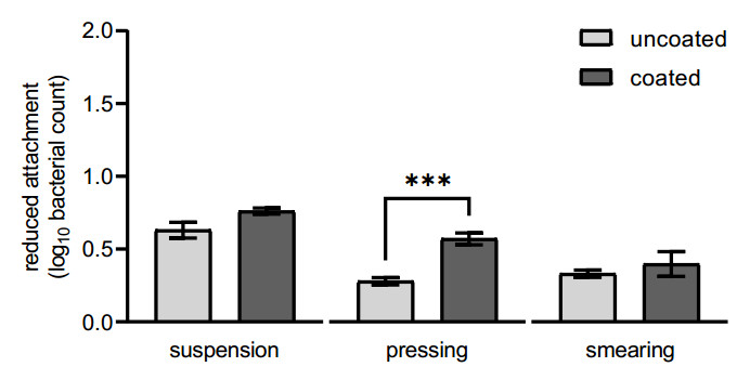
Campylobacteriosis is the most reported gastrointestinal zoonotic disease worldwide and is caused by the consumption of inadequately heated and contaminated food, especially poultry meat. This may result from cross-contamination events during poultry slaughtering and cutting processes. Carcass contact surfaces in slaughterhouses, such as plucking fingers of rubber or stainless-steel surfaces, are high-risk points for contamination, with intestinal contents likely containing Campylobacter bacteria that may result in the cross-contamination of subsequent carcasses. Modification of these food contact surfaces by coating can be beneficial in combating bacterial contamination, as already applied in the packaging materials of the food industry. The aim of this study was to compare the attachment, growth and detachment of Campylobacter jejuni on uncoated and nanoscale silicon dioxide coated stainless steel and plucking fingers during laboratory experiments. The coating partly resulted in significantly reduced attachment and an improved detachment of the target organism on stainless steel. In contrast, there was no significant decrease in Campylobacter adherence to the coated plucking fingers as compared to the uncoated ones. However, a significantly higher reduction of recultivable bacteria on the coated plucking fingers was observed during a five-hour period. In future studies, specific coating parameters should be investigated to further support development, and thus a better adaptation of the coating to the environmental conditions.
Citation: Victoria Blaeske, Felicitas Maria Schumann-Muck, Ahmad Hamedy, Peggy G. Braun, Martin Koethe. Campylobacter colonisation of slaughterhouse surfaces may be affected by ultra-thin silica coating[J]. AIMS Agriculture and Food, 2024, 9(1): 52-68. doi: 10.3934/agrfood.2024004
Campylobacteriosis is the most reported gastrointestinal zoonotic disease worldwide and is caused by the consumption of inadequately heated and contaminated food, especially poultry meat. This may result from cross-contamination events during poultry slaughtering and cutting processes. Carcass contact surfaces in slaughterhouses, such as plucking fingers of rubber or stainless-steel surfaces, are high-risk points for contamination, with intestinal contents likely containing Campylobacter bacteria that may result in the cross-contamination of subsequent carcasses. Modification of these food contact surfaces by coating can be beneficial in combating bacterial contamination, as already applied in the packaging materials of the food industry. The aim of this study was to compare the attachment, growth and detachment of Campylobacter jejuni on uncoated and nanoscale silicon dioxide coated stainless steel and plucking fingers during laboratory experiments. The coating partly resulted in significantly reduced attachment and an improved detachment of the target organism on stainless steel. In contrast, there was no significant decrease in Campylobacter adherence to the coated plucking fingers as compared to the uncoated ones. However, a significantly higher reduction of recultivable bacteria on the coated plucking fingers was observed during a five-hour period. In future studies, specific coating parameters should be investigated to further support development, and thus a better adaptation of the coating to the environmental conditions.

| [1] | World Health Organization (WHO) (2020) Campylobacter. Available from: https://www.who.int/news-room/fact-sheets/detail/campylobacter. |
| [2] |
Igwaran A, Okoh AI (2019) Human campylobacteriosis: A public health concern of global importance. Heliyon 5: e02814. https://doi.org/10.1016/j.heliyon.2019.e02814 doi: 10.1016/j.heliyon.2019.e02814

|
| [3] | Centers for Disease Control and Prevention (2023) Information for Health Professionals | Campylobacter | CDC. |
| [4] | Windhorst HW (2022) Patterns and dynamics of global egg and poultry meat trade. Chicken and Turkey meat trade. Zootecnica International, Available from: https://zootecnicainternational.com/focus-on/market-trends/patterns-and-dynamics-of-global-egg-and-poultry-meat-trade-part-3/. |
| [5] |
Lawes JR, Vidal A, Clifton-Hadley FA, et al. (2012) Investigation of prevalence and risk factors for Campylobacter in broiler flocks at slaughter: results from a UK survey. Epidemiol Infect 140: 1725–1737. https://doi.org/10.1017/S0950268812000982 doi: 10.1017/S0950268812000982
|
| [6] |
Seliwiorstow T, Baré J, Berkvens D, et al. (2016) Identification of risk factors for Campylobacter contamination levels on broiler carcasses during the slaughter process. Int J Food Microbiol 226: 26–32. https://doi.org/10.1016/j.ijfoodmicro.2016.03.010 doi: 10.1016/j.ijfoodmicro.2016.03.010
|
| [7] |
Sasaki Y, Maruyama N, Zou B, et al. (2013) Campylobacter cross-contamination of chicken products at an abattoir. Zoonoses Public Health 60: 134–140. https://doi.org/10.1111/j.1863-2378.2012.01509.x doi: 10.1111/j.1863-2378.2012.01509.x
|
| [8] |
Allen VM, Bull SA, Corry J, et al. (2007) Campylobacter spp. contamination of chicken carcasses during processing in relation to flock colonisation. Int J Food Microbiol 113: 54–61. https://doi.org/10.1016/j.ijfoodmicro.2006.07.011 doi: 10.1016/j.ijfoodmicro.2006.07.011
|
| [9] |
Rasschaert G, de Zutter L, Herman L, et al. (2020) Campylobacter contamination of broilers: the role of transport and slaughterhouse. Int J Food Microbiol 322: 108564. https://doi.org/10.1016/j.ijfoodmicro.2020.108564 doi: 10.1016/j.ijfoodmicro.2020.108564
|
| [10] |
Berrang ME, Buhr RJ, Cason JA, et al. (2001) Broiler carcass contamination with Campylobacter from feces during defeathering. J Food Prot 64: 2063–2066. https://doi.org/10.4315/0362-028x-64.12.2063 doi: 10.4315/0362-028x-64.12.2063
|
| [11] |
Borck B, Pedersen K (2005) Pulsed-field gel electrophoresis types of Campylobacter spp. in Danish turkeys before and after slaughter. Int J Food Microbiol 101: 63–72. https://doi.org/10.1016/j.ijfoodmicro.2004.10.044 doi: 10.1016/j.ijfoodmicro.2004.10.044
|
| [12] |
Peyrat MB, Soumet C, Maris P, et al. (2008) Recovery of Campylobacter jejuni from surfaces of poultry slaughterhouses after cleaning and disinfection procedures: Analysis of a potential source of carcass contamination. Int J Food Microbiol 124: 188–194. https://doi.org/10.1016/j.ijfoodmicro.2008.03.030 doi: 10.1016/j.ijfoodmicro.2008.03.030
|
| [13] |
Lindqvist R, Lindblad M (2008) Quantitative risk assessment of thermophilic Campylobacter spp. and cross-contamination during handling of raw broiler chickens evaluating strategies at the producer level to reduce human campylobacteriosis in Sweden. Int J Food Microbiol 121: 41–52. https://doi.org/10.1016/j.ijfoodmicro.2007.10.008 doi: 10.1016/j.ijfoodmicro.2007.10.008
|
| [14] |
European Food Safety Authority (EFSA) (2011) A quantitative microbiological risk assessment of Campylobacter in the broiler meat chain. EFSA Supporting Publ 8: 132E. https://doi.org/10.2903/sp.efsa.2011.EN-132. doi: 10.2903/sp.efsa.2011.EN-132
|
| [15] |
Vinueza-Burgos C, Cevallos M, Cisneros M, et al. (2018) Quantification of the Campylobacter contamination on broiler carcasses during the slaughter of Campylobacter positive flocks in semi-industrialized slaughterhouses. Int J Food Microbiol 269: 75–79. https://doi.org/10.1016/j.ijfoodmicro.2018.01.021 doi: 10.1016/j.ijfoodmicro.2018.01.021
|
| [16] |
Arnold JW, Silvers S (2000) Comparison of poultry processing equipment surfaces for susceptibility to bacterial attachment and biofilm formation. Poult Sci 79: 1215–1221. https://doi.org/10.1093/ps/79.8.1215 doi: 10.1093/ps/79.8.1215
|
| [17] |
Kelleher SM, Habimana O, Lawler J, et al. (2016) Cicada Wing Surface Topography: An Investigation into the Bactericidal Properties of Nanostructural Features. ACS Appl Mater Interfaces 8: 14966–14974. https://doi.org/10.1021/acsami.5b08309 doi: 10.1021/acsami.5b08309
|
| [18] |
Graham M, Cady N (2014) Nano and Microscale Topographies for the Prevention of Bacterial Surface Fouling. Coatings 4: 37–59. https://doi.org/10.3390/coatings4010037 doi: 10.3390/coatings4010037
|
| [19] |
Feng G, Cheng Y, Wang S-Y, et al. (2015) Bacterial attachment and biofilm formation on surfaces are reduced by small-diameter nanoscale pores: how small is small enough? NPJ Biofilms Microbiomes 1: 15022. https://doi.org/10.1038/npjbiofilms.2015.22 doi: 10.1038/npjbiofilms.2015.22
|
| [20] |
Hsu LC, Fang J, Borca-Tasciuc DA, et al. (2013) Effect of micro- and nanoscale topography on the adhesion of bacterial cells to solid surfaces. Appl Environ Microbiol 79: 2703–2712. https://doi.org/10.1128/AEM.03436-12 doi: 10.1128/AEM.03436-12
|
| [21] |
Simchi A, Tamjid E, Pishbin F, et al. (2011) Recent progress in inorganic and composite coatings with bactericidal capability for orthopaedic applications. Nanomedicine 7: 22–39. https://doi.org/10.1016/j.nano.2010.10.005 doi: 10.1016/j.nano.2010.10.005
|
| [22] |
Ahmed J, Arfat YA, Bher A, et al. (2018) Active Chicken Meat Packaging Based on Polylactide Films and Bimetallic Ag–Cu Nanoparticles and Essential Oil. J Food Sci 83: 1299–1310. https://doi.org/10.1111/1750-3841.14121 doi: 10.1111/1750-3841.14121
|
| [23] |
Gallocchio F, Cibin V, Biancotto G, et al. (2016) Testing nano-silver food packaging to evaluate silver migration and food spoilage bacteria on chicken meat. Food Addit Contam: Part A 33: 1063–1071. https://doi.org/10.1080/19440049.2016.1179794 doi: 10.1080/19440049.2016.1179794
|
| [24] |
Di Cerbo A, Mescola A, Rosace G, et al. (2021) Antibacterial effect of stainless steel surfaces treated with a nanotechnological coating approved for food contact. Microorganisms 9: 248. https://doi.org/10.3390/microorganisms9020248 doi: 10.3390/microorganisms9020248
|
| [25] |
Gu T, Meerisom A, Luo Y, et al. (2021) Listeria monocytogenes biofilm formation as affected by stainless steel surface topography and coating composition. Food Control 130: 108275. https://doi.org/10.1016/j.foodcont.2021.108275 doi: 10.1016/j.foodcont.2021.108275
|
| [26] |
Rao KS, El-Hami K, Kodaki T, et al. (2005) A novel method for synthesis of silica nanoparticles. J Colloid Interface Sci 289: 125–131. https://doi.org/10.1016/j.jcis.2005.02.019 doi: 10.1016/j.jcis.2005.02.019
|
| [27] |
Barros CHN, Fulaz S, Vitale S, et al. (2020) Interactions between functionalised silica nanoparticles and Pseudomonas fluorescens biofilm matrix: A focus on the protein corona. PLoS ONE 15: e0236441. https://doi.org/10.1371/journal.pone.0236441. doi: 10.1371/journal.pone.0236441
|
| [28] |
El-Shetehy M, Moradi A, Maceroni M, et al. (2021) Silica nanoparticles enhance disease resistance in Arabidopsis plants. Nat Nanotechnol 16: 344–353. https://doi.org/10.1038/s41565-020-00812-0 doi: 10.1038/s41565-020-00812-0
|
| [29] |
Schumann‐Muck FM, Hillig N, Braun PG, et al. (2023a) Impact of nanoscale coating of stainless steel on Salmonella Enteritidis and Escherichia coli. J Food Safety 43: e13075. https://doi.org/10.1111/jfs.13075 doi: 10.1111/jfs.13075
|
| [30] |
Arnold JW (2007) Bacterial contamination on rubber picker fingers before, during, and after processing. Poult Sci 86: 2671–2675. https://doi.org/10.3382/ps.2007-00187 doi: 10.3382/ps.2007-00187
|
| [31] | Schmidt R, Erickson D, Sims S, et al. (2012) Characteristics of food contact surface materials: Stainless steel. Food Prot Trends 32: 574–584. |
| [32] |
Flint SH, Brooks JD, Bremer PJ (2000) Properties of the stainless steel substrate, influencing the adhesion of thermo-resistant streptococci. J Food Eng 43: 235–242. https://doi.org/10.1016/S0260-8774(99)00157-0 doi: 10.1016/S0260-8774(99)00157-0
|
| [33] |
Verma J, Khanna AS, Sahney R, et al. (2020) Super protective anti-bacterial coating development with silica-titania nano core-shells. Nanoscale Adv 2: 4093–4105. https://doi.org/10.1039/d0na00387e doi: 10.1039/d0na00387e
|
| [34] |
Hori K, Matsumoto S (2010) Bacterial adhesion: From mechanism to control. Biochem Eng J 48: 424–434. https://doi.org/10.1016/j.bej.2009.11.014 doi: 10.1016/j.bej.2009.11.014
|
| [35] |
Nguyen VT, Turner MS, Dykes GA (2011) Influence of cell surface hydrophobicity on attachment of Campylobacter to abiotic surfaces. Food Microbiol 28: 942–950. https://doi.org/10.1016/j.fm.2011.01.004 doi: 10.1016/j.fm.2011.01.004
|
| [36] |
Joseph B, Otta SK, Karunasagar I, et al. (2001) Biofilm formation by Salmonella spp. on food contact surfaces and their sensitivity to sanitizers. Int J Food Microbiol 64: 367–372. https://doi.org/10.1016/s0168-1605(00)00466-9 doi: 10.1016/s0168-1605(00)00466-9
|
| [37] |
Teh AHT, Lee SM, Dykes GA (2019) Association of some Campylobacter jejuni with Pseudomonas aeruginosa biofilms increases attachment under conditions mimicking those in the environment. PLoS ONE 14: e0215275. https://doi.org/10.1371/journal.pone.0215275 doi: 10.1371/journal.pone.0215275
|
| [38] |
Buswell CM, Herlihy YM, Lawrence LM, et al. (1998) Extended survival and persistence of Campylobacter spp. in water and aquatic biofilms and their detection by immunofluorescent-antibody and -rRNA staining. Appl Environ Microbiol 64: 733–741. https://doi.org/10.1128/AEM.64.2.733-741.1998 doi: 10.1128/AEM.64.2.733-741.1998
|
| [39] |
Park SF (2002) The physiology of Campylobacter species and its relevance to their role as foodborne pathogens. Int J Food Microbiol 74: 177–188. https://doi.org/10.1016/S0168-1605(01)00678-X doi: 10.1016/S0168-1605(01)00678-X
|
| [40] |
Kusumaningrum H (2003) Survival of foodborne pathogens on stainless steel surfaces and cross-contamination to foods. Int J Food Microbiol 85: 227–236. https://doi.org/10.1016/S0168-1605(02)00540-8 doi: 10.1016/S0168-1605(02)00540-8
|
| [41] |
Doyle MP, Roman DJ (1982) Sensitivity of Campylobacter jejuni to Drying. J Food Prot 45: 507–510. https://doi.org/10.4315/0362-028X-45.6.507 doi: 10.4315/0362-028X-45.6.507
|
| [42] |
Oosterom J, Wilde GJA de, Boer E de, et al. (1983) Survival of Campylobacter jejuni during Poultry Processing and Pig Slaughtering. J Food Prot 46: 702–706. https://doi.org/10.4315/0362-028X-46.8.702 doi: 10.4315/0362-028X-46.8.702
|
| [43] |
Zakarienė G, Novoslavskij A, Meškinis Š, et al. (2018) Diamond like carbon Ag nanocomposites as a control measure against Campylobacter jejuni and Listeria monocytogenes on food preparation surfaces. Diam Relat Mater 81: 118–126. https://doi.org/10.1016/j.diamond.2017.12.007 doi: 10.1016/j.diamond.2017.12.007
|
| [44] |
Sterniša M, Gradišar Centa U, Drnovšek A, et al. (2023) Pseudomonas fragi biofilm on stainless steel (at low temperatures) affects the survival of Campylobacter jejuni and Listeria monocytogenes and their control by a polymer molybdenum oxide nanocomposite coating. Int J Food Microbiol 394: 110159. https://doi.org/10.1016/j.ijfoodmicro.2023.110159 doi: 10.1016/j.ijfoodmicro.2023.110159
|
| [45] |
Nguyen DHK, Pham VTH, Truong VK, et al. (2018) Role of topological scale in the differential fouling of Pseudomonas aeruginosa and Staphylococcus aureus bacterial cells on wrinkled gold-coated polystyrene surfaces. Nanoscale 10: 5089–5096. https://doi.org/10.1039/c7nr08178b doi: 10.1039/c7nr08178b
|
| [46] |
Ivanova EP, Hasan J, K. Webb H, et al. (2012) Natural Bactericidal Surfaces: Mechanical Rupture of Pseudomonas aeruginosa Cells by Cicada Wings. Small 8: 2489–2494. https://doi.org/10.1002/smll.201200528 doi: 10.1002/smll.201200528
|
| [47] |
Bremer PJ, Fillery S, McQuillan AJ (2006) Laboratory scale Clean-In-Place (CIP) studies on the effectiveness of different caustic and acid wash steps on the removal of dairy biofilms. Int J Food Microbiol 106: 254–262. https://doi.org/10.1016/j.ijfoodmicro.2005.07.004 doi: 10.1016/j.ijfoodmicro.2005.07.004
|
| [48] |
Singh AV, Vyas V, Patil R, et al. (2011) Quantitative characterization of the influence of the nanoscale morphology of nanostructured surfaces on bacterial adhesion and biofilm formation. PLoS ONE 6: e25029. https://doi.org/10.1371/journal.pone.0025029 doi: 10.1371/journal.pone.0025029
|
| [49] |
Oh E, Chui L, Bae J, et al. (2018) Frequent Implication of Multistress-Tolerant Campylobacter jejuni in Human Infections. Emerg Infect Dis 24: 1037–1044. https://doi.org/10.3201/eid2406.171587 doi: 10.3201/eid2406.171587
|
| [50] |
Revez J, Rossi M, Ellström P, et al. (2011) Finnish Campylobacter jejuni strains of multilocus sequence type ST-22 complex have two lineages with different characteristics. PLoS ONE 6: e26880. https://doi.org/10.1371/journal.pone.0026880 doi: 10.1371/journal.pone.0026880
|
| [51] |
Rollins DM, Colwell RR (1986) Viable but nonculturable stage of Campylobacter jejuni and its role in survival in the natural aquatic environment. Appl Environ Microbiol 52: 531–538. https://doi.org/10.1128/aem.52.3.531-538.1986 doi: 10.1128/aem.52.3.531-538.1986
|
| [52] |
Dykes GA, Sampathkumar B, Korber DR (2003) Planktonic or biofilm growth affects survival, hydrophobicity and protein expression patterns of a pathogenic Campylobacter jejuni strain. Int J Food Microbiol 89: 1–10. https://doi.org/10.1016/S0168-1605(03)00123-5 doi: 10.1016/S0168-1605(03)00123-5
|
| [53] |
Chaisowwong W, Kusumoto A, Hashimoto M, Harada T, Maklon K, Kawamoto K (2012) Physiological characterization of Campylobacter jejuni under cold stresses conditions: its potential for public threat. J Vet Med Sci 74: 43–50. https://doi.org/10.1292/jvms.11-0305 doi: 10.1292/jvms.11-0305
|
| [54] |
Klančnik A, Guzej B, Jamnik P, Vučković D, Abram M, Smole Možina S (2009) Stress response and pathogenic potential of Campylobacter jejuni cells exposed to starvation. Res Microbiol 160: 345–352. https://doi.org/10.1016/j.resmic.2009.05.002 doi: 10.1016/j.resmic.2009.05.002
|
| [55] |
Li L, Mendis N, Trigui H, Oliver JD, Faucher SP (2014) The importance of the viable but non-culturable state in human bacterial pathogens. Front Microbiol 5: 258. https://doi.org/10.3389/fmicb.2014.00258 doi: 10.3389/fmicb.2014.00258
|


Figures(5) / Tables(1)

Victoria Blaeske, Felicitas Maria Schumann-Muck, Ahmad Hamedy, Peggy G. Braun, Martin Koethe. Campylobacter colonisation of slaughterhouse surfaces may be affected by ultra-thin silica coating[J]. AIMS Agriculture and Food, 2024, 9(1): 52-68. doi: 10.3934/agrfood.2024004


 DownLoad: