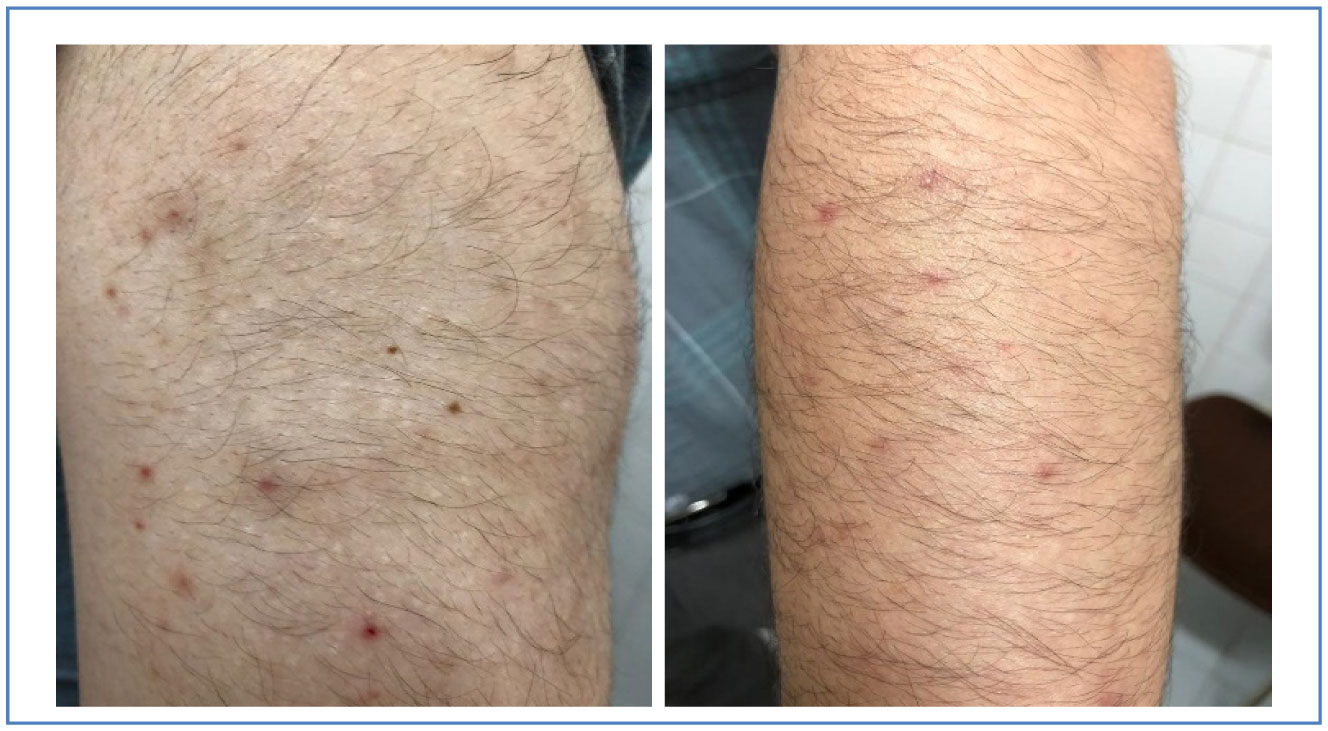
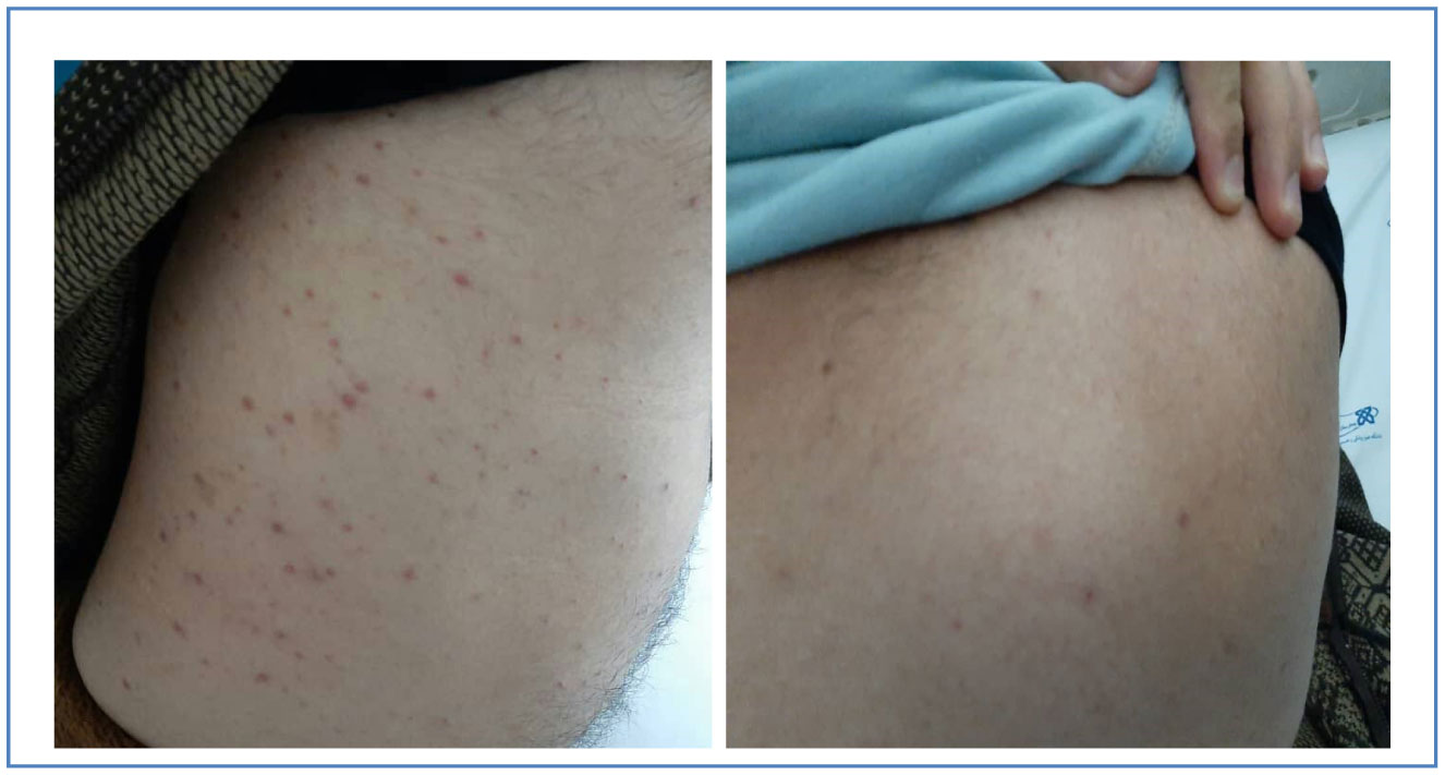
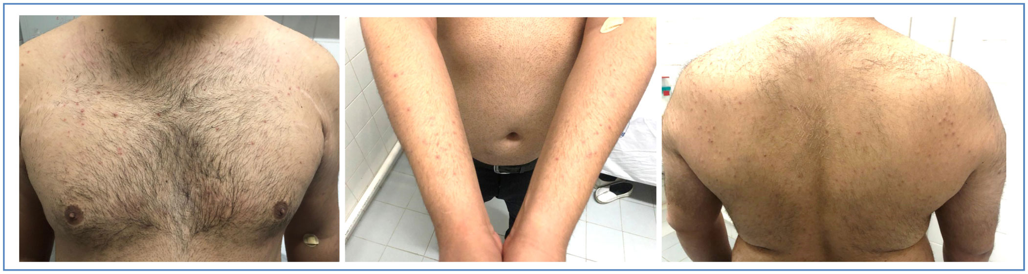
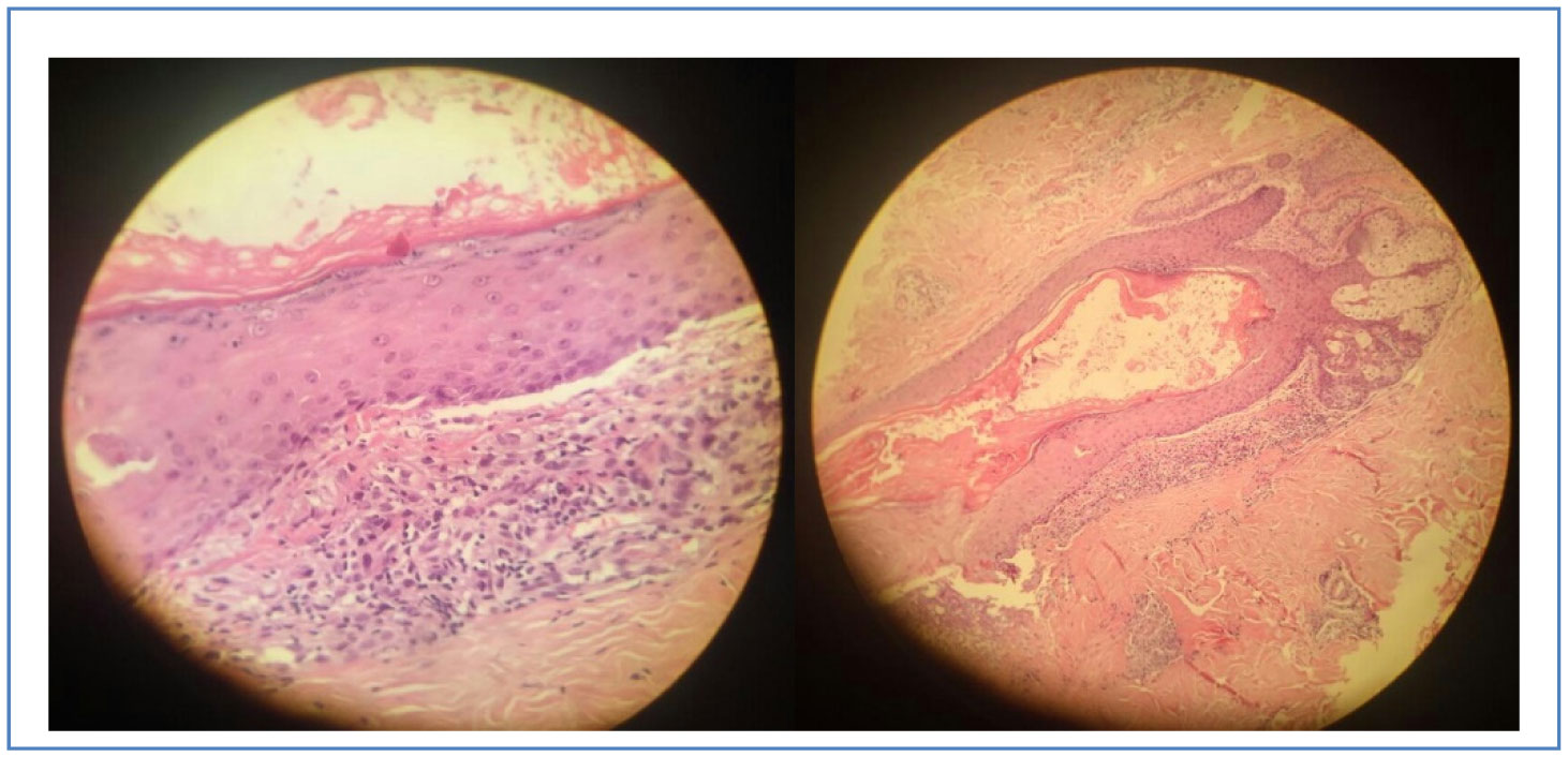
COVID-19 can be associated with varying degrees of cutaneous lesions. Although these manifestations were rare, they can be considered as a specific symptom of COVID-19. Here, we present 3 patients for whom the diagnosis of COVID-19 was made by RT-PCR and a few days after the onset of systemic symptoms, papular cutaneous lesions appeared and on average after 6 days, the lesions started to disappear without any specific treatments. Demographic and clinical features of the 3 patients are summarized in
Citation: Hossein Sheibani, Elham Azmoodeh, Amirhessam Kheirieh, Khadijeh Aerab Sheibani. COVID-19 and papular manifestations pattern in 3 patients: a retrospective case series[J]. AIMS Medical Science, 2021, 8(1): 36-41. doi: 10.3934/medsci.2021004
COVID-19 can be associated with varying degrees of cutaneous lesions. Although these manifestations were rare, they can be considered as a specific symptom of COVID-19. Here, we present 3 patients for whom the diagnosis of COVID-19 was made by RT-PCR and a few days after the onset of systemic symptoms, papular cutaneous lesions appeared and on average after 6 days, the lesions started to disappear without any specific treatments. Demographic and clinical features of the 3 patients are summarized in
Coronavirus disease 2019
Severe acute respiratory syndrome
Middle east respiratory syndrome
World Health Organization
Reverse transcription polymerase chain reaction
Polymorphonuclear leukocytes
Severe acute respiratory syndrome coronavirus 2

| [1] |
Huang C, Wang Y, Li X, et al. (2020) Clinical features of patients infected with 2019 novel coronavirus in Wuhan, China. Lancet 395: 497-506. 
|
| [2] |
Zhu N, Zhang D, Wang W, et al. (2020) A novel coronavirus from patients with pneumonia in China, 2019. N Engl J Med 382: 727-733.
|
| [3] | World Health OrganizationWeekly operational update on COVID-19—21 December 2020 (2020). Available from: https://www.who.int/docs/default-source/coronaviruse/wou_21-dec_cleared.pdf?sfvrsn=a7575c1f_1&download=true |
| [4] | Recalcati S (2020) Cutaneous manifestations in COVID-19: a first perspective. J Eur Acad Dermatol Venereol 34: e212-e213. |
| [5] |
Suchonwanit P, Leerunyakul K, Kositkuljorn C (2020) Cutaneous manifestations in COVID-19: lessons learned from current evidence. J Am Acad Dermatol 83: e57-e60.
|
| [6] |
Hoenig LJ, Pereira FA (2020) Eruption as a clinical manifestation of COVID-19: photographs of a patient. Clin Dermatol 38: 502-505.
|
| [7] |
Kaya G, Kaya A, Saurat JH (2020) Clinical and histopathological features and potential pathological mechanisms of skin lesions in COVID-19: review of the literature. Dermatopathology 7: 3-16.
|
| [8] |
Marzano AV, Genovese G, Fabbrocini G, et al. (2020) Varicella-like exanthem as a specific COVID-19–associated skin manifestation: multicenter case series of 22 patients. J Am Acad Dermatol 83: 280-285.
|
| [9] |
Lechien JR, Chiesa-Estomba CM, De Siati DR, et al. (2020) Olfactory and gustatory dysfunctions as a clinical presentation of mild-to-moderate forms of the coronavirus disease (COVID-19): a multicenter European study. Eur Arch Otorhinolaryngol 277: 2251-2261.
|
| [10] |
Sameni F, Hajikhani B, Yaslianifard S, et al. (2020) COVID-19 and skin manifestations: an overview of case reports/case series and meta-analysis of prevalence studies. Front Med 7.
|
Figures(4) / Tables(1)

Hossein Sheibani, Elham Azmoodeh, Amirhessam Kheirieh, Khadijeh Aerab Sheibani. COVID-19 and papular manifestations pattern in 3 patients: a retrospective case series[J]. AIMS Medical Science, 2021, 8(1): 36-41. doi: 10.3934/medsci.2021004


 DownLoad: