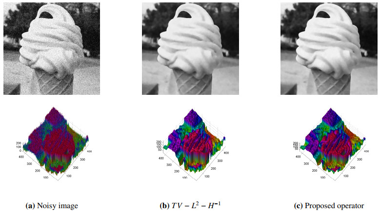
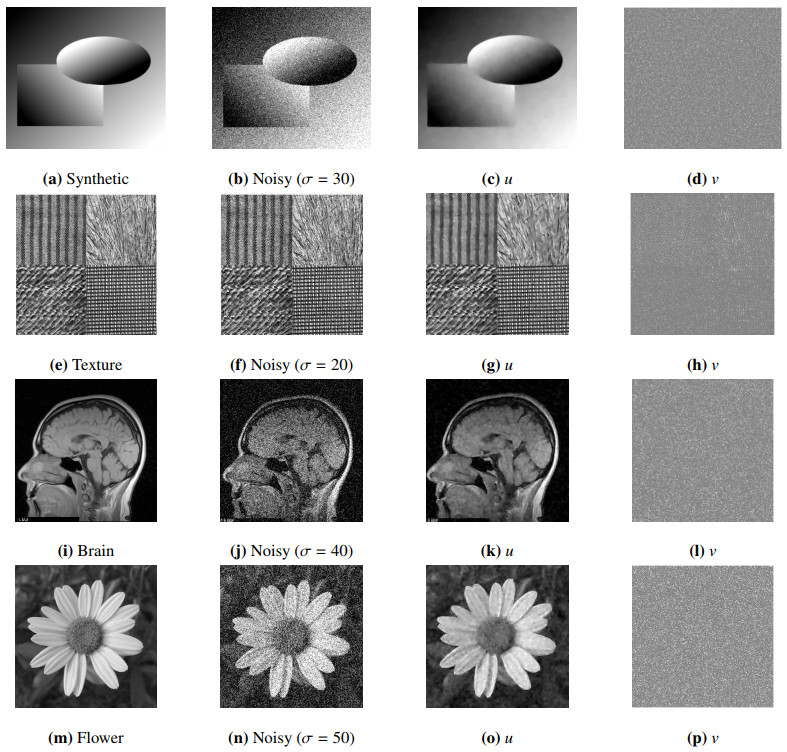
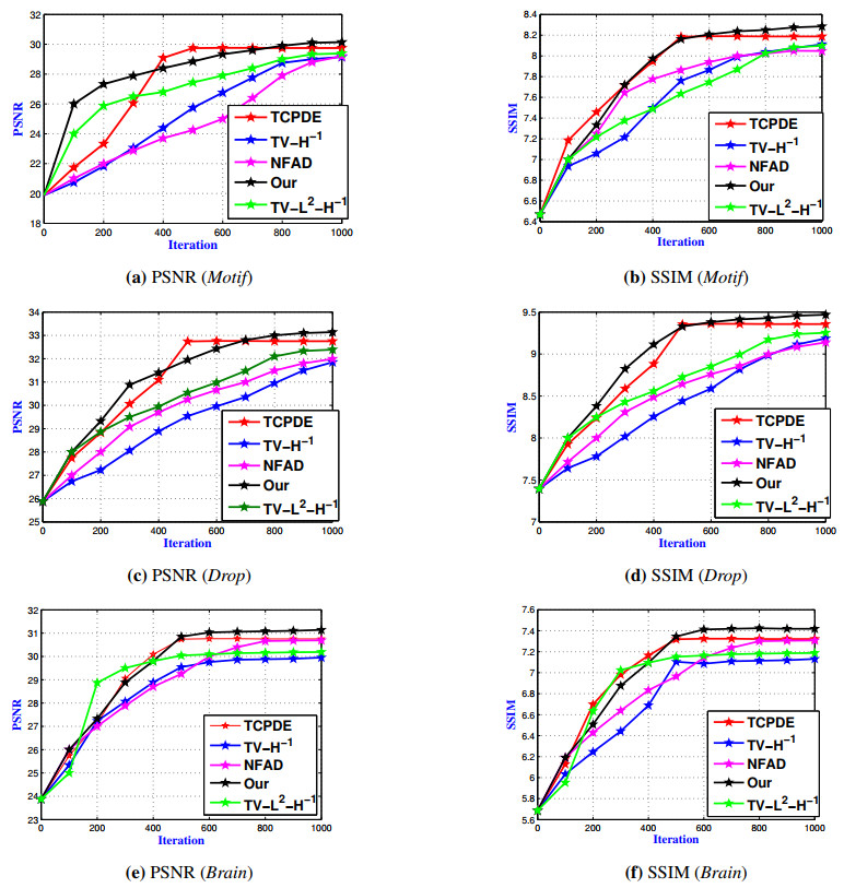
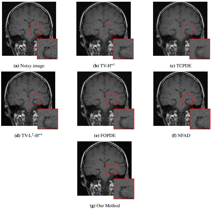
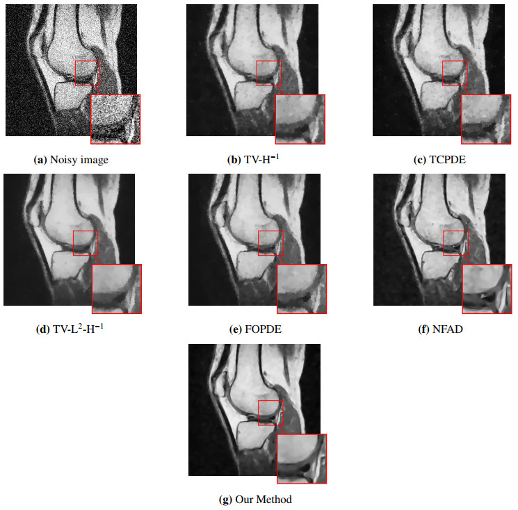
The problem of interest in this paper is the mathematical and numerical analysis of a new non-variational model based on a high order non-linear PDE system resulting from image denoising. This model is motivated by involving the decomposition approach of $ H^{-1} $ norm suggested by Guo et al. [
Citation: Abdelmajid El Hakoume, Lekbir Afraites, Amine Laghrib. An improved coupled PDE system applied to the inverse image denoising problem[J]. Electronic Research Archive, 2022, 30(7): 2618-2642. doi: 10.3934/era.2022134
The problem of interest in this paper is the mathematical and numerical analysis of a new non-variational model based on a high order non-linear PDE system resulting from image denoising. This model is motivated by involving the decomposition approach of $ H^{-1} $ norm suggested by Guo et al. [

| [1] |
Z. Guo, J. Yin, Q. Liu, On a reaction-diffusion system applied to image decomposition and restoration, Math. Comput. Model., 53 (2011), 1336–1350. https://doi.org/10.1016/j.mcm.2010.12.031 doi: 10.1016/j.mcm.2010.12.031

|
| [2] |
Z. Guo, Q. Liu, J. Sun, B. Wu, Reaction-diffusion systems with $p(x)$-growth for image denoising, Nonlinear Anal. Real World Appl., 12 (2011), 2904–2918. https://doi.org/10.1016/j.nonrwa.2011.04.015 doi: 10.1016/j.nonrwa.2011.04.015
|
| [3] |
A. Hadri, H. Khalfi, A. Laghrib, M. Nachaoui, An improved spatially controlled reaction–diffusion equation with a non-linear second order operator for image super-resolution, Nonlinear Anal. Real World Appl., 62 (2021), 103352. https://doi.org/10.1016/j.nonrwa.2021.103352 doi: 10.1016/j.nonrwa.2021.103352
|
| [4] |
M. Lysaker, A. lundervold, X. Tai, Noise removal using fourth-order partial differential equation with applications to medical magnetic resonance images in space and time, IEEE Trans. Image Process, 12 (2003), 1579–1590. https://doi.org/10.1109/TIP.2003.819229 doi: 10.1109/TIP.2003.819229
|
| [5] |
S. G. Chang, B. Yu, M. Vetterli, Adaptive wavelet thresholding for image denoising and compression, IEEE Trans. Image Process, 9 (2000), 1532–1546. https://doi.org/10.1109/83.862633 doi: 10.1109/83.862633
|
| [6] | G. Gimel'farb, Image Textures and Gibbs Random Fields, Kluwer Academic Publishers, 1999. |
| [7] |
A. Buades, B. Coll, J. Morel, A non-local algorithm for image denoising, IEEE Comput. Vis. Pattern Recognit, 2 (2005), 60–65. https://doi.org/10.1109/CVPR.2005.38 doi: 10.1109/CVPR.2005.38
|
| [8] |
L. Rudin, S. Osher, E. Fatemi, Nonlinear total variation based noise removal algorithms, Physica D, 60 (1992), 259–268. https://doi.org/10.1016/0167-2789(92)90242-F doi: 10.1016/0167-2789(92)90242-F
|
| [9] |
R. Acar, C. Vogel, Analysis of bounded variation penalty methods for ill-posed problems, Inverse Probl., 10 (1994), 1217–1229. https://doi.org/10.1088/0266-5611/10/6/003 doi: 10.1088/0266-5611/10/6/003
|
| [10] |
A. Chambolle, P. Lions, Image recovery via total variation minimization and related problems, Numer. Math., 76 (1997), 167–188. https://doi.org/10.1007/s002110050258 doi: 10.1007/s002110050258
|
| [11] |
A. Hadri, A. Laghrib, H. Oummi, An optimal variable exponent model for magnetic resonance images denoising, Pattern Recognit. Lett., 151 (2021), 302–309. https://doi.org/10.1016/j.patrec.2021.08.031 doi: 10.1016/j.patrec.2021.08.031
|
| [12] | L. Vese, S. Osher, Modeling textures with total variation minimization and oscillating patterns in image processing, J. Sci. Comput., 19 (2003), 553–572. |
| [13] |
S. Osher, A. Solé, L. Vese, Image decomposition and restoration using total variation minimization and the H-1 norm, Multiscale Model. Simul., 1 (2003), 349–370. https://doi.org/10.1137/S1540345902416247 doi: 10.1137/S1540345902416247
|
| [14] | Y. Meyer, Oscillating Patterns in Image Processing and Nonlinear Evolution Equations, in: Univ. Lecture Ser., AMS, 2002. |
| [15] |
A. Atlas, M. Bendahmane, F. Karami, D. Meskine, O. Oubbih, A nonlinear fractional reaction-diffusion system applied to image denoising and decomposition, Discrete Contin. Dyn. Syst. Ser. B, 26 (2021), 4963. https://doi.org/10.3934/dcdsb.2020321 doi: 10.3934/dcdsb.2020321
|
| [16] |
M. M. Y. Giga, P. Rybka, A duality based approach to the minimizing total variation flow in the space $H^s$, Jpn. J. Ind. Appl. Math., 36 (2019), 261–286. https://doi.org/10.1007/s13160-018-00340-4 doi: 10.1007/s13160-018-00340-4
|
| [17] |
A. Halim, B. R. Kumar, A TV- L2- H- 1 PDE model for effective denoising, Comput. Math. with Appl., 80 (2020), 2176–2193. https://doi.org/10.1016/j.camwa.2020.09.009 doi: 10.1016/j.camwa.2020.09.009
|
| [18] |
K. Papafitsoros, C. B. Schoenlieb, B. Sengul, Combined first and second order total variation inpainting using split bregman, Image Process. Line, 3 (2013), 112–136. https://doi.org/10.5201/ipol.2013.40 doi: 10.5201/ipol.2013.40
|
| [19] | J. Weickert, Anisotropic Diffusion in Image Processing, Teubner, Stuttgart, 1998. |
| [20] |
P. Perona, J. Malik, Scale-space and edge detection using anisotropic diffusion, IEEE Trans. Pattern Anal. Mach. Intell., 12 (1990), 629–639. https://doi.org/10.1109/34.56205 doi: 10.1109/34.56205
|
| [21] |
I. El Mourabit, M. El Rhabi, A. Hakim, A. Laghrib, E. Moreau, A new denoising model for multi-frame super-resolution image reconstruction, Signal Process., 132 (2017), 51–65. https://doi.org/10.1016/j.sigpro.2016.09.014 doi: 10.1016/j.sigpro.2016.09.014
|
| [22] |
F. Catté, P.-L. Lions, J.-M. Morel, T. Coll, Image selective smoothing and edge detection by nonlinear diffusion, SIAM J. Numer. Anal., 29 (1992), 182–193. https://doi.org/10.1137/0729012 doi: 10.1137/0729012
|
| [23] | E. Zeidler, Nonlinear Functional Analysis Vol.1: Fixed-Point Theorems, Springer-Verlag Berlin and Heidelberg GmbH and Co. K, 1986. |
| [24] | L. C. Evans, Partial differential equations, volume 19. Rhode Island, USA, 1998. |
| [25] | H. Attouch, G. Buttazzo, G. Michaille, Variational analysis in Sobolev and BV spaces: applications to PDEs and optimization, SIAM, 2014. |
| [26] | J.-P. Aubin, Un théoreme de compacité, CR Acad. Sci. Paris, 256 (1963), 5042–5044. |
| [27] |
S. Majee, S. K. Jain, R. K. Ray, A. K. Majee, On the development of a coupled nonlinear telegraph-diffusion model for image restoration, Comput. Math. with Appl., 80 (2020), 1745–1766. https://doi.org/10.1016/j.camwa.2020.08.010 doi: 10.1016/j.camwa.2020.08.010
|
| [28] |
A. Laghrib, A. Hadri, A. Hakim, An edge preserving high-order PDE for multiframe image super-resolution, J. Franklin Inst., 356 (2019), 5834–5857. https://doi.org/10.1016/j.jfranklin.2019.02.032 doi: 10.1016/j.jfranklin.2019.02.032
|
Figures(11) / Tables(1)
Abdelmajid El Hakoume, Lekbir Afraites, Amine Laghrib. An improved coupled PDE system applied to the inverse image denoising problem[J]. Electronic Research Archive, 2022, 30(7): 2618-2642. doi: 10.3934/era.2022134


 DownLoad: