Research article Special Issues

Improved growth and harvesting of microalgae Chlorella vulgaris on textile fabrics as 2.5D substrates

  • The green microalgae Chlorella vulgaris can be used in diverse applications from food to biofuel production. Growing them in suspension leads to challenging harvesting and processing. One possibility to overcome these problems is growing them as biofilms, i.e. adhering on a surface. While previous experiments of several research groups concentrated on flat, rigid surfaces, partly chemically modified, here the possibility to grow them on different textile substrates was investigated which were shown to be suitable as substrates for germination and growth of higher plants. Microalgae were counted after one week, subdivided into adhered and suspended ones, to evaluate the ideal substrate for cultivation and harvesting. The results show clear differences between the different woven and knitted fabrics from diverse materials, indicating that especially an open-pore jute woven fabric increased the overall algae concentration by approx. a factor of 2 and increased the adhesion of C. vulgaris by a factor of 5-10, as compared to most other textile substrates under investigation, followed by two other hairy knitted fabrics. Such textile fabrics can thus be suggested as possible substrates for improved growth and harvesting of this microalga.

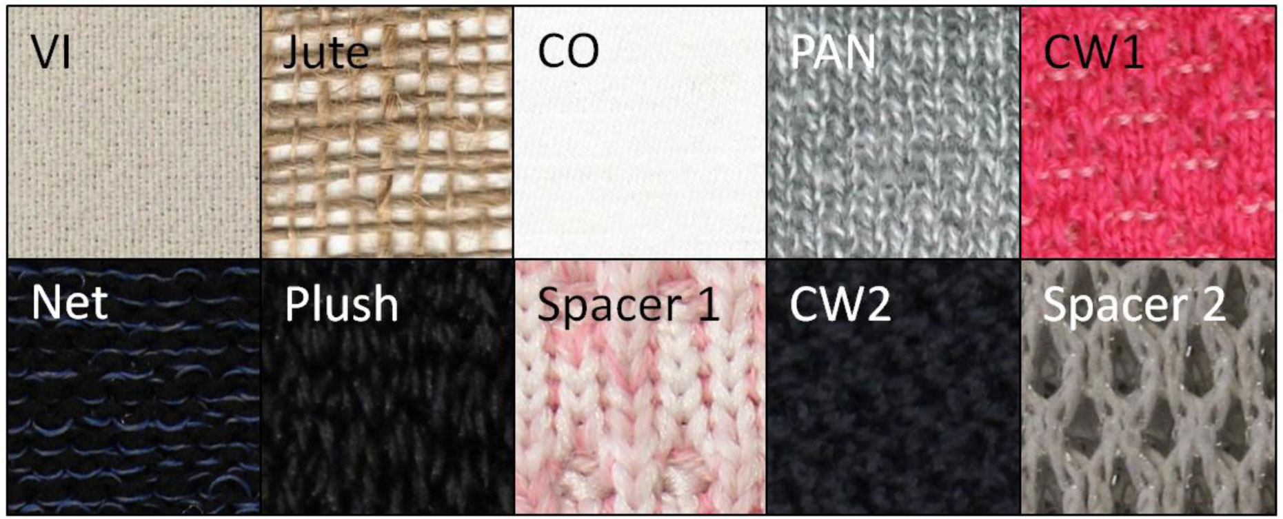
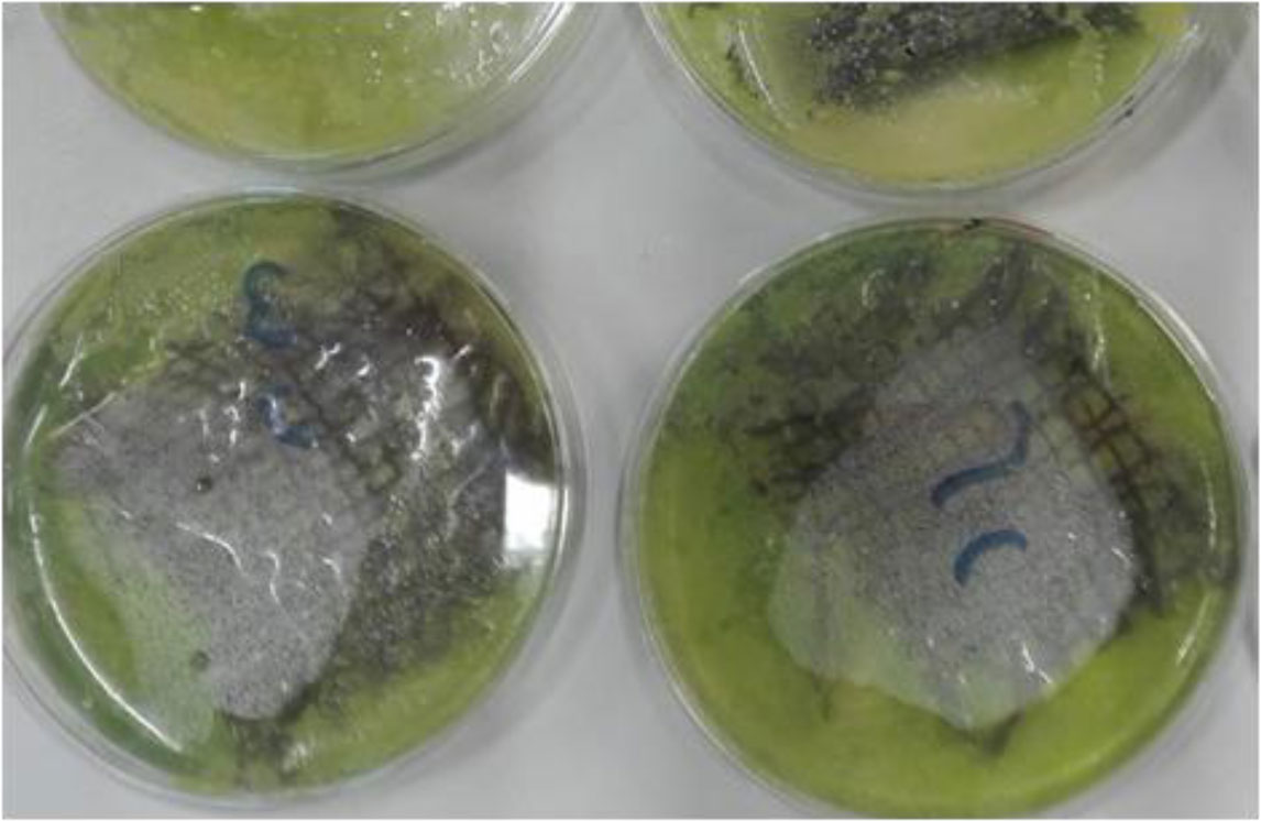
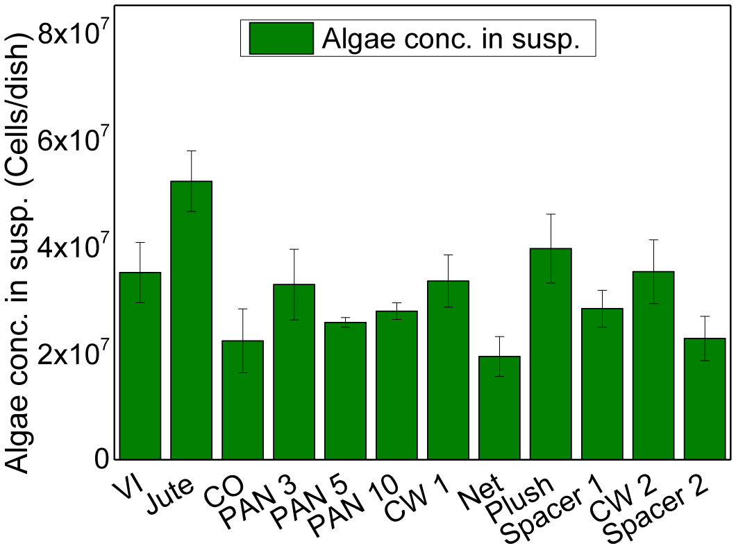
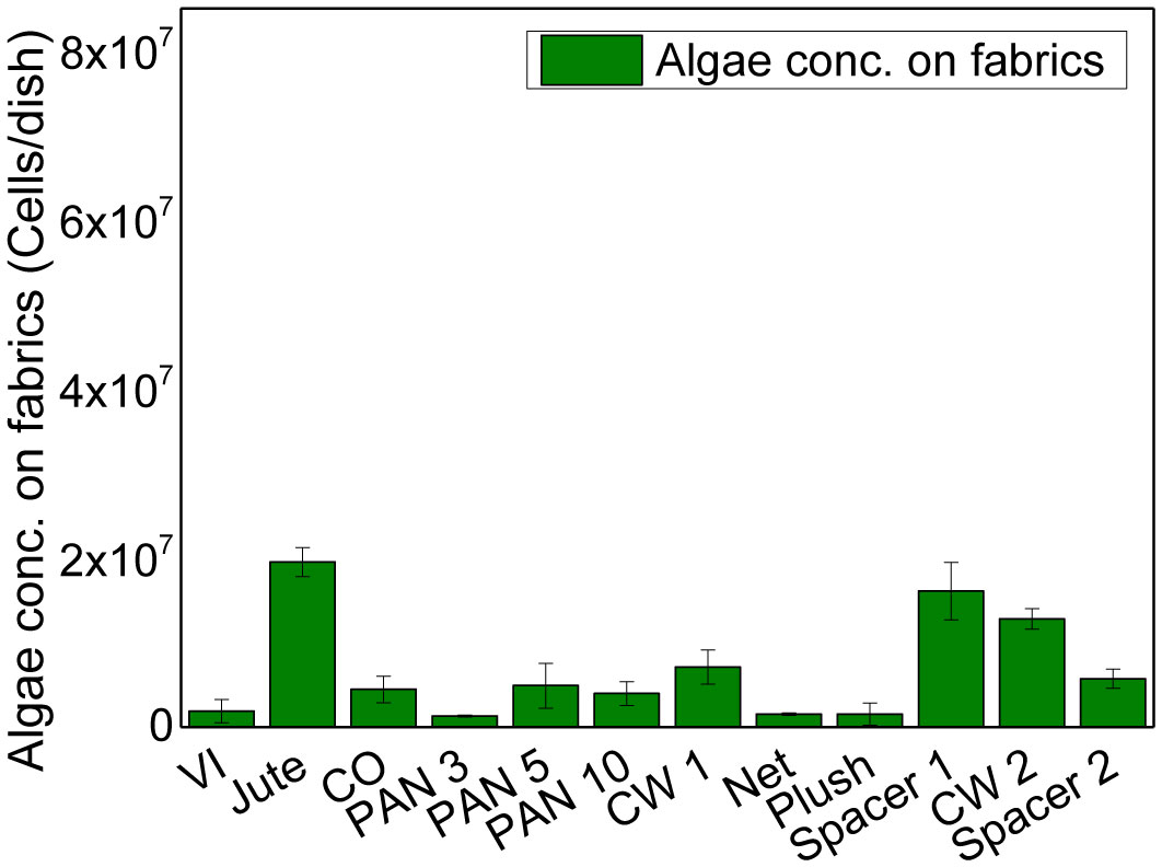
    Citation: Bennet Brockhagen, Jan Lukas Storck, Timo Grothe, Robin Böttjer, Andrea Ehrmann. Improved growth and harvesting of microalgae Chlorella vulgaris on textile fabrics as 2.5D substrates[J]. AIMS Bioengineering, 2021, 8(1): 16-24. doi: 10.3934/bioeng.2021003

    Related Papers:

    [1] Christina Großerhode, Daria Wehlage, Timo Grothe, Nils Grimmelsmann, Sandra Fuchs, Jessica Hartmann, Patrycja Mazur, Vanessa Reschke, Helena Siemens, Anke Rattenholl, Sarah Vanessa Homburg, Andrea Ehrmann . Investigation of microalgae growth on electrospun nanofiber mats. AIMS Bioengineering, 2017, 4(3): 376-385. doi: 10.3934/bioeng.2017.3.376
    [2] Bennet Brockhagen, Fabian Schoden, Jan Lukas Storck, Timo Grothe, Christian Eßelmann, Robin Böttjer, Anke Rattenholl, Frank Gudermann . Investigating minimal requirements for plants on textile substrates in low-cost hydroponic systems. AIMS Bioengineering, 2021, 8(2): 173-191. doi: 10.3934/bioeng.2021016
    [3] Daria Wehlage, Hannah Blattner, Al Mamun, Ines Kutzli, Elise Diestelhorst, Anke Rattenholl, Frank Gudermann, Dirk Lütkemeyer, Andrea Ehrmann . Cell growth on electrospun nanofiber mats from polyacrylonitrile (PAN) blends. AIMS Bioengineering, 2020, 7(1): 43-54. doi: 10.3934/bioeng.2020004
    [4] Daniel Pelaez, John H. Michel, Herman S. Cheung . Growth on elastic silicone substrate elicits a partial myogenic response in periodontal ligament derived stem cells. AIMS Bioengineering, 2016, 3(4): 515-527. doi: 10.3934/bioeng.2016.4.515
    [5] Chiara Ceresa, Francesco Tessarolo, Devid Maniglio, Iole Caola, Giandomenico Nollo, Maurizio Rinaldi, Letizia Fracchia . Inhibition of Candida albicans biofilm by lipopeptide AC7 coated medical-grade silicone in combination with farnesol. AIMS Bioengineering, 2018, 5(3): 192-208. doi: 10.3934/bioeng.2018.3.192
    [6] Sven Sölmann, Anke Rattenholl, Hannah Blattner, Guido Ehrmann, Frank Gudermann, Dirk Lütkemeyer, Andrea Ehrmann . Mammalian cell adhesion on different 3D printed polymers with varying sterilization methods and acidic treatment. AIMS Bioengineering, 2021, 8(1): 25-35. doi: 10.3934/bioeng.2021004
    [7] Wiebke Wesseling . Beneficial biofilms in marine aquaculture? Linking points of biofilm formation mechanisms in Pseudomonas aeruginosa and Pseudoalteromonas species. AIMS Bioengineering, 2015, 2(3): 104-125. doi: 10.3934/bioeng.2015.3.104
    [8] Frank R. Bengelsdorf, Christina Gabris, Lisa Michel, Manuel Zak, Marian Kazda . Syntrophic microbial communities on straw as biofilm carrier increase the methane yield of a biowaste-digesting biogas reactor. AIMS Bioengineering, 2015, 2(3): 264-276. doi: 10.3934/bioeng.2015.3.264
    [9] Anirudh Gupta, Bhairavi Kale, Deepika Masurkar, Priyanka Jaiswal . Etiology of dental implant complication and failure—an overview. AIMS Bioengineering, 2023, 10(2): 141-152. doi: 10.3934/bioeng.2023010
    [10] Ana Y. Rioja, Maritza Muniz-Maisonet, Thomas J. Koob, Nathan D. Gallant . Effect of nordihydroguaiaretic acid cross-linking on fibrillar collagen: in vitro evaluation of fibroblast adhesion strength and migration. AIMS Bioengineering, 2017, 4(2): 300-317. doi: 10.3934/bioeng.2017.2.300
  • The green microalgae Chlorella vulgaris can be used in diverse applications from food to biofuel production. Growing them in suspension leads to challenging harvesting and processing. One possibility to overcome these problems is growing them as biofilms, i.e. adhering on a surface. While previous experiments of several research groups concentrated on flat, rigid surfaces, partly chemically modified, here the possibility to grow them on different textile substrates was investigated which were shown to be suitable as substrates for germination and growth of higher plants. Microalgae were counted after one week, subdivided into adhered and suspended ones, to evaluate the ideal substrate for cultivation and harvesting. The results show clear differences between the different woven and knitted fabrics from diverse materials, indicating that especially an open-pore jute woven fabric increased the overall algae concentration by approx. a factor of 2 and increased the adhesion of C. vulgaris by a factor of 5-10, as compared to most other textile substrates under investigation, followed by two other hairy knitted fabrics. Such textile fabrics can thus be suggested as possible substrates for improved growth and harvesting of this microalga.


    Microalgae like Chlorella vulgaris are not only edible, but can also be used for diverse applications such as pharmaceutics, cosmetic, dyes and even biofuel, making them interesting for research and industry since decades [1][6]. This is why large-scale commercial cultivation of these and other microalgae began already in the 1960s and is still going on [7][10].

    C. vulgaris is a unicellular microalga growing in fresh water with a high protein content of more than 55% of its dry weight, anti-cancer and immune-modulating properties as well as protection against age-related diseases [3],[11][15]. Typically, these microalgae are cultivated under photoautotrophic conditions in open ponds or closed bioreactors [16]. Growth in photobioreactors, however, is economically not efficient since only a part of the applied light is used, and production costs are high. Another important factor is the harvesting and dewatering process if algae are grown in relatively strongly diluted suspensions, which is thus investigated by many research groups.

    The most straightforward way to make harvesting easier is letting the microalgae grow adhering to a substrate, i.e. as a biofilm [17]. Some studies reported on the general adhesion properties of C. vulgaris on rigid substrates [18][20]. Gao et al. investigated a membrane photobioreactor with solid carriers for C. vulgaris growth in adhesion, with a relatively complicated harvesting process [21]. Melo et al. built a rotating flat plate photobioreactor and found good adhesion to different polymer flat planes [22]. Shen et al. investigated a porous substrate to enable attached C. vulgaris culture in a photobioreactor [23]. Here, the substrate was a not nearer defined canvas, i.e. a woven textile fabric of unclear material, areal weight, thickness etc. In an earlier study, the group used also a cotton rope and Spandex, finding the canvas to be the optimum substrate [24]. In that study, rotating drum and plate biofilm reactors were used of which only the bottom was submerged in the medium reservoir, thus making the fluid retaining capacity of these substrates highly important.

    Previous studies of this group found, on the one hand, good adhesion and growth of the green microalga Chlamydomonas reinhardtii on nanofiber mats and macroscopic nonwovens [25], and on the other hand, a positive influence of knitted fabrics as possible substrates for growth of higher plants [26],[27]. No investigation was found in the literature about the growth of C. vulgaris on different textile substrates in common flat-bed reactors where only textile material and structure can influence the microalgae growth. Here for the first time an experimental investigation of different textile materials is shown, finding that especially an open-pore jute woven fabric as well as similarly hairy knitted fabrics resulted in significantly higher adhesion than flatter, denser fabrics, thus suggesting new experiments with more textile fabrics beyond the canvas used in [23] and [24].

    The paper is structured as follows: After describing the diverse textile substrates and the algae cultivation, results of algae concentration in suspension and on substrates are reported. At the end, an estimation is given which textile fabrics should be preferred.

    The textile fabrics under examination are presented in Table 1, images of the surfaces are shown in Figure 1. The fabrics were chosen to give a broad overview of possible fabric constructions (woven/knitted), man-made and natural fibers, thicknesses, areal weights, open (Jute) and dense fabrics.

    Table 1.  Description of the fabrics under investigation.
    Fabric name Material Production Thickness Areal weight
    VI Viscose Plain weave 0.55 mm 224 g/m²
    Jute Jute Plain weave 1.59 mm 153 g/m²
    CO Cotton Single jersey 0.89 mm 165 g/m²
    PAN 3/5/10 Polyacrylic Single jersey with stitch dimensions 3/5/10 1.78/1.82/1.90 mm 211/196/159 g/m²
    CW 1 Cashwool Double jersey 2.42 mm 469 g/m²
    Net Polyester Knitted fabric 2.78 mm 261 g/m²
    Plush Polyester Knitted fabric 6.36 mm 977 g/m²
    Spacer 1 Polyamide/cashwool Weft knitted spacer fabric 5.82 mm 1191 g/m²
    CW 2 Cashwool Knitted fabric 2.86 mm 530 g/m²
    Spacer 2 Polyester Warp knitted spacer fabric 15.1 mm 966 g/m²

     | Show Table
    DownLoad: CSV
    Figure 1.  Surfaces of textile fabrics under investigation.

    Microalgae C. vulgaris (Interaquaristi.de Shop, Biedenkopf-Breidenstein, Germany) were cultivated on the textile fabrics, cut in samples of 5 cm × 5 cm (n = 3) and cultured in petri dishes with TAP (tris-acetate-phosphate) medium [28], in which they were found to grow well in pre-experiments. Colonization was started with 40 ml medium including approx. 10,000 algae cells/ml, i.e. with approx. 400,000 algae cells per petri dish.

    The petri dishes were illuminated with warm-white LED lights (CRI930 linear flex band, purchased from ISOLED, Schwoich/Austria) with an average intensity of 14–15 W/m² during a photoperiod of 10 h/d. An impression of some petri dishes with textile fabrics after 6 d is given in Figure 2.

    After cultivation for 7 d, the textile fabrics were carefully taken out of the petri dishes, drained above the respective dish and afterwards put into a Greiner tube where 20 ml of distilled water were added. The adhered algae were separated from the textile fabrics using an ultrasonic device (UP200Ht, hielscher, Teltow, Germany). This method was chosen due to its high reliability, enabling completely detaching the microalgae from the textiles, which is necessary for the recent basic investigation, while less important in large-scale cultivation.

    Figure 2.  Algae growth in petri dishes with Jute fabrics, photographed on day 6.

    Algae suspensions were counted in an Improved Neubauer ruled hemocytometer (Paul Marienfeld GmbH & Co. KG, Lauda Königshofen, Germany) (undiluted, i.e. with a concentration of approx. 10,000 algae cells/ml, and in dilutions of 1:1, 1:2, 1:3, 1:5 and 1:10). The same algae concentrations were investigated in a UV/Vis spectrophotometer Genesys 10S (Thermo Fisher Scientific, Waltham, MA, USA), using a wavelength of 750 nm [29],[30], to evaluate the absorbance. The correlation of both values was used to develop a straight calibration line. To investigate the concentration of algae in the suspension and in the supernatant after ultrasonic treatment, both methods were applied, thus improving the accuracy of the results.

    The concentration of algae measured in the supernatant is shown in Figure 3. Generally, comparing these values with the seeding value of 400,000 algae cells per petri dish shows a strong increase of the concentration of algae per petri dish, approximately by a factor of 55–130, depending on the textile fabric in the respective petri dish. This shows clearly that none of the textile substrates inhibited algae proliferation. Since here only parts of the overall algae in the petri dishes are measured, a more quantitative evaluation will be given later.

    Comparing the impact of the different textile materials, Jute shows the highest concentration of algae in suspension; however, most differences are not significant, as visible by the error bars.

    Figure 3.  Concentration of algae in suspension, measured after 7 days.

    Next, Figure 4 depicts the algae adhered on the fabric. Opposite to the concentration of algae in suspension, here a clear difference is visible between Jute and most other fabrics. While the Jute fabric already showed a slightly advantageous growth of the algae in the suspension around the fabric, here up to one order of magnitude more algae can be found on the Jute fabric than on most others, followed by Spacer 1 and CW 2. This finding clearly shows that a hairy, open-pore textile like the jute woven fabric or the knitted fabrics Spacer 1 and CW 2 are not only supportive for growth of macroalgae, as investigated in a previous study [31], but also for the microalgae under examination here.

    Finally, Figure 5 depicts the overall concentration of algae after 7 d. Here, the concentration of microalgae grown in the petri dishes with Jute fabrics is approx. twice as large as the concentration of microalgae found in most other samples, clearly underlining the possibilities of using simple and inexpensive jute woven fabrics to support microalgae growth and harvesting.

    Figure 4.  Concentration of algae on textile substrates, measured after 7 days.

    Quantitatively, the overall concentration of algae per petri dish increased by a factor between (67 ± 19) for cotton and (179 ± 18) for the jute woven fabric during one week of cultivation. In comparison with typical literature values of C. vulgaris growth in suspension, showing an increase by approx. a factor of 2–6 for 7 d of cultivation [32][34], these values are significantly higher. Growing C. vulgaris as a biofilm on rigid substrates was reported to result in a biomass increase by approx. a factor of 30 [35], which is still lower than the values found here. This quantitative comparison suggests further investigations of jute and other hairy fabrics as possible substrates for better growth and easier harvesting of C. vulgaris and other green microalgae.

    Figure 5.  Overall concentration of algae, measured after 7 days.

    For the first time, microalgae C. vulgaris were cultivated on different textile fabrics. For a woven fabric from jute, approximately twice the concentration of algae was found at the end of the cultivation than in the other samples, with approximately one order of magnitude more algae adhered on the jute fabric than on most others.

    These first results clearly show the great potential of jute and other hairy, open-pore fabric for microalgae growth and harvesting, thus offering a solution for the problematic cultivation and harvesting of green microalgae. Nevertheless, more research is necessary to investigate how the adhesion on the fabric can further be increased, to support the harvesting process, e.g. by using more fabric per fluid volume. The time-dependent daily growth rate is also an important factor which will be examined in the near future, concentrating on the most promising substrates. Alternatively, it has to be investigated whether steady microalgae harvesting from a continuous culture, harvested daily or in similar short time intervals, is enabled by the recent percentage of adhered algae.


    Acknowledgments


    The project was funded by the Federal Ministry for Economic Affairs and Energy in the scope of the ZIM project ZF4036107. The APC is funded by the Open Access Publication Fund of Bielefeld University of Applied Sciences and the Deutsche Forschungsgemeinschaft (DFG, German Research Foundation)–414001623. The authors acknowledge Inken Blanca Post, Thorsten Bache and Rumen Korchev for producing some of the weft-knitted fabrics under examination.

    Conflict of interest


    The authors declare no conflict of interest.

    Author contributions:


    Bennet Brockhagen: investigation, evaluation, conceptualization; Jan Lukas Storck, Timo Grothe, Robin Böttjer: conceptualization; Andrea Ehrmann: writing – first draft. All authors read and substantially modified the manuscript.

    [1] González-Fernández C, Sialve B, Bernet N, et al. (2012) Impact of microalgae characteristics on their conversion to biofuel. Part I: focus on cultivation and biofuel production. Biofuels Bioprod Biorefin 6: 105-113. doi: 10.1002/bbb.338
    [2] Fradique M, Batista AP, Nunes MC, et al. (2010) Incorporation of Chlorella vulgaris and Spirulina maxima biomass in pasta products. Part 1: preparation and evaluation. J Sci Food Agric 90: 1656-1664. doi: 10.1002/jsfa.3999
    [3] Becker W (2004) Microalgae in human and animal nutrition. Handbook of Microalgal Culture: Biotechnology and Applied Phycology Oxford: Blackwell, 312-351.
    [4] Borowitzka MA (1999) Commercial production of microalgae: ponds, tanks, tubes and fermenters. J Biotechnol 70: 313-321. doi: 10.1016/S0168-1656(99)00083-8
    [5] Pulz O, Scheibenbogen K (1998) Photobioreactors: design and performance with respect to light energy input. Bioprocess and Algae Reactor Technology, Apoptosis Heidelberg: Springer, 123-151. doi: 10.1007/BFb0102298
    [6] Chaumont D (1993) Biotechnology of algal biomass production: a review of systems for outdoor mass culture. J Appl Phycol 5: 593-604. doi: 10.1007/BF02184638
    [7] Day JG, Gong YC, Hu Q (2017) Microzooplanktonic grazers—A potentially devastating threat to the commercial success of microalgal mass culture. Algal Res 27: 356-365. doi: 10.1016/j.algal.2017.08.024
    [8] Iwamoto H (2004) Industrial production of microalgal cell-mass and secondary products—major industrial species: ChlorellaHandbook of Microalgal Culture: Biotechnology and Applied Phycology Oxford: Blackwell, 253-263.
    [9] Pulz O, Gross W (2004) Valuable products from biotechnology of microalgae. Appl Microbiol Biotechnol 65: 635-648. doi: 10.1007/s00253-004-1647-x
    [10] Lee YK (2001) Microalgal mass culture systems and methods: Their limitation and potential. J Appl Phycol 13: 307-315. doi: 10.1023/A:1017560006941
    [11] Souza Queiroz J, Barbosa CMV, da Rocha MC, et al. (2012) Chlorella vulgaris treatment ameliorates the suppressive effects of single and repeated stressors on hematopoiesis. Brain Behav Immun 29: 39-50. doi: 10.1016/j.bbi.2012.12.001
    [12] Morris HJ, Carillo OV, Almarales Á, et al. (2009) Protein hydrolysates from the alga Chlorella vulgaris 87/1 with potentialities in immunonutrition. Biotechnol Appl 26: 162-165.
    [13] Justo GZ, Silva MR, Queiroz ML (2001) Effects of the green algae Chlorella vulgaris on the response of the host hematopoietic system to intraperitoneal ehrlich ascites tumor transplantation in mice. Immunopharmacol Immunotoxicol 23: 119-132. doi: 10.1081/IPH-100102573
    [14] Morimoto T, Nagatsu A, Murakami N, et al. (1995) Anti-tumour-promoting glyceroglycolipids from the green alga Chlorella vulgarisPhytochemistry 40: 1433-1437. doi: 10.1016/0031-9422(95)00458-J
    [15] Sano T, Tanaka Y (1987) Effect of dried, powdered Chlorella vulgaris on experimental atherosclerosis and alimentary hypercholesterolemia in cholesterol-fed rabbits. Artery 14: 76-84.
    [16] Borowitzka MA (2013) High-value products from microalgae—their development and commercialisation. J Appl Phycol 25: 743-756. doi: 10.1007/s10811-013-9983-9
    [17] Ozkan A, Kinney K, Katz L, et al. (2012) Reduction of water and energy requirement of algae cultivation using an algae biofilm photobioreactor. Bioresour Technol 114: 542-548. doi: 10.1016/j.biortech.2012.03.055
    [18] Johnson MB, Wen Z (2010) Development of an attached microalgal growth system for biofuel production. Appl Microbiol Biotechnol 85: 525-534. doi: 10.1007/s00253-009-2133-2
    [19] Sekar R, Venugopalan V, Satpathy K, et al. (2004) Laboratory studies on adhesion of microalgae to hard substrates. Hydrobiologia 512: 109-116. doi: 10.1023/B:HYDR.0000020315.40349.38
    [20] Ozkan A, Berberoglu H (2013) Adhesion of algal cells to surfaces. Biofouling 29: 469-482. doi: 10.1080/08927014.2013.782397
    [21] Gao F, Yang Z-H, Li C, et al. (2015) A novel algal biofilm membrane photobioreactor for attached microalgae growth and nutrients removal from secondary effluent. Bioresour Technol 179: 8-12. doi: 10.1016/j.biortech.2014.11.108
    [22] Melo M, Fernandes S, Caetano N, et al. (2018) Chlorella vulgaris (SAG 211-12) biofilm formation capacity and proposal of a rotating flat plate photobioreactor for more sustainable biomass production. J Appl Phycol 30: 887-899. doi: 10.1007/s10811-017-1290-4
    [23] Shen Y, Yang T, Zhu W, et al. (2017) Wastewater treatment and biofuel production through attached culture of Chlorella vulgaris in a porous substratum biofilm reactor. J Appl Phycol 29: 833-841. doi: 10.1007/s10811-016-0981-6
    [24] Shen Y, Zhu W, Chen C, et al. (2016) Biofilm formation in attached microalgal reactors. Bioproc Biosyst Eng 39: 1281-1288. doi: 10.1007/s00449-016-1606-9
    [25] Großerhode C, Wehlage D, Grothe D, et al. (2017) Investigation of microalgae growth on electrospun nanofiber mats. AIMS Bioengineering 4: 376-385. doi: 10.3934/bioeng.2017.3.376
    [26] Böttjer R, Storck JL, Vahle D, et al. (2019) Influence of textile and environmental parameters on plant growth on vertically mounted knitted fabrics. Tekstilec 62: 200-207. doi: 10.14502/Tekstilec2019.62.200-207
    [27] Storck JL, Böttjer R, Vahle D, et al. (2019) Seed germination and seedling growth on knitted fabrics as new substrates for hydroponic systems. Horticulturae 5: 73. doi: 10.3390/horticulturae5040073
    [28] Gorman DS, Levine RP (1965) Cytochrome f and plastocyanin: their sequence in the photosynthetic electron transport chain of Chlamydomonas reinhardiProc Natl Acad Sci USA 54: 1665-1669. doi: 10.1073/pnas.54.6.1665
    [29] Latala A, Nędzi M, Stepnowski P (2009) Acute toxicity assessment of perfluorinated carboxylic acids towards the Baltic microalgae. Environment Toxicol Pharmacol 28: 167-171. doi: 10.1016/j.etap.2009.03.010
    [30] Jacobi A, Steinweg C, Sastre RR, et al. (2012) Advanced photobioreactor LED illumination system: Scale-down approach to study microalgal growth kinetics. Eng Life Sci 12: 621-630. doi: 10.1002/elsc.201200004
    [31] Sebök S, Brockhagen B, Storck JL, et al. (2020)  Growth of marine macroalgae Ectocarpus sp. on various textile substrates. doi: 10.1080/09593330.2020.1829086
    [32] Atta M, Idris A, Bukhari A, et al. (2013) Intensity of blue LED light: A potential stimulus for biomass and lipid content in fresh water microalgae Chlorella vulgarisBioresour Technol 148: 373-378. doi: 10.1016/j.biortech.2013.08.162
    [33] Bhola V, Desikan R, Santosh SH, et al. (2011) Effects of parameters affecting biomass yield and thermal behaviour of Chlorella vulgarisJ Biosci Bioeng 111: 377-382. doi: 10.1016/j.jbiosc.2010.11.006
    [34] Sasi D, Mitra P, Vigueras A, et al. (2011) Growth kinetics and lipid production using Chlorella vulgaris in a circulating loop photobioreactor. J Chem Technol Biotechnol 86: 875-880. doi: 10.1002/jctb.2603
    [35] Zhang Q, Yu ZG, Jin SP, et al. (2019) Lignocellulosic residue as bio-carrier for algal biofilm growth: Effects of carrier physicochemical proprieties and toxicity on algal biomass production and composition. Bioresour Technol 293: 122091. doi: 10.1016/j.biortech.2019.122091
  • Reader Comments
  • © 2021 the Author(s), licensee AIMS Press. This is an open access article distributed under the terms of the Creative Commons Attribution License (http://creativecommons.org/licenses/by/4.0)
通讯作者: 陈斌, bchen63@163.com
  • 1. 

    沈阳化工大学材料科学与工程学院 沈阳 110142

  1. 本站搜索
  2. 百度学术搜索
  3. 万方数据库搜索
  4. CNKI搜索

Metrics

Article views(3784) PDF downloads(180) Cited by(0)

Figures and Tables

Figures(5)  /  Tables(1)

/

DownLoad:  Full-Size Img  PowerPoint
Return
Return

Catalog