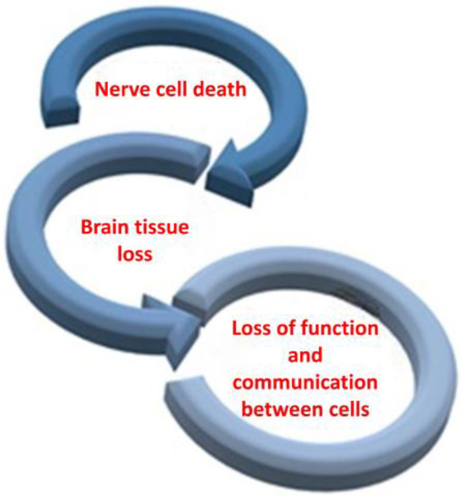
Postsynaptic protein neurogranin (Ng), which plays a role in synaptic plasticity, learning, and memory, has been identified as the candidate biomarker of Alzheimer's disease (AD). Cortical Amyloid β pathology seems to accelerate the onset of clinical symptoms; therefore, it is potentially valuable for early diagnosis of AD and therapeutic intervention. Synaptic pathology was shown to be an early feature of AD. Thus, proteins involved in synaptic function, such as Ng, are of great interest in studying the disease. Some prior human studies have found that Ng, a protein involved in the regulation of synaptic function, is present at greater levels in the cerebrospinal fluid of people with AD compared with those without the disease. High levels of neurogranin are associated with increased levels of synaptic vulnerability and decreased cognitive function in AD patients. This review, therefore, looked at the functionality of Ng in the brain, its association with other synaptic proteins, and its applicability as a diagnostic marker in AD. This study, therefore, sought to expand the knowledge on Ng changes in AD as it relates to synaptic dysfunction and enhanced the search for a better diagnostic and therapeutic approach.
Citation: Rajkumar Bavaharini, Chaitanya Sree Somala, Konda Mani Saravanan, Thirunavukarasou Anand. Neurogranin in Alzheimer's Disease: Roles in synaptic function, pathology, and potential as a diagnostic biomarker[J]. AIMS Molecular Science, 2024, 11(4): 330-350. doi: 10.3934/molsci.2024020
Postsynaptic protein neurogranin (Ng), which plays a role in synaptic plasticity, learning, and memory, has been identified as the candidate biomarker of Alzheimer's disease (AD). Cortical Amyloid β pathology seems to accelerate the onset of clinical symptoms; therefore, it is potentially valuable for early diagnosis of AD and therapeutic intervention. Synaptic pathology was shown to be an early feature of AD. Thus, proteins involved in synaptic function, such as Ng, are of great interest in studying the disease. Some prior human studies have found that Ng, a protein involved in the regulation of synaptic function, is present at greater levels in the cerebrospinal fluid of people with AD compared with those without the disease. High levels of neurogranin are associated with increased levels of synaptic vulnerability and decreased cognitive function in AD patients. This review, therefore, looked at the functionality of Ng in the brain, its association with other synaptic proteins, and its applicability as a diagnostic marker in AD. This study, therefore, sought to expand the knowledge on Ng changes in AD as it relates to synaptic dysfunction and enhanced the search for a better diagnostic and therapeutic approach.

| [1] | Alzheimer's Association.2023 Alzheimer's disease facts and figures. Alzheimers Dement (2023) 19: 1598-1695. https://doi.org/10.1002/alz.13016 |
| [2] |
Diogo VS, Ferreira HA, Prata D, et al. (2022) Early diagnosis of Alzheimer's disease using machine learning: A multi-diagnostic, generalizable approach. Alz Res Therapy 14: 107. https://doi.org/10.1186/s13195-022-01047-y 
|
| [3] |
Jakob-Roetne R, Jacobsen H (2009) Alzheimer's disease: From pathology to therapeutic approaches. Angew Chem Int Ed Engl 48: 3030-3059. https://doi.org/10.1002/anie.200802808
|
| [4] |
Ferrari-Souza JP, Bellaver B, Ferreira PCL, et al. (2023) APOEϵ4 potentiates amyloid β effects on longitudinal tau pathology. Nat Aging 3: 1210-1218. https://doi.org/10.1038/s43587-023-00490-2
|
| [5] |
Saravanan KM, Kannan M, Meera P, et al. (2022) E3 ligases: A potential multi-drug target for different types of cancers and neurological disorders. Future Med Chem 14: 187-201. https://doi.org/10.4155/fmc-2021-0157
|
| [6] |
Moore KBE, Hung TJ, Fortin JS (2023) Hyperphosphorylated tau (p-tau) and drug discovery in the context of Alzheimer's disease and related tauopathies. Drug Discov Today 28: 103487. https://doi.org/10.1016/j.drudis.2023.103487
|
| [7] |
Saravanan KM, Zhang H, Zhang H, et al. (2020) On the conformational dynamics of β-amyloid forming peptides: A computational perspective. Front Bioeng Biotechnol 8: 532. https://doi.org/10.3389/fbioe.2020.00532
|
| [8] |
Agnello L, Lo Sasso B, Vidali M, et al. (2021) Neurogranin as a reliable biomarker for synaptic dysfunction in Alzheimer's disease. Diagnostics 11: 2339. https://doi.org/10.3390/diagnostics11122339
|
| [9] |
Pelucchi S, Gardoni F, Di Luca M, et al. (2022) Chapter 28 - Synaptic dysfunction in early phases of Alzheimer's Disease. Handbook of Clinical Neurology . Elsevier 417-438. https://doi.org/10.1016/B978-0-12-819410-2.00022-9
|
| [10] |
Saunders T, Gunn C, Blennow K, et al. (2023) Neurogranin in Alzheimer's disease and ageing: A human post-mortem study. Neurobiol Dis 177: 105991. https://doi.org/10.1016/j.nbd.2023.105991
|
| [11] |
Rani S, Dhar SB, Khajuria A, et al. (2023) Advanced overview of biomarkers and techniques for early diagnosis of Alzheimer's disease. Cell Mol Neurobiol 43: 2491-2523. https://doi.org/10.1007/s10571-023-01330-y
|
| [12] |
Abraham WC, Jones OD, Glanzman DL (2019) Is plasticity of synapses the mechanism of long-term memory storage?. npj Sci Learn 4: 9. https://doi.org/10.1038/s41539-019-0048-y
|
| [13] |
Xue M, Sun FR, Ou YN, et al. (2020) Association of cerebrospinal fluid neurogranin levels with cognition and neurodegeneration in Alzheimer's disease. Aging 12: 9365-9379. https://doi.org/10.18632/aging.103211
|
| [14] |
Toader C, Dobrin N, Brehar FM, et al. (2023) From recognition to remedy: The significance of biomarkers in neurodegenerative disease pathology. Int J Mol Sci 24: 16119. https://doi.org/10.3390/ijms242216119
|
| [15] |
Scheltens P, De Strooper B, Kivipelto M, et al. (2021) Alzheimer's disease. Lancet 397: 1577-1590. https://doi.org/10.1016/S0140-6736(20)32205-4
|
| [16] |
Cardoso BR, Roberts BR, Malpas CB, et al. (2019) Supranutritional sodium selenate supplementation delivers selenium to the central nervous system: Results from a randomized controlled pilot trial in Alzheimer's disease. Neurotherapeutics 16: 192-202. https://doi.org/10.1007/s13311-018-0662-z
|
| [17] |
DeTure MA, Dickson DW (2019) The neuropathological diagnosis of Alzheimer's disease. Mol Neurodegener 14: 32. https://doi.org/10.1186/s13024-019-0333-5
|
| [18] | Zhang XX, Tian Y, Wang ZT, et al. (2021) The Epidemiology of Alzheimer's disease modifiable risk factors and prevention. J Prev Alzheimers Dis 8: 313-321. https://doi.org/10.14283/jpad.2021.15 |
| [19] |
Dubois B, von Arnim CAF, Burnie N, et al. (2023) Biomarkers in Alzheimer's disease: Role in early and differential diagnosis and recognition of atypical variants. Alz Res Therapy 15: 175. https://doi.org/10.1186/s13195-023-01314-6
|
| [20] |
Jellinger KA (2022) Recent update on the heterogeneity of the Alzheimer's disease spectrum. J Neural Transm 129: 1-24. https://doi.org/10.1007/s00702-021-02449-2
|
| [21] |
Dubois B, Villain N, Frisoni GB, et al. (2021) Clinical diagnosis of Alzheimer's disease: Recommendations of the International Working Group. Lancet Neurol 20: 484-496. https://doi.org/10.1016/S1474-4422(21)00066-1
|
| [22] |
Camporesi E, Nilsson J, Brinkmalm A, et al. (2020) Fluid biomarkers for synaptic dysfunction and loss. Biomark Insights 15: 1177271920950319. https://doi.org/10.1177/1177271920950319
|
| [23] |
Liu W, Lin H, He X, et al. (2020) Neurogranin as a cognitive biomarker in cerebrospinal fluid and blood exosomes for Alzheimer's disease and mild cognitive impairment. Transl Psychiatry 10: 125. https://doi.org/10.1038/s41398-020-0801-2
|
| [24] |
Hindley N, Sanchez Avila A, Henstridge C (2023) Bringing synapses into focus: Recent advances in synaptic imaging and mass-spectrometry for studying synaptopathy. Front Synaptic Neurosci 15: 1130198. https://doi.org/10.3389/fnsyn.2023.1130198
|
| [25] |
Mravinacová S, Alanko V, Bergström S, et al. (2024) CSF protein ratios with enhanced potential to reflect Alzheimer's disease pathology and neurodegeneration. Mol Neurodegener 19: 15. https://doi.org/10.1186/s13024-024-00705-z
|
| [26] | Milos T, Vuic B, Balic N, et al. (2024) Cerebrospinal fluid in the differential diagnosis of Alzheimer's disease: An update of the literature. Expert Rev Neurother 1–17. https://doi.org/10.1080/14737175.2024.2400683 |
| [27] |
Pereira JB, Janelidze S, Ossenkoppele R, et al. (2021) Untangling the association of amyloid-β and tau with synaptic and axonal loss in Alzheimer's disease. Brain 144: 310-324. https://doi.org/10.1093/brain/awaa395
|
| [28] |
Kester MI, Teunissen CE, Crimmins DL, et al. (2015) Neurogranin as a cerebrospinal fluid biomarker for synaptic loss in symptomatic Alzheimer disease. JAMA Neurol 72: 1275-1280. https://doi.org/10.1001/jamaneurol.2015.1867
|
| [29] |
Willemse EAJ, Sieben A, Somers C, et al. (2021) Neurogranin as biomarker in CSF is non-specific to Alzheimer's disease dementia. Neurobiol Aging 108: 99-109. https://doi.org/10.1016/j.neurobiolaging.2021.08.002
|
| [30] |
van Dyck CH, Swanson CJ, Aisen P, et al. (2023) Lecanemab in early Alzheimer's disease. N Engl J Med 388: 9-21. https://doi.org/10.1056/NEJMoa2212948
|
| [31] |
Bergström S, Remnestål J, Yousef J, et al. (2021) Multi-cohort profiling reveals elevated CSF levels of brain-enriched proteins in Alzheimer's disease. Ann Clin Transl Neurol 8: 1456-1470. https://doi.org/10.1002/acn3.51402
|
| [32] |
Remnestål J, Just D, Mitsios N, et al. (2016) CSF profiling of the human brain enriched proteome reveals associations of neuromodulin and neurogranin to Alzheimer's disease. Proteomics Clin Appl 10: 1242-1253. https://doi.org/10.1002/prca.201500150
|
| [33] |
Sandelius Å, Portelius E, Källén Å, et al. (2019) Elevated CSF GAP-43 is Alzheimer's disease specific and associated with tau and amyloid pathology. Alzheimers Dement 15: 55-64. https://doi.org/10.1016/j.jalz.2018.08.006
|
| [34] |
McGrowder DA, Miller F, Vaz K, et al. (2021) Cerebrospinal fluid biomarkers of Alzheimer's disease: Current evidence and future perspectives. Brain Sci 11: 215. https://doi.org/10.3390/brainsci11020215
|
| [35] |
Remnestål J, Bergström S, Olofsson J, et al. (2021) Association of CSF proteins with tau and amyloid β levels in asymptomatic 70-year-olds. Alz Res Therapy 13: 54. https://doi.org/10.1186/s13195-021-00789-5
|
| [36] |
Rosenberg A, Öhlund-Wistbacka U, Hall A, et al. (2022) β-amyloid, tau, neurodegeneration classification and eligibility for anti-amyloid treatment in a memory clinic population. Neurology 99: e2102-e2113. https://doi.org/10.1212/WNL.0000000000201043
|
| [37] |
Cummings J (2019) The national institute on aging—Alzheimer's association framework on Alzheimer's disease: Application to clinical trials. Alzheimers Dement 15: 172-178. https://doi.org/10.1016/j.jalz.2018.05.006
|
| [38] |
Tahami Monfared AA, Byrnes MJ, White LA, et al. (2022) Alzheimer's disease: Epidemiology and clinical progression. Neurol Ther 11: 553-569. https://doi.org/10.1007/s40120-022-00338-8
|
| [39] |
Xiang Y, Xin J, Le W, et al. (2020) Neurogranin: A potential biomarker of neurological and mental diseases. Front Aging Neurosci 12: 584743. https://doi.org/10.3389/fnagi.2020.584743
|
| [40] |
van der Flier WM, de Vugt ME, Smets EMA, et al. (2023) Towards a future where Alzheimer's disease pathology is stopped before the onset of dementia. Nat Aging 3: 494-505. https://doi.org/10.1038/s43587-023-00404-2
|
| [41] | Rasmussen J, Langerman H (2019) Alzheimer's disease–why we need early diagnosis. Degener Neurol Neuromuscul Dis 123–130. https://doi.org/10.2147/DNND.S228939 |
| [42] |
Liang Y, Kang X, Zhang H, et al. (2023) Knockdown and inhibition of hippocampal GPR17 attenuates lipopolysaccharide-induced cognitive impairment in mice. J Neuroinflammation 20. https://doi.org/271.10.1186/s12974-023-02958-9
|
| [43] |
Schindler SE, Bollinger JG, Ovod V, et al. (2019) High-precision plasma β-amyloid 42/40 predicts current and future brain amyloidosis. Neurology 93: e1647-e1659. https://doi.org/10.1212/WNL.0000000000008081
|
| [44] |
Klyucherev TO, Olszewski P, Shalimova AA, et al. (2022) Advances in the development of new biomarkers for Alzheimer's disease. Transl Neurodegener 11: 25. https://doi.org/10.1186/s40035-022-00296-z
|
| [45] |
Janelidze S, Mattsson N, Palmqvist S, et al. (2020) Plasma P-tau181 in Alzheimer's disease: Relationship to other biomarkers, differential diagnosis, neuropathology and longitudinal progression to Alzheimer's dementia. Nat Med 26: 379-386. https://doi.org/10.1038/s41591-020-0755-1
|
| [46] |
O'Bryant SE, Petersen M, Hall J, et al. (2022) Characterization of mild cognitive impairment and dementia among community-dwelling Mexican Americans and non-hispanic whites. J Alzheimers Dis 90: 905-915. https://doi.org/10.3233/JAD-220300
|
| [47] |
Pohlan J, Kress W, Hermann K-G, et al. (2020) Computed tomography thermography for ablation zone prediction in microwave ablation and cryoablation: Advantages and challenges in an ex vivo porcine liver model. J Comput Assist Tomogr 44: 744-749. https://doi.org/10.1097/RCT.0000000000001081
|
| [48] |
Agnello L, Gambino CM, Lo Sasso B, et al. (2021) Neurogranin as a novel biomarker in Alzheimer's disease. Lab Med 52: 188-196. https://doi.org/10.1093/labmed/lmaa062
|
| [49] |
Nashine S, Kenney MC (2024) Effects of Humanin G (HNG) on angiogenesis and neurodegeneration markers in Age-related Macular Degeneration (AMD). Mitochondrion 74: 101818. https://doi.org/10.1016/j.mito.2023.11.001
|
| [50] | Vontell RT, Gober R, Dallmeier J, et al. (2024) Association of region-specific hippocampal reduction of neurogranin with inflammasome proteins in post mortem brains of Alzheimer's disease. Alzheimers Dement 10: e12444. https://doi.org/10.1002/trc2.12444 |
| [51] |
Li L, Lai M, Cole S, et al. (2020) Neurogranin stimulates Ca2+/calmodulin-dependent kinase II by suppressing calcineurin activity at specific calcium spike frequencies. PLoS Comput Biol 16: e1006991. https://doi.org/10.1371/journal.pcbi.1006991
|
| [52] |
Kvartsberg H, Lashley T, Murray CE, et al. (2019) The intact postsynaptic protein neurogranin is reduced in brain tissue from patients with familial and sporadic Alzheimer's disease. Acta Neuropathol 137: 89-102. https://doi.org/10.1007/s00401-018-1910-3
|
| [53] |
Svirsky SE, Henchir J, Li Y, et al. (2024) Temporal-specific sex and injury-dependent changes on neurogranin-associated synaptic signaling after controlled cortical impact in rats. Mol Neurobiol 61: 7256-7268. https://doi.org/10.1007/s12035-024-04043-5
|
| [54] |
Zhong L, Gerges NZ (2020) Neurogranin regulates metaplasticity. Front Mol Neurosci 12: 322. https://doi.org/10.3389/fnmol.2019.00322
|
| [55] |
Rueda-García V, Rondón-Barragán IS (2024) Molecular characterization of neurogranin (NRGN) gene from Red‑Bellied Pacu (Piaractus brachypomus). Mol Neurobiol 61: 2620-2630. https://doi.org/10.1007/s12035-023-03700-5
|
| [56] |
Casaletto KB, Elahi FM, Bettcher BM, et al. (2017) Neurogranin, a synaptic protein, is associated with memory independent of Alzheimer biomarkers. Neurology 89: 1782-1788. https://doi.org/10.1212/WNL.0000000000004569
|
| [57] |
Vasung L, Abaci Turk E, Ferradal SL, et al. (2019) Exploring early human brain development with structural and physiological neuroimaging. Neuroimage 187: 226-254. https://doi.org/10.1016/j.neuroimage.2018.07.041
|
| [58] |
Subramanian L, Calcagnotto ME, Paredes MF (2020) Cortical malformations: Lessons in human brain development. Front Cell Neurosci 13: 576. https://doi.org/10.3389/fncel.2019.00576
|
| [59] |
Bick J, Nelson CA (2016) Early adverse experiences and the developing brain. Neuropsychopharmacology 41: 177-196. https://doi.org/10.1038/npp.2015.252
|
| [60] |
Lista S, Santos-Lozano A, Emanuele E, et al. (2024) Monitoring synaptic pathology in Alzheimer's disease through fluid and PET imaging biomarkers: a comprehensive review and future perspectives. Mol Psychiatry 29: 847-857. https://doi.org/10.1038/s41380-023-02376-6
|
| [61] |
Svirsky S, Henchir J, Li Y, et al. (2019) Neurogranin protein expression is reduced after controlled cortical impact in rats. J Neurotrauma 37: 939-949. https://doi.org/10.1089/neu.2019.6759
|
| [62] |
Garrido-García A, de Andrés R, Jiménez-Pompa A, et al. (2019) Neurogranin expression is regulated by synaptic activity and promotes synaptogenesis in cultured hippocampal neurons. Mol Neurobiol 56: 7321-7337. https://doi.org/10.1007/s12035-019-1593-3
|
| [63] |
Hwang H, Szucs MJ, Ding LJ, et al. (2021) Neurogranin, encoded by the schizophrenia risk gene NRGN, bidirectionally modulates synaptic plasticity via calmodulin-dependent regulation of the neuronal phosphoproteome. Biol Psychiatry 89: 256-269. https://doi.org/10.1016/j.biopsych.2020.07.014
|
| [64] |
Cheriyan VT, Alfaidi M, Jorgensen AN, et al. (2020) Neurogranin regulates eNOS function and endothelial activation. Redox Biol 34: 101487. https://doi.org/10.1016/j.redox.2020.101487
|
| [65] |
Lin H, Zhang J, Dai Y, et al. (2023) Neurogranin as an important regulator in swimming training to improve the spatial memory dysfunction of mice with chronic cerebral hypoperfusion. J Sport Heal Sci 12: 116-129. https://doi.org/10.1016/j.jshs.2022.01.008
|
| [66] |
Gribaudo S, Saraulli D, Nato G, et al. (2021) Neurogranin regulates adult-born olfactory granule cell spine density and odor-reward associative memory in mice. Int J Mol Sci 22: 4269. https://doi.org/10.3390/ijms22084269
|
| [67] |
Cardozo LP, de Lima BQI, Maciel MAE, et al. (2019) Synaptic elimination in neurological disorders. Curr Neuropharmacol 17: 1071-1095. https://doi.org/10.2174/1570159X17666190603170511
|
| [68] |
Ordyan M, Bartol T, Kennedy M, et al. (2020) Interactions between calmodulin and neurogranin govern the dynamics of CaMKII as a leaky integrator. PLoS Comput Biol 16: e1008015. https://doi.org/10.1371/journal.pcbi.1008015
|
| [69] |
Putkey JA, Hoffman L, Berka V, et al. (2024) Neurogranin modulates the rate of association between calmodulin and target peptides. Biophys J 123: 1676-1689. https://doi.org/10.1016/j.bpj.2024.05.010
|
| [70] |
O'Day DH (2020) Calmodulin binding proteins and Alzheimer's disease: Biomarkers, regulatory enzymes and receptors that are regulated by calmodulin. Int J Mol Sci 21: 7344. https://doi.org/10.3390/ijms21197344
|
| [71] |
Moradi F, Copeland EN, Baranowski RW, et al. (2020) Calmodulin-binding proteins in muscle: A minireview on nuclear receptor interacting protein, neurogranin, and growth-associated protein 43. Int J Mol Sci 21: 1016. https://doi.org/10.3390/ijms21031016
|
| [72] |
Dulewicz M, Kulczyńska-Przybik A, Słowik A, et al. (2021) Neurogranin and neuronal pentraxin receptor as synaptic dysfunction biomarkers in Alzheimer's disease. J Clin Med 10: 4575. https://doi.org/10.3390/jcm10194575
|
| [73] |
Poejo J, Salazar J, Mata AM, et al. (2021) The relevance of amyloid β-calmodulin complexation in neurons and brain degeneration in Alzheimer's disease. Int J Mol Sci 22: 4976. https://doi.org/10.3390/ijms22094976
|
| [74] |
Kumar V, Chichili VPR, Zhong L, et al. (2013) Structural basis for the interaction of unstructured neuron specific substrates neuromodulin and neurogranin with calmodulin. Sci Rep 3: 1392. https://doi.org/10.1038/srep01392
|
| [75] |
Szklarczyk D, Kirsch R, Koutrouli M, et al. (2023) The STRING database in 2023: Protein–protein association networks and functional enrichment analyses for any sequenced genome of interest. Nucleic Acids Res 51: D638-D646. https://doi.org/10.1093/nar/gkac1000
|
| [76] |
Nazir FH, Camporesi E, Brinkmalm G, et al. (2021) Molecular forms of neurogranin in cerebrospinal fluid. J Neurochem 157: 816-833. https://doi.org/10.1111/jnc.15252
|
| [77] | Joglekar AP A cell-type centric view of alternative splicing in the mammalian brain (2022). Available from: https://research.weill.cornell.edu/about-us/news-updates/cell-type-centric-view-alternative-splicing-mammalian-brain |
| [78] |
Beghi S, Furmanik M, Jaminon A, et al. (2022) Calcium signalling in heart and vessels: Role of calmodulin and downstream calmodulin-dependent protein kinases. Int J Mol Sci 23: 16139. https://doi.org/10.3390/ijms232416139
|
| [79] |
O'Day DH (2022) Calmodulin binding domains in critical risk proteins involved in neurodegeneration. Curr Issues Mol Biol 44: 5802-5814. https://doi.org/10.3390/cimb44110394
|
| [80] |
Zhang H, Jiang X, Ma L, et al. (2022) Role of Aβ in Alzheimer's-related synaptic dysfunction. Front Cell Dev Biol 10: 964075. https://doi.org/10.3389/fcell.2022.964075
|
| [81] |
Abi-Dargham A, Moeller SJ, Ali F, et al. (2023) Candidate biomarkers in psychiatric disorders: State of the field. World Psychiatry 22: 236-262. https://doi.org/10.1002/wps.21078
|
| [82] |
Blennow K, Diaz-Lucena D, Zetterberg H, et al. (2019) CSF neurogranin as a neuronal damage marker in CJD: A comparative study with AD. J Neurol Neurosurg Psychiatry 90: 846-853. https://doi.org/10.1136/jnnp-2018-320155
|
| [83] |
Rabbito A, Dulewicz M, Kulczyńska-Przybik A, et al. (2020) Biochemical markers in Alzheimer's disease. Int J Mol Sci 21: 1989. https://doi.org/10.3390/ijms21061989
|
| [84] |
Mavroudis IA, Petridis F, Chatzikonstantinou S, et al. (2020) A meta-analysis on CSF neurogranin levels for the diagnosis of Alzheimer's disease and mild cognitive impairment. Aging Clin Exp Res 32: 1639-1646. https://doi.org/10.1007/s40520-019-01326-z
|
| [85] |
Siddappaji KK, Gopal S (2021) Molecular mechanisms in Alzheimer's disease and the impact of physical exercise with advancements in therapeutic approaches. AIMS Neurosci 8: 357-389. https://doi.org/10.3934/Neuroscience.2021020
|
| [86] |
Vaughn MN, Winston CN, Levin N, et al. (2022) Developing biomarkers of mild traumatic brain injury: Promise and progress of CNS-derived exosomes. Front Neurol 12: 698206. https://doi.org/10.3389/fneur.2021.698206
|
| [87] |
Kivisäkk P, Carlyle BC, Sweeney T, et al. (2022) Increased levels of the synaptic proteins PSD-95, SNAP-25, and neurogranin in the cerebrospinal fluid of patients with Alzheimer's disease. Alzheimers Res Ther 14: 58. https://doi.org/10.1186/s13195-022-01002-x
|
| [88] |
Kent SA, Spires-Jones TL, Durrant CS (2020) The physiological roles of tau and Aβ: Implications for Alzheimer's disease pathology and therapeutics. Acta Neuropathol 140: 417-447. https://doi.org/10.1007/s00401-020-02196-w
|
| [89] | Bouwman FH, Frisoni GB, Johnson SC, et al. (2022) Clinical application of CSF biomarkers for Alzheimer's disease: From rationale to ratios. Alzheimers Dement 14: e12314. https://doi.org/10.1002/dad2.12314 |
| [90] |
Saunders TS, Gadd DA, Spires-Jones TL, et al. (2022) Associations between cerebrospinal fluid markers and cognition in ageing and dementia: A systematic review. Eur J Neurosci 56: 5650-5713. https://doi.org/10.1111/ejn.15656
|
| [91] |
Lepeta K, Lourenco MV, Schweitzer BC, et al. (2016) Synaptopathies: Synaptic dysfunction in neurological disorders–A review from students to students. J Neurochem 138: 785-805. https://doi.org/10.1111/jnc.13713
|


Figures(5) / Tables(2)

Rajkumar Bavaharini, Chaitanya Sree Somala, Konda Mani Saravanan, Thirunavukarasou Anand. Neurogranin in Alzheimer's Disease: Roles in synaptic function, pathology, and potential as a diagnostic biomarker[J]. AIMS Molecular Science, 2024, 11(4): 330-350. doi: 10.3934/molsci.2024020


 DownLoad: