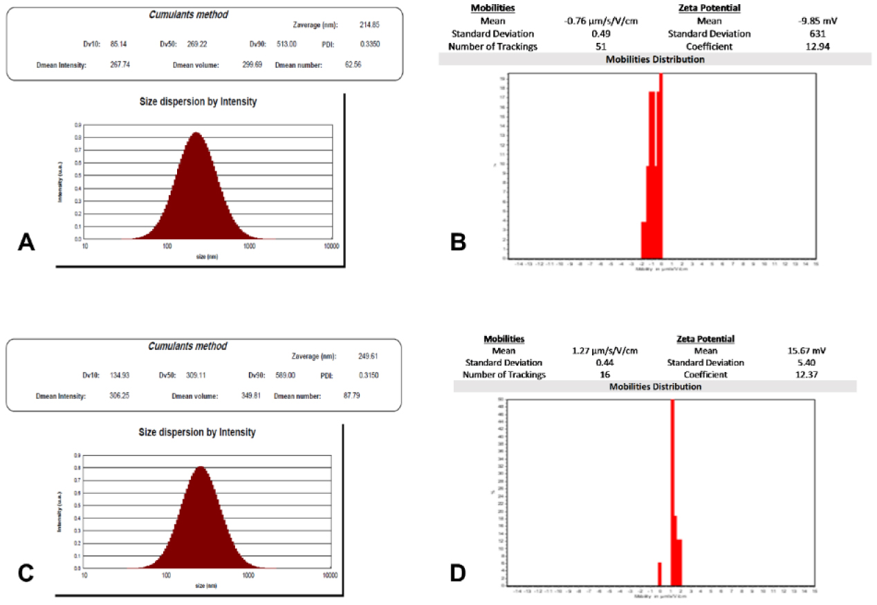
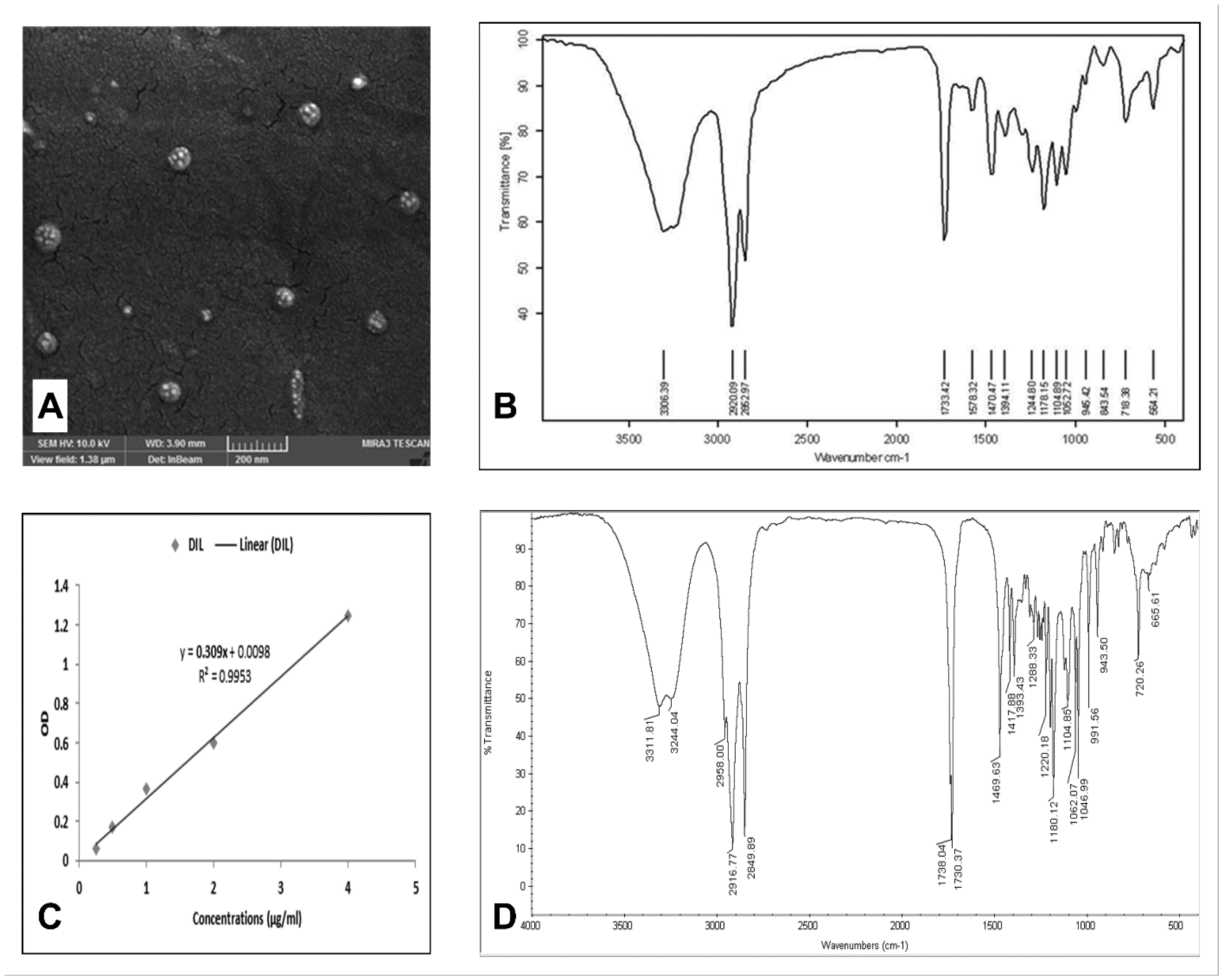
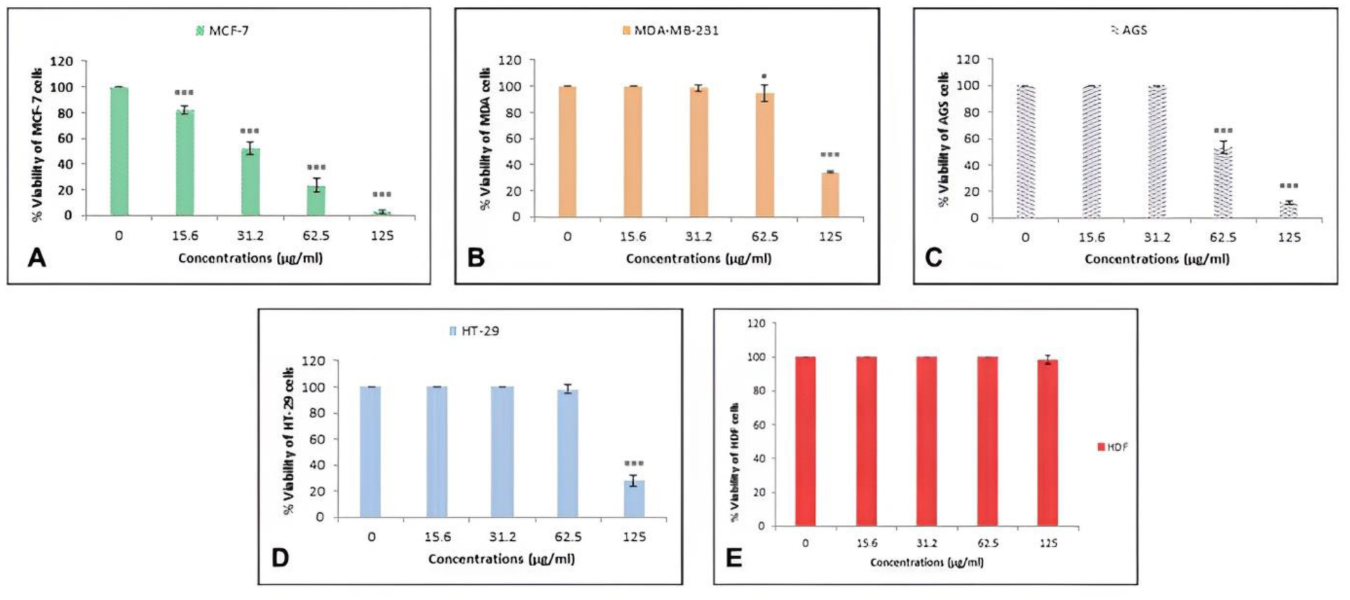
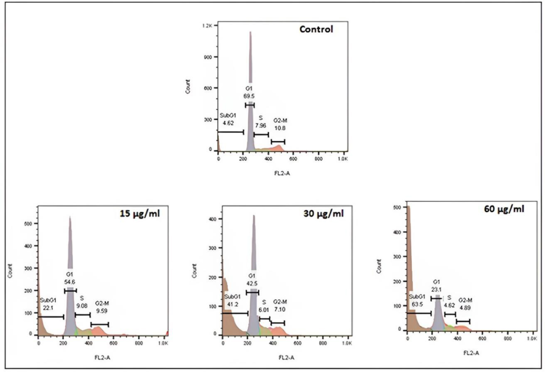
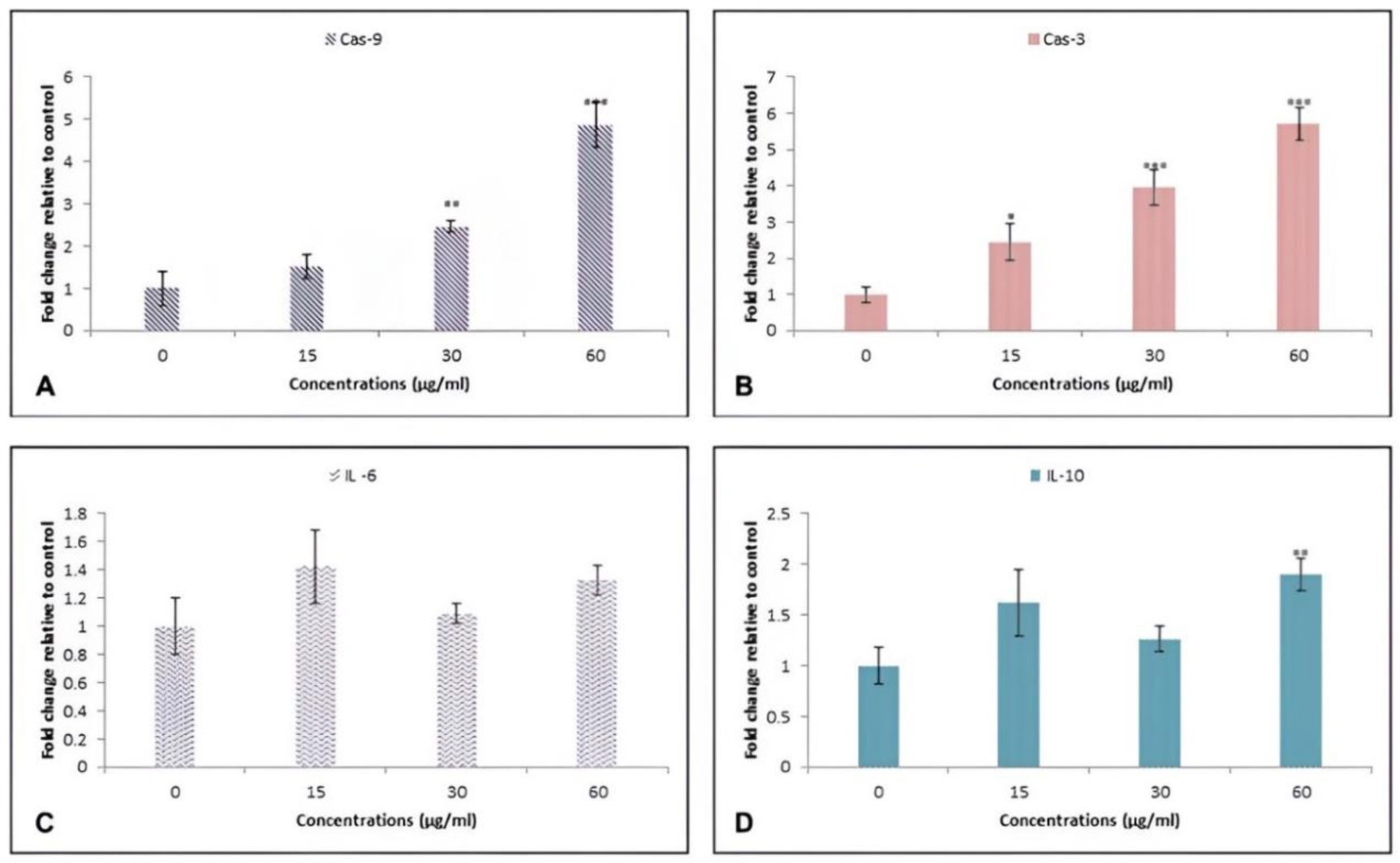
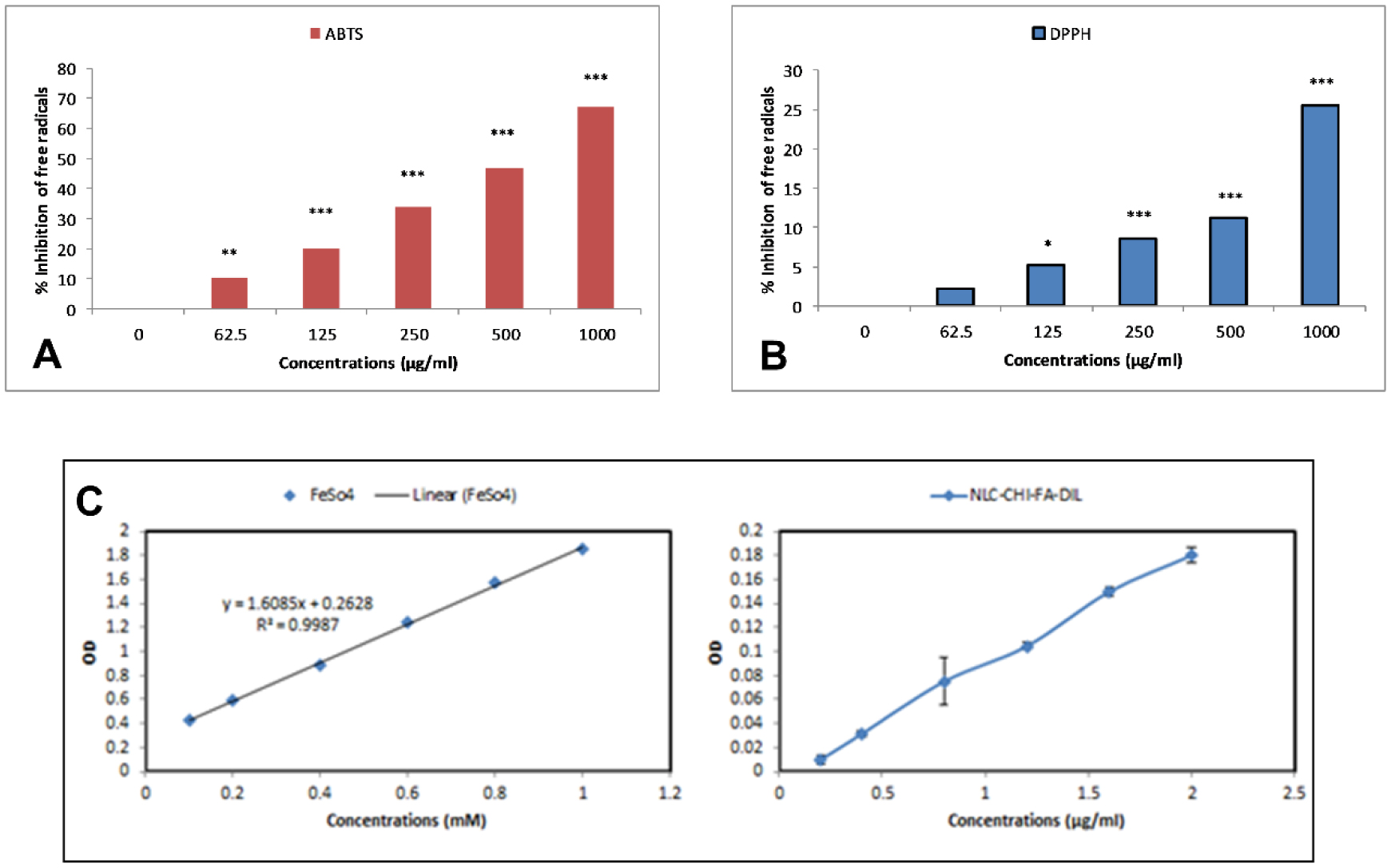
The main goal of cancer treatment is to ensure that the drug reaches the tumor tissue and to reduce the side effects of the drug. This study was conducted to synthesize a novel nanostructured lipid carrier modified with chitosan-folate to deliver diltiazem to cancer cell lines and to evaluate its anticancer effect. Dynamic light scattering (DLS), field emission scanning electron microscopy (FESEM), and Fourier transform infrared spectrometer (FTIR) methods were used to characterize the nanoparticles. The cytotoxicity effect on cancer cell lines variants was measured. Flow cytometry was used for cell cycle analysis, and reverse transcription polymerase chain reaction (RT-PCR) was performed to assess the induction of apoptosis. The inflammatory effects were evaluated by molecular analysis, and the 2,2′-azino-bis(3-ethylbenzothiazoline-6-sulfonic acid, 2,2-Diphenyl-1-picrylhydrazyl, and Ferric reducing antioxidant power methods were used to measure the antioxidant power of the nanoparticles. The results reported the mean of the real and hydrodynamic diameter of the nanoparticles as 87.7 and 249 nm, respectively, and the encapsulation efficiency of diltiazem was reported to be 86.6%. The cytotoxicity results revealed that the breast cancer cells were more sensitive to treatment, with a median concentration of 33.5µg/ml. Additionally, the nanoparticle treatment led to the arrest of cells in the SubG1 phase while increasing the expression of caspases 3 and 9, which indicates the activation of the internal pathway of apoptosis. Additionally, the increase in the expression of interleukins 6 and 10 suggests an effect of the nanoparticles on inflammation. In addition, the ability to inhibit 2,2′-azino-bis (3-ethylbenzothiazoline-6-sulfonic acid and 2,2-Diphenyl-1-picrylhydrazyl free radicals with an average concentration of 577, which is more significant than 1000 µg/ml, and the ability of the nanoparticles to reduce iron ions confirmed the antioxidant effect of diltiazem-loaded nanostructured lipid carriers. These results suggest that the nanoparticles have an excellent potential to treat breast cancer.
Citation: Vahid Pouresmaeil, Marwa Mawlood Salman Al-zand, Aida Pouresmaeil, Seyedeh Samira Saghravanian, Masoud Homayouni Tabrizi. Loading diltiazem onto surface-modified nanostructured lipid carriers to evaluate its apoptotic, cytotoxic, and inflammatory effects on human breast cancer cells[J]. AIMS Molecular Science, 2024, 11(3): 231-250. doi: 10.3934/molsci.2024014
The main goal of cancer treatment is to ensure that the drug reaches the tumor tissue and to reduce the side effects of the drug. This study was conducted to synthesize a novel nanostructured lipid carrier modified with chitosan-folate to deliver diltiazem to cancer cell lines and to evaluate its anticancer effect. Dynamic light scattering (DLS), field emission scanning electron microscopy (FESEM), and Fourier transform infrared spectrometer (FTIR) methods were used to characterize the nanoparticles. The cytotoxicity effect on cancer cell lines variants was measured. Flow cytometry was used for cell cycle analysis, and reverse transcription polymerase chain reaction (RT-PCR) was performed to assess the induction of apoptosis. The inflammatory effects were evaluated by molecular analysis, and the 2,2′-azino-bis(3-ethylbenzothiazoline-6-sulfonic acid, 2,2-Diphenyl-1-picrylhydrazyl, and Ferric reducing antioxidant power methods were used to measure the antioxidant power of the nanoparticles. The results reported the mean of the real and hydrodynamic diameter of the nanoparticles as 87.7 and 249 nm, respectively, and the encapsulation efficiency of diltiazem was reported to be 86.6%. The cytotoxicity results revealed that the breast cancer cells were more sensitive to treatment, with a median concentration of 33.5µg/ml. Additionally, the nanoparticle treatment led to the arrest of cells in the SubG1 phase while increasing the expression of caspases 3 and 9, which indicates the activation of the internal pathway of apoptosis. Additionally, the increase in the expression of interleukins 6 and 10 suggests an effect of the nanoparticles on inflammation. In addition, the ability to inhibit 2,2′-azino-bis (3-ethylbenzothiazoline-6-sulfonic acid and 2,2-Diphenyl-1-picrylhydrazyl free radicals with an average concentration of 577, which is more significant than 1000 µg/ml, and the ability of the nanoparticles to reduce iron ions confirmed the antioxidant effect of diltiazem-loaded nanostructured lipid carriers. These results suggest that the nanoparticles have an excellent potential to treat breast cancer.
Nanostructured lipid carriers
Diltiazem
Dynamic Light Scattering
Field emission scanning electron microscope
Fourier transform infrared
3-(4,5-dimethylthiazol-2-yl)-2,5-diphenyltetrazolium bromide
Acridine orange
DAPI or Propidium iodide
2,2-azinobis (3-ethylbenzothiazoline-6-sulfonic acid)
2,2-diphenyl-1-picrylhydrazyl
Polydispersity Index
1-Ethyl-3-(3-dimethylaminopropyl) carbodiimide
N-Hydroxysuccinimide
Breast cancer cell lines
Gastric cancer cell lines
Colon cancer cell lines
Human dermal fibroblasts

| [1] |
Jeevanandam J, Barhoum A, Chan YS, et al. (2018) Review on nanoparticles and nanostructured materials: History, sources, toxicity and regulations. Beilstein J Nanotechnol 9: 1050-1074. https://doi.org/10.3762/bjnano.9.98 
|
| [2] |
Rudramurthy GR, Swamy MK (2018) Potential applications of engineered nanoparticles in medicine and biology: An update. J Biol Inorg Chem 23: 1185-1204. https://doi.org/10.1007/s00775-018-1600-6
|
| [3] |
Liu T, Bai R, Zhou H, et al. (2020) The effect of size and surface ligands of iron oxide nanoparticles on blood compatibility. RSC Adv 10: 7559-7569. https://doi.org/10.1039/C9RA10969B
|
| [4] |
Emami F, Banstola A, Vatanara A, et al. (2019) Doxorubicin and anti-PD-L1 antibody conjugated gold nanoparticles for colorectal cancer photochemotherapy. Mol Pharmaceutics 16: 1184-1199. https://doi.org/10.1021/acs.molpharmaceut.8b01157
|
| [5] |
Kiplagat A, Martin DR, Onani MO, et al. (2020) Aptamer-conjugated magnetic nanoparticles for the efficient capture of cancer biomarker proteins. J Magn Magn Mater 497: 166063. https://doi.org/10.1016/j.jmmm.2019.166063
|
| [6] |
Tagami T, Ozeki T (2017) Recent trends in clinical trials related to carrier-based drugs. J Pharm Sci 106: 2219-2226. https://doi.org/10.1016/j.xphs.2017.02.026
|
| [7] |
Varela-Fernández R, García-Otero X, Díaz-Tomé V, et al. (2022) Lactoferrin-loaded nanostructured lipid carriers (NLCs) as a new formulation for optimized ocular drug delivery. Eur J Pharm Biopharm 172: 144-156. https://doi.org/10.1016/j.ejpb.2022.02.010
|
| [8] |
Abinaya B, Prasith TP, Ashwin B, et al. (2019) Chitosan in surface modification for bone tissue engineering applications. Biotechnology J 14: 1900171. https://doi.org/10.1002/biot.201900171
|
| [9] |
Chen MC, Mi FL, Liao ZX, et al. (2013) Recent advances in chitosan-based nanoparticles for oral delivery of macromolecules. Adv Drug Deliver Rev 65: 865-879. https://doi.org/10.1016/j.addr.2012.10.010
|
| [10] |
Amini A, Kamali M, Amini B, et al. (2019) Bio-barcode technology for detection of Staphylococcus aureus protein A based on gold and iron nanoparticles. Int J Biol Macromol 124: 1256-1263. https://doi.org/10.1016/j.ijbiomac.2018.11.123
|
| [11] |
Zhong S, Zhang H, Liu Y, et al. (2017) Folic acid functionalized reduction-responsive magnetic chitosan nanocapsules for targeted delivery and triggered release of drugs. Carbohyd Polym 168: 282-289. https://doi.org/10.1016/j.carbpol.2017.03.083
|
| [12] | Ruiz-Torres A, Lozano R, Melon J, et al. (2003) L-calcium channel blockade induced by diltiazem inhibits proliferation, migration and F-actin membrane rearrangements in human vascular smooth muscle cells stimulated with insulin and IGF-1. Int J Clin Pharmacol Ther 41: 386-391. |
| [13] |
Kaddour-Djebbar I, Choudhary V, Lakshmikanthan V, et al. (2012) Diltiazem enhances the apoptotic effects of proteasome inhibitors to induce prostate cancer cell death. J Pharmacol Exp Ther 341: 646-655. https://doi.org/10.1124/jpet.111.188151
|
| [14] | Yıldız C, Cetin A, Demirci F, et al. (2013) Anti-angiogenic effects of diltiazem, imatinib, and bevacizumab in the CAM assay. Int J Sci Res Publ 3: 1-8. |
| [15] |
Chen YC, Wu CT, Chen JH, et al. (2022) Diltiazem inhibits breast cancer metastasis via mediating growth differentiation factor 15 and epithelial-mesenchymal transition. Oncogenesis 11: 48. https://doi.org/10.1038/s41389-022-00423-5
|
| [16] |
Li C, Wei C, Zhao G, et al. (2023) Cancer cells remodeling and quality control are inextricably linked to autophagy. AIMS Mol Sci 10: 109-126. https://doi.org/10.3934/molsci.2023009
|
| [17] |
Li P, Zhou L, Zhao T, et al. (2017) Caspase-9: Structure, mechanisms and clinical application. Oncotarget 8: 23996-24008. https://doi.org/10.18632/oncotarget.15098
|
| [18] |
Huang KH, Fang WL, Li AFY, et al. (2018) Caspase-3, a key apoptotic protein, as a prognostic marker in gastric cancer after curative surgery. Int J Surg 52: 258-263. https://doi.org/10.1016/j.ijsu.2018.02.055
|
| [19] |
Held C, White HD, Stewart RAH, et al. (2017) Inflammatory biomarkers interleukin-6 and C-reactive protein and outcomes in stable coronary heart disease: Experiences from the STABILITY (stabilization of atherosclerotic plaque by initiation of darapladib therapy) trial. J Am Heart Assoc 6: e005077. https://doi.org/10.1161/JAHA.116.005077
|
| [20] |
Iyer SS, Cheng G (2012) Role of interleukin 10 transcriptional regulation in inflammation and autoimmune disease. Crit Rev Immunol 32: 23-63. https://doi.org/10.1615/critrevimmunol.v32.i1.30
|
| [21] |
Dirican E, Özcan H, Uzunçakmak SK, et al. (2023) Evaluation expression of the caspase-3 and caspase-9 apoptotic genes in schizophrenia patients. Clin Psychopharmacol Neurosci 21: 171-178. https://doi.org/10.9758/cpn.2023.21.1.171
|
| [22] |
Vainer N, Dehlendorff C, Johansen JS (2018) Systematic literature review of IL-6 as a biomarker or treatment target in patients with gastric, bile duct, pancreatic and colorectal cancer. Oncotarget 9: 29820-29841. https://doi.org/10.18632/oncotarget.25661
|
| [23] |
Zhou L, Tang C, Li X, et al. (2022) IL-6/IL-10 mRNA expression ratio in tumor tissues predicts prognosis in gastric cancer patients without distant metastasis. Sci Rep 12: 19427. https://doi.org/10.1038/s41598-022-24189-3
|
| [24] |
Sadeghi S, Tabrizi MH, Farhadi A (2023) Folic acid-chitosan coated stylosin nanostructured lipid carriers: Fabrication, in vitro–in vivo assessment in breast malignant cells. J Biomater Sci Polym Ed 34: 791-809. https://doi.org/10.1080/09205063.2022.2145868
|
| [25] | Al-Hasnawi HNG, Pouresmaeil V, Davoodi-Dehaghani F, et al. (2023) Synthesis folate-linked chitosan-coated quetiapine/BSA nano-carriers as the efficient targeted anti-cancer drug delivery system. Mol Biotechnol 1–11. https://doi.org/10.1007/s12033-023-00858-0 |
| [26] |
Alkwedhim MAH, Pouresmaeil V, Davoodi-Dehaghani F, et al. (2023) Synthesis and evaluation of biological effects of modified graphene oxide nanoparticles containing Lawson (Henna extract) on gastric cancer cells. Mol Biol Rep 50: 8971-8983. https://doi.org/10.1007/s11033-023-08797-4
|
| [27] |
Tuekaew J, Siriwatanametanon N, Wongkrajang Y, et al. (2014) Evaluation of the antioxidant activities of Ya-hom Intajak, a Thai herbal formulation, and its component plants. Trop J Pharm Res 13: 1477-1485. http://doi.org/10.4314/tjpr.v13i9.14
|
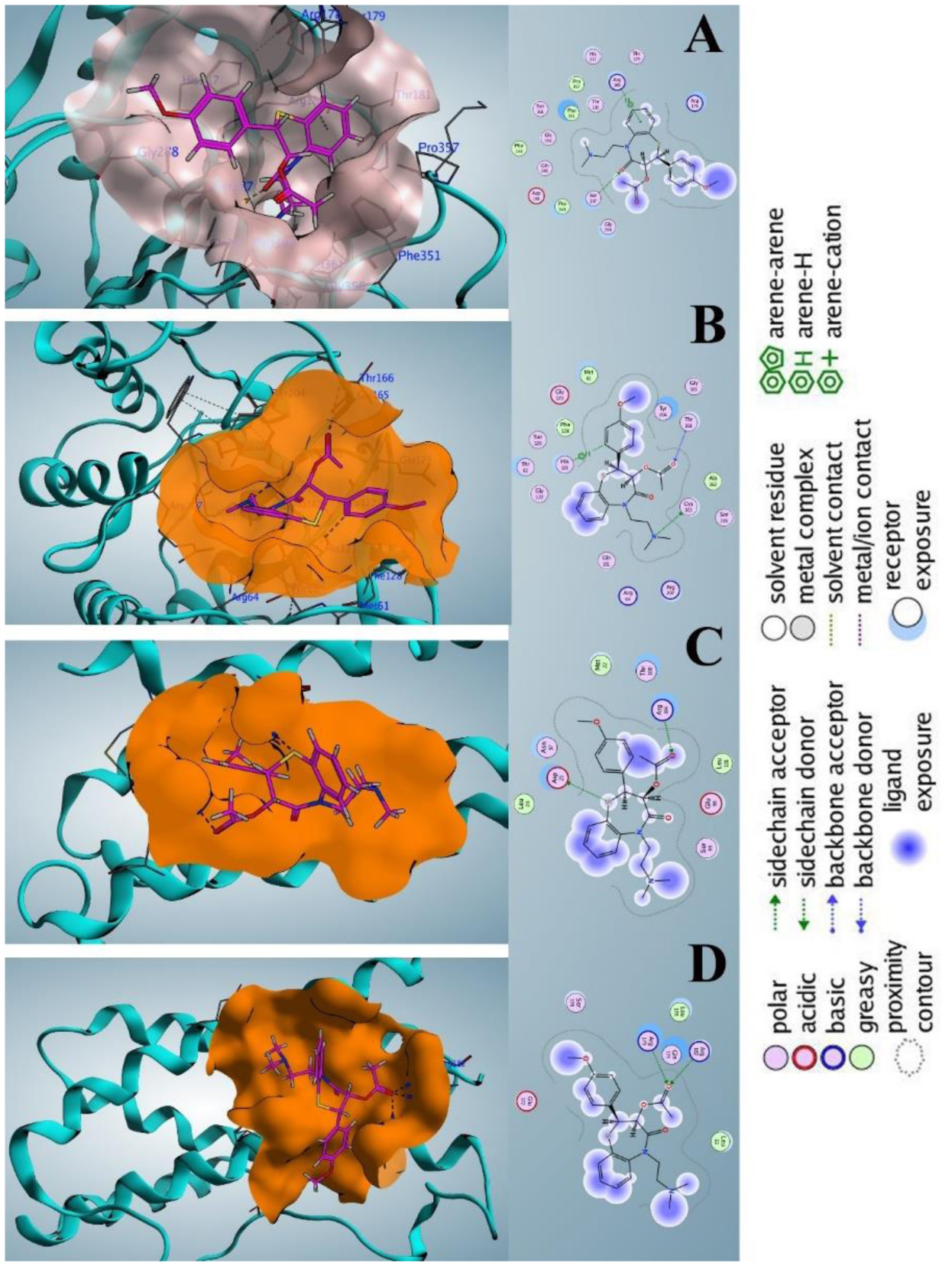
| [28] | Inc CC (2016) Molecular operating environment (MOE). Chemical Computing Group Inc. |
| [29] |
Chao Y, Shiozaki EN, Srinivasula SM, et al. (2005) Engineering a dimeric caspase-9: A re-evaluation of the induced proximity model for caspase activation. PLOS Biol 3: e183. https://doi.org/10.1371/journal.pbio.0030183
|
| [30] |
Ni CZ, Li C, Wu JC, et al. (2003) Conformational restrictions in the active site of unliganded human caspase-3. J Mol Recognit 16: 121-124. https://doi.org/10.1002/jmr.615
|
| [31] |
Somers W, Stahl M, Seehra JS (1997) 1.9 Å crystal structure of interleukin 6: Implications for a novel mode of receptor dimerization and signaling. EMBO J 16: 989-997. https://doi.org/10.1093/emboj/16.5.989
|
| [32] |
Yoon SI, Logsdon NJ, Sheikh F, et al. (2006) Conformational changes mediate interleukin-10 receptor 2 (IL-10R2) binding to IL-10 and assembly of the signaling complex. J Biol Chem 281: 35088-35096. https://doi.org/10.1074/jbc.M606791200
|
| [33] |
Eid RK, Ashour DS, Essa EA, et al. (2020) Chitosan coated nanostructured lipid carriers for enhanced in vivo efficacy of albendazole against Trichinella spiralis. Carbohyd Polym 232: 115826. https://doi.org/10.1016/j.carbpol.2019.115826
|
| [34] |
Lu B, Lv X, Le Y (2019) Chitosan-modified PLGA nanoparticles for control-released drug delivery. Polymers 11: 304. https://doi.org/10.3390/polym11020304
|
| [35] | Liyanage PY, Hettiarachchi SD, Zhou Y, et al. (2019) Nanoparticle-mediated targeted drug delivery for breast cancer treatment. BBA-Rev Cancer 1871: 419-433. https://doi.org/10.1016/j.bbcan.2019.04.006 |
| [36] |
Rizwanullah M, Amin S, Ahmad J (2017) Improved pharmacokinetics and antihyperlipidemic efficacy of rosuvastatin-loaded nanostructured lipid carriers. J Drug Target 25: 58-74. https://doi.org/10.1080/1061186X.2016.1191080
|
| [37] |
Aibani N, Rai R, Patel P, et al. (2021) Chitosan nanoparticles at the biological interface: Implications for drug delivery. Pharmaceutics 13: 1686. https://doi.org/10.3390/pharmaceutics13101686
|
| [38] |
Danhier F, Feron O, Préat V (2010) To exploit the tumor microenvironment: Passive and active tumor targeting of nanocarriers for anti-cancer drug delivery. J Control Release 148: 135-146. https://doi.org/10.1016/j.jconrel.2010.08.027
|
| [39] |
Neves AR, Martins S, Segundo MA, et al. (2016) Nanoscale delivery of resveratrol towards enhancement of supplements and nutraceuticals. Nutrients 8: 131. https://doi.org/10.3390/nu8030131
|
| [40] | Truong Công TTC (2012) Nanoformulations pour la protection de flavonoïdes instables: Exemple de la quercétine: Médecine humaine et pathologie. Université Paris Sud - Paris XI, 2012. Français.ffNNT: 2012PA114850ff . |
| [41] |
Sou K, Inenaga S, Takeoka S, et al. (2008) Loading of curcumin in macrophages using lipid based nanoparticles. Int J pharm 352: 287-293. https://doi.org/10.1016/j.ijpharm.2007.10.033
|
| [42] |
Feliciano CP, Cammas-Marion S, Nagasaki Y (2023) Recent advances in self-assembling redox nanoparticles as a radiation protective agent. AIMS Mol Sci 10: 52-69. https://doi.org/10.3934/molsci.2023005
|
| [43] |
Padial LR, Baron-Esquivias G, Madrid AH, et al. (2016) Clinical experience with diltiazem in the treatment of cardiovascular diseases. Cardiol Ther 5: 75-82. https://doi.org/10.1007/s40119-016-0059-1
|
| [44] |
Sukhanova A, Bozrova S, Sokolov P, et al. (2018) Dependence of nanoparticle toxicity on their physical and chemical properties. Nanoscale research letters 13: 44. https://doi.org/10.1186/s11671-018-2457-x
|
| [45] |
Marshalek JP, Sheeran PS, Ingram P, et al. (2016) Intracellular delivery and ultrasonic activation of folate receptor-targeted phase-change contrast agents in breast cancer cells in vitro. J Control Release 243: 69-77. https://doi.org/10.1016/j.jconrel.2016.09.010
|
| [46] |
Monteiro CAP, Oliveira ADPR, Silva RC, et al. (2020) Evaluating internalization and recycling of folate receptors in breast cancer cells using quantum dots. J Photochem Photobiol B 209: 111918. https://doi.org/10.1016/j.jphotobiol.2020.111918
|
| [47] |
Sabzichi M, Samadi N, Mohammadian J, et al. (2016) Sustained release of melatonin: A novel approach in elevating efficacy of tamoxifen in breast cancer treatment. Colloids and Surfaces B: Biointerfaces 145: 64-71. https://doi.org/10.1016/j.colsurfb.2016.04.042
|
| [48] |
Aghazadeh T, Bakhtiari N, Rad IA, et al. (2021) Formulation of kaempferol in nanostructured lipid carriers (NLCs): a delivery platform to sensitization of MDA-MB468 breast cancer cells to paclitaxel. Biointerface Res Appl Chem 11: 14591-14601. https://doi.org/10.33263/BRIAC116.1459114601
|
| [49] |
Ni CZ, Li C, Wu JC, et al. (2003) Conformational restrictions in the active site of unliganded human caspase-3. J Mol Recognit 16: 121-124. https://doi.org/10.1002/jmr.615
|
Figures(7) / Tables(2)

Vahid Pouresmaeil, Marwa Mawlood Salman Al-zand, Aida Pouresmaeil, Seyedeh Samira Saghravanian, Masoud Homayouni Tabrizi. Loading diltiazem onto surface-modified nanostructured lipid carriers to evaluate its apoptotic, cytotoxic, and inflammatory effects on human breast cancer cells[J]. AIMS Molecular Science, 2024, 11(3): 231-250. doi: 10.3934/molsci.2024014


 DownLoad: