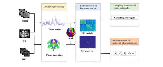
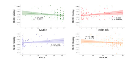
The coupling between functional and structural brain networks is difficult to clarify due to the complicated alterations in gray matter and white matter for the development of Alzheimer's disease (AD). A cohort of 112 participants [normal control group (NC, 62 cases), mild cognitive impairment group (MCI, 31 cases) and AD group (19 cases)], was recruited in our study. The brain networks of rsfMRI functional connectivity (rsfMRI-FC) and diffusion tensor imaging structural connectivity (DTI-SC) across the three groups were constructed, and their correlations were evaluated by Pearson's correlation analyses and multiple comparison with Bonferroni correction. Furthermore, the correlations between rsfMRI-SC/DTI-FC coupling and four neuropsychological scores of mini-mental state examination (MMSE), clinical dementia rating-sum of boxes (CDR-SB), functional activities questionnaire (FAQ) and montreal cognitive assessment (MoCA) were inferred by partial correlation analyses, respectively. The results demonstrated that there existed significant correlation between rsfMRI-FC and DTI-SC (p < 0.05), and the coupling of rsfMRI-FC/DTI-SC showed negative correlation with MMSE score (p < 0.05), positive correlations with CDR-SB and FAQ scores (p < 0.05), and no correlation with MoCA score (p > 0.05). It was concluded that there existed FC/SC coupling and varied network characteristics for rsfMRI and DTI, and this would provide the clues to understand the underlying mechanisms of cognitive deficits of AD.
Citation: Xia Xu, Song Xu, Liting Han, Xufeng Yao. Coupling analysis between functional and structural brain networks in Alzheimer's disease[J]. Mathematical Biosciences and Engineering, 2022, 19(9): 8963-8974. doi: 10.3934/mbe.2022416
The coupling between functional and structural brain networks is difficult to clarify due to the complicated alterations in gray matter and white matter for the development of Alzheimer's disease (AD). A cohort of 112 participants [normal control group (NC, 62 cases), mild cognitive impairment group (MCI, 31 cases) and AD group (19 cases)], was recruited in our study. The brain networks of rsfMRI functional connectivity (rsfMRI-FC) and diffusion tensor imaging structural connectivity (DTI-SC) across the three groups were constructed, and their correlations were evaluated by Pearson's correlation analyses and multiple comparison with Bonferroni correction. Furthermore, the correlations between rsfMRI-SC/DTI-FC coupling and four neuropsychological scores of mini-mental state examination (MMSE), clinical dementia rating-sum of boxes (CDR-SB), functional activities questionnaire (FAQ) and montreal cognitive assessment (MoCA) were inferred by partial correlation analyses, respectively. The results demonstrated that there existed significant correlation between rsfMRI-FC and DTI-SC (p < 0.05), and the coupling of rsfMRI-FC/DTI-SC showed negative correlation with MMSE score (p < 0.05), positive correlations with CDR-SB and FAQ scores (p < 0.05), and no correlation with MoCA score (p > 0.05). It was concluded that there existed FC/SC coupling and varied network characteristics for rsfMRI and DTI, and this would provide the clues to understand the underlying mechanisms of cognitive deficits of AD.

| [1] |
N. Hao, Z. Wang, P. Liu, R. Becker, S. Yang, K. Yang, et al., Acoustofluidic multimodal diagnostic system for Alzheimer's disease, Biosens. Bioelectron., 196 (2022), 113730. https://doi.org/10.1016/j.bios.2021.113730 doi: 10.1016/j.bios.2021.113730

|
| [2] |
B. T. Hyman, C. H. Phelps, T. G. Beach, E. H. Bigio, N. J. Cairns, M. C. Carrillo, et al., National institute on aging-Alzheimer's association guidelines for the neuropathologic assessment of Alzheimer's disease, Alzheimer's Dementia, 8 (2012), 1-13. https://doi.org/10.1016/j.jalz.2011.10.007 doi: 10.1016/j.jalz.2011.10.007
|
| [3] |
V. L. Villemagne, S. Burnham, P. Bourgeat, B. Brown, K. A. Ellis, O. Salvado, et al., Amyloid β deposition, neurodegeneration, and cognitive decline in sporadic Alzheimer's disease: a prospective cohort study, Lancet Neurol., 12 (2013), 357-367. https://doi.org/10.1016/s1474- 4422(13)70044-9 doi: 10.1016/s1474-4422(13)70044-9
|
| [4] |
O. Sporns, G. Tononi, R. Kötter, The human connectome: A structural description of the human brain, PLoS Comput. Biol., 1 (2005), e42. https://doi.org/10.1371/journal.pcbi.0010042 doi: 10.1371/journal.pcbi.0010042
|
| [5] |
B. B. Biswal, M. Mennes, X. N. Zuo, S. Gohel, C. Kelly, S. M. Smith, et al., Toward discovery science of human brain function, PNAS, 107 (2010), 4734-4739. https://doi.org/10.1073/pnas.0911855107 doi: 10.1073/pnas.0911855107
|
| [6] |
F. Agosta, S. Galantucci, M. Filippi, Advanced magnetic resonance imaging of neurodegenerative diseases, Neurol. Sci., 38 (2017), 41-51. https://doi.org/10.1007/s10072-016-2764-x doi: 10.1007/s10072-016-2764-x
|
| [7] |
M. W. Cho, M. Y. Choi, Brain networks: Graph theoretical analysis and development models, Int. J. Imaging Syst. Technol., 20 (2010), 108-116. https://doi.org/https://doi.org/10.1002/ima.20229 doi: 10.1002/ima.20229
|
| [8] |
J. delEtoile, H. Adeli, Graph ttheory and brain connectivity in Alzheimer's disease, Neuroscientist, 23 (2017), 616-626. https://doi.org/10.1177/1073858417702621 doi: 10.1177/1073858417702621
|
| [9] |
A. M. Tuladhar, I. W. M. Van Uden, L. C. A. Rutten-Jacobs, A. Lawrence, H. Van Der Holst, A. Van Norden, et al., Structural network efficiency predicts conversion to dementia, Neurology, 86 (2016), 1112-1119. https://doi.org/10.1212/wnl.0000000000002502 doi: 10.1212/wnl.0000000000002502
|
| [10] |
F. U. Fischer, D. Wolf, A. Scheurich, A. Fellgiebel, Altered whole-brain white matter networks in preclinical Alzheimer's disease, NeuroImage: Clin., 8 (2015), 660-666. https://doi.org/10.1016/j.nicl.2015.06.007 doi: 10.1016/j.nicl.2015.06.007
|
| [11] |
A. Wada, O. Abe, Graph theoretic analysis of structural connectivity of Alzheimer's disease by using diffusion MR imaging, Med. Imaging Technol., 34 (2016), 18-21. https://doi.org/10.11409/mit.34.18 doi: 10.11409/mit.34.18
|
| [12] |
C. Yang, S. Zhong, X. Zhou, L. Wei, L. Wang, S. Nie, The abnormality of topological asymmetry between hemispheric brain white matter networks in Alzheimer's disease and mild cognitive impairment, Front. Aging Neurosci., 9 (2017), 261. https://doi.org/10.3389/fnagi.2017.00261 doi: 10.3389/fnagi.2017.00261
|
| [13] |
L. Zajac, B. B. Koo, C. Bauer, R. Killiany, Seed location impacts whole-brain structural network comparisons between healthy elderly and individuals with Alzheimer's disease, Brain Sci., 7 (2017), 37. https://doi.org/10.3390/brainsci7040037 doi: 10.3390/brainsci7040037
|
| [14] |
E. S. Lee, K. Yoo, Y. B. Lee, J. Chung, J. E. Lim, B. Yoon, et al., Default mode network functional connectivity in early and late mild cognitive impairment: Results from the Alzheimer's disease neuroimaging initiative, Alzheimer Dis. Assoc. Disord., 30 (2016), 289-296. https://doi.org/10.1097/wad.0000000000000143 doi: 10.1097/wad.0000000000000143
|
| [15] |
L. Chuanming, Z. Jian, W. Jian, G. Li, L. Chuan, An fMRI stroop task study of prefrontal cortical function in normal aging, mild cognitive impairment, and Alzheimer's disease, Curr. Alzheimer Res., 6 (2009), 525-530. https://doi.org/10.2174/156720509790147142 doi: 10.2174/156720509790147142
|
| [16] | K. Çiftçi, Graph theoretical analysis of functional brain networks during Alzheimer's disease, in 2010 IEEE 18th Signal Processing and Communications Applications Conference, (2010), 925-928. https://doi.org/10.1109/SIU.2010.5651274. |
| [17] |
K. Hahn, N. Myers, S. Prigarin, K. Rodenacker, A. Kurz, H. Förstl, et al., Selectively and progressively disrupted structural connectivity of functional brain networks in Alzheimer's disease-Revealed by a novel framework to analyze edge distributions of networks detecting disruptions with strong statistical evidence, NeuroImage, 81 (2013), 96-109. https://doi.org/10.1016/j.neuroimage.2013.05.011 doi: 10.1016/j.neuroimage.2013.05.011
|
| [18] |
R. Balachandar, J. P. John, J. Saini, K. J. Kumar, H. Joshi, S. Sadanand, et al., A study of structural and functional connectivity in early Alzheimer's disease using rest fMRI and diffusion tensor imaging, Int. J. Geriatr. Psychiatry, 30 (2015), 497-504. https://doi.org/10.1002/gps.4168 doi: 10.1002/gps.4168
|
| [19] |
Z. Liu, Y. Zhang, H. Yan, L. Bai, R. Dai, W. Wei, et al., Altered topological patterns of brain networks in mild cognitive impairment and Alzheimer's disease: A resting-state fMRI study, Psychiatry Res.: Neuroimaging, 202 (2012), 118-125. https://doi.org/10.1016/j.pscychresns.2012.03.002 doi: 10.1016/j.pscychresns.2012.03.002
|
| [20] |
K. Mevel, G. Chételat, F. Eustache, B. Desgranges, The default mode network in healthy aging and Alzheimer's disease, Int. J. Alzheimers Dis., 2011 (2011), 535816. https://doi.org/10.4061/2011/535816 doi: 10.4061/2011/535816
|
| [21] |
J. S. Lim, D. W. Kang, Stroke connectome and its implications for cognitive and behavioral sequela of stroke, J. Stroke, 17 (2015), 256-267. https://doi.org/10.5853/jos.2015.17.3.256 doi: 10.5853/jos.2015.17.3.256
|
| [22] |
S. Khalsa, S. D. Mayhew, M. Chechlacz, M. Bagary, A. P. Bagshaw, The structural and functional connectivity of the posterior cingulate cortex: Comparison between deterministic and probabilistic tractography for the investigation of structure-function relationships, NeuroImage, 102 (2014), 118-127. https://doi.org/10.1016/j.neuroimage.2013.12.022 doi: 10.1016/j.neuroimage.2013.12.022
|
| [23] |
Y. Sun, Q. Yin, R. Fang, X. Yan, Y. Wang, A. Bezerianos, et al., Disrupted functional brain connectivity and its association to structural connectivity in amnestic mild cognitive impairment and Alzheimer's disease, PloS one, 9 (2014), e96505. https://doi.org/10.1371/journal.pone.0096505 doi: 10.1371/journal.pone.0096505
|
| [24] |
Z. Dai, Q. Lin, T. Li, X. Wang, H. Yuan, X. Yu, et al., Disrupted structural and functional brain networks in Alzheimer's disease, Neurobiol. Aging, 75 (2019), 71-82. https://doi.org/10.1016/j.neurobiolaging.2018.11.005 doi: 10.1016/j.neurobiolaging.2018.11.005
|
| [25] |
J. J. Crofts, M. Forrester, R. D. O'Dea, Structure-function clustering in multiplex brain networks, Europhys. Lett., 116 (2016), 18003. https://doi.org/10.1209/0295-5075/116/18003 doi: 10.1209/0295-5075/116/18003
|
| [26] |
J. Zhao, X. Ding, Y. Du, X. Wang, G. Men, Functional connectivity between white matter and gray matter based on fMRI for Alzheimer's disease classification, Brain Behav., 9 (2019), https://doi.org/10.1002/brb3.1407 doi: 10.1002/brb3.1407
|
| [27] |
C. Yan, Y. Zang, DPARSF: a MATLAB toolbox for "pipeline" data analysis of resting-state fMRI, Front. Syst. Neurosci., 4 (2010), 13. https://doi.org/10.3389/fnsys.2010.00013 doi: 10.3389/fnsys.2010.00013
|
| [28] |
Z. Cui, S. Zhong, P. Xu, G. Gong, Y. He, PANDA: a pipeline toolbox for analyzing brain diffusion images, Front. Hum. Neurosci., 7 (2013), 42. https://doi.org/10.3389/fnhum.2013.00042 doi: 10.3389/fnhum.2013.00042
|
| [29] |
S. Mori, W. E. Kaufmann, C. Davatzikos, B. Stieltjes, L. Amodei, K. Fredericksen, et al., Imaging cortical association tracts in the human brain using diffusion-tensor-based axonal tracking, Magn. Reson. Med., 47 (2002), 215-223. https://doi.org/10.1002/mrm.10074 doi: 10.1002/mrm.10074
|
| [30] |
N. Tzourio-Mazoyer, B. Landeau, D. Papathanassiou, F. Crivello, O. Etard, N. Delcroix, et al., Automated anatomical labeling of activations in SPM using a macroscopic anatomical parcellation of the MNI MRI single-subject brain, NeuroImage., 15 (2002), 273-289. https://doi.org/https://doi.org/10.1006/nimg.2001.0978 doi: 10.1006/nimg.2001.0978
|
| [31] |
C. F. Bond, K. Richardson, Seeing the FisherZ-transformation, Psychometrika., 69 (2004), 291-303. https://doi.org/10.1007/bf02295945 doi: 10.1007/bf02295945
|
| [32] |
J. Wang, L. Wang, Y. Zang, H. Yang, H. Tang, Q. Gong, et al., Parcellation-dependent small-world brain functional networks: A resting-state fMRI study, Hum. Brain Mapp., 30 (2009), 1511-1523. https://doi.org/10.1002/hbm.20623 doi: 10.1002/hbm.20623
|
| [33] | P. Hinton, C. Brownlow, I. McMurray, B. Charlotte, SPSS Explained, 1st Edition, 2004. https://doi.org/10.4324/9780203642597 |
| [34] |
J. Wang, X. Wang, M. Xia, X. Liao, A. Evans, Y. He, GRETNA: a graph theoretical network analysis toolbox for imaging connectomics, Front. Hum. Neurosci., 9 (2015), 386. https://doi.org/10.3389/fnhum.2015.00386 doi: 10.3389/fnhum.2015.00386
|
| [35] |
X. Liao, A. V. Vasilakos, Y. He, Small-world human brain networks: Perspectives and challenges, Neurosci. Biobehav. Rev., 77 (2017), 286-300. https://doi.org/10.1016/j.neubiorev.2017.03.018 doi: 10.1016/j.neubiorev.2017.03.018
|
| [36] |
L. J. Zhang, G. Zheng, L. Zhang, J. Zhong, Q. Li, T. Z. Zhao, et al., Disrupted small world networks in patients without overt hepatic encephalopathy: A resting state fMRI study, Eur. J. Radiol., 83 (2014), 1890-1899. https://doi.org/10.1016/j.ejrad.2014.06.019 doi: 10.1016/j.ejrad.2014.06.019
|
| [37] |
Y. Li, Y. Liu, J. Li, W. Qin, K. Li, C. Yu, et al., Brain anatomical network and intelligence, PLoS Comput. Biol., 5 (2009), e1000395. https://doi.org/10.1371/journal.pcbi.1000395 doi: 10.1371/journal.pcbi.1000395
|
| [38] |
P. Hagmann, L. Cammoun, X. Gigandet, R. Meuli, C. J. Honey, V. J. Wedeen, et al., Mapping the structural core of human cerebral cortex, PLoS Biol., 6 (2008), e159. https://doi.org/10.1371/journal.pbio.0060159 doi: 10.1371/journal.pbio.0060159
|
| [39] |
Z. Wang, Z. Dai, G. Gong, C. Zhou, Y. He, Understanding structural-functional relationships in the human brain: A large-scale network perspective, Neuroscientist, 21 (2014), 290-305. https://doi.org/10.1177/1073858414537560 doi: 10.1177/1073858414537560
|
| [40] |
J. Wang, R. Khosrowabadi, K. K. Ng, Z. Hong, J. S. X. Chong, Y. Wang, et al., Alterations in brain network topology and structural-functional connectome coupling relate to cognitive impairment, Front. Aging Neurosci., 10 (2018), 404. https://doi.org/10.3389/fnagi.2018.00404 doi: 10.3389/fnagi.2018.00404
|
| [41] |
Z. Dai, Y. He, Disrupted structural and functional brain connectomes in mild cognitive impairment and Alzheimer's disease, Neurosci. Bull., 30 (2014), 217-232. https://doi.org/10.1007/s12264-013-1421-0 doi: 10.1007/s12264-013-1421-0
|
| [42] |
S. Gardini, A. Venneri, F. Sambataro, F. Cuetos, F. Fasano, M. Marchi, et al., Increased functional connectivity in the default mode network in mild cognitive impairment: A maladaptive compensatory mechanism associated with poor semantic memory performance, J. Alzheimers Dis., 45 (2015), 457-470. https://doi.org/10.3233/JAD-142547 doi: 10.3233/JAD-142547
|
| [43] |
J. Zimmermann, P. Ritter, K. Shen, S. Rothmeier, M. Schirner, A. R. McIntosh, Structural architecture supports functional organization in the human aging brain at a regionwise and network level, Hum. Brain Mapp., 37 (2016), 2645-2661. https://doi.org/10.1002/hbm.23200 doi: 10.1002/hbm.23200
|
| [44] |
R. Cao, X. Wang, Y. Gao, T. Li, H. Zhang, W. Hussain, et al., Abnormal anatomical rich-club organization and structural-functional coupling in mild cognitive impairment and Alzheimer's disease, Front. Neurol., 11 (2020), 53. https://doi.org/10.3389/fneur.2020.00053 doi: 10.3389/fneur.2020.00053
|
| [45] |
M. P. Van Den Heuvel, O. Sporns, G. Collin, T. Scheewe, R. C. W. Mandl, W. Cahn, et al., Abnormal rich club organization and functional brain dynamics in schizophrenia, JAMA Psychiatry, 70 (2013), 783. https://doi.org/10.1001/jamapsychiatry.2013.1328 doi: 10.1001/jamapsychiatry.2013.1328
|
Figures(2) / Tables(3)

Xia Xu, Song Xu, Liting Han, Xufeng Yao. Coupling analysis between functional and structural brain networks in Alzheimer's disease[J]. Mathematical Biosciences and Engineering, 2022, 19(9): 8963-8974. doi: 10.3934/mbe.2022416


 DownLoad: