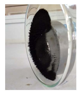
We synthesized and characterized amidoxime-modified Fe3O4/SiO2 core-shell magnetic microspheres tailored for maximal U(VI) sorption efficiency from seawater. Through meticulous structure and spectroscopy analyses, the microspheres, which were designed with amidoxime functionality, exhibited remarkable U(VI) sorption capabilities compared to raw silica-coated Fe3O4 counterparts. The maximum percent uranium adsorption (98.57%) was achieved at 60 minutes with 0.05 g of adsorbent, using a synthetic solution of 25 mg L−1 UO2(CH3COO)2. 2H2O at pH 7 and 25 º C (298 K). The kinetic studies highlighted rapid equilibrium achieved within 1 hours. Following the pseudo-second-order model, the microspheres reflected a maximum sorption capacity of 24.286 mg g-1 at pH 7 and 298 K. The U(VI)-loaded microspheres could be efficiently separated via an external magnetic field with adsorption efficiency of 91.67% at pH 6.5 and efficiently regenerated by HCl, indicating their potential for U(VI) preconcentration and separation from seawater. This research contributed to the development of high-performance sorbents for U(VI) removal and holds promise for solving the radioactive element elimination and enrichment, performing its stability, selectivity, and reusability across multiple cycles.
Citation: Alif Alfarisyi Syah, Anugrah Ricky Wijaya, Irma Kartika Kusumaningrum. Preparation and development of amidoxime-modified Fe3O4/SiO2 core-shell magnetic microspheres for enhancing U(VI) adsorption efficiency from seawater[J]. AIMS Environmental Science, 2024, 11(1): 21-37. doi: 10.3934/environsci.2024002
We synthesized and characterized amidoxime-modified Fe3O4/SiO2 core-shell magnetic microspheres tailored for maximal U(VI) sorption efficiency from seawater. Through meticulous structure and spectroscopy analyses, the microspheres, which were designed with amidoxime functionality, exhibited remarkable U(VI) sorption capabilities compared to raw silica-coated Fe3O4 counterparts. The maximum percent uranium adsorption (98.57%) was achieved at 60 minutes with 0.05 g of adsorbent, using a synthetic solution of 25 mg L−1 UO2(CH3COO)2. 2H2O at pH 7 and 25 º C (298 K). The kinetic studies highlighted rapid equilibrium achieved within 1 hours. Following the pseudo-second-order model, the microspheres reflected a maximum sorption capacity of 24.286 mg g-1 at pH 7 and 298 K. The U(VI)-loaded microspheres could be efficiently separated via an external magnetic field with adsorption efficiency of 91.67% at pH 6.5 and efficiently regenerated by HCl, indicating their potential for U(VI) preconcentration and separation from seawater. This research contributed to the development of high-performance sorbents for U(VI) removal and holds promise for solving the radioactive element elimination and enrichment, performing its stability, selectivity, and reusability across multiple cycles.

| [1] |
Plant JA, Simpson PR, Smith B, et al. (1999) Uranium ore deposits - Products of the radioactive earth. Rev Mineral Geochem 38: 254–319. https://doi.org/10.1515/9781501509193-011 doi: 10.1515/9781501509193-011

|
| [2] | Waltar AE, Reynolds AB (1983) Fast Reactors. Nucl Power Technol 1: 297–333. |
| [3] |
Wijaya AR, Ouchi AK, Tanaka K, et al. (2012) Metal contents and Pb isotopes in road-side dust and sediment of Japan. J Geochem Explor 118: 68–76. https://doi.org/10.1016/j.gexplo.2012.04.009 doi: 10.1016/j.gexplo.2012.04.009
|
| [4] |
Wijaya AR, Kusumaningrum IK, Hakim L, et al. (2022) Road-side dust from central Jakarta, Indonesia: Assessment of metal(loid) content, mineralogy, and bioaccessibility. Environ Technol Innov 28: 102934. https://doi.org/10.1016/j.eti.2022.102934 doi: 10.1016/j.eti.2022.102934
|
| [5] |
Rao TP, Metilda P, Gladis JM (2006) Preconcentration techniques for uranium(VI) and thorium(IV) prior to analytical determination-an overview. Talanta 68: 1047–1064. https://doi.org/10.1016/j.talanta.2005.07.021 doi: 10.1016/j.talanta.2005.07.021
|
| [6] |
Buszewski B, Szultka M (2012) Past, Present, and Future of Solid Phase Extraction: A Review. Crit Rev Anal Chem 42: 198–213. https://doi.org/10.1080/07373937.2011.645413 doi: 10.1080/07373937.2011.645413
|
| [7] | Wijaya AR, Khoerunnisa F, Armid A, et al. (2022) The best-modified BCR and Tessier with microwave-assisted methods for leaching of Cu/Zn and their δ65Cu/δ66Zn for tracing sources in marine sediment fraction. Environ Technol Innov 28. |
| [8] |
Khatamian M, Divband B, Shahi R (2019) Ultrasound assisted co-precipitation synthesis of Fe3O4/ bentonite nanocomposite: Performance for nitrate, BOD and COD water treatment. J Water Process Eng 31: 100870. https://doi.org/10.1016/j.jwpe.2019.100870 doi: 10.1016/j.jwpe.2019.100870
|
| [9] | Nanlohy F, Wijaya AR, Semedi B (2021) Synthesis of Fe3O4/MnO2/Humic acid nanocomposite for strontium ion adsorption and its interferences, AIP Conference Proceedings, 030108. https://doi.org/10.1063/5.0052981 |
| [10] |
Yang S, Zong P, Ren X, et al. (2012) Rapid and highly efficient preconcentration of Eu(Ⅲ) by core-shell structured Fe3O4/Humic acid magnetic nanoparticles. ACS Appl Mater Inter 4: 6891–6900. https://doi.org/10.1021/am3020372 doi: 10.1021/am3020372
|
| [11] |
Liu JF, Zhao ZS, Jiang G Bin (2008) Coating Fe3O4 magnetic nanoparticles with humic acid for high efficient removal of heavy metals in water. Environ Sci Technol 42: 6949–6954. https://doi.org/10.1021/es800924c doi: 10.1021/es800924c
|
| [12] |
Chang YC, Chen DH (2005) Preparation and adsorption properties of monodisperse chitosan-bound Fe3O4 magnetic nanoparticles for removal of Cu(Ⅱ) ions. J Colloid Interface Sci 283: 446–451. https://doi.org/10.1016/j.jcis.2004.09.010 doi: 10.1016/j.jcis.2004.09.010
|
| [13] |
Chae HS, Kim SD, Piao SH, et al. (2016) Core-shell Structured Fe3O4/SiO2 Nanoparticles Fabricated by Sol–gel Method and Their Magnetorheology. Colloid Polym Sci 294: 647–655. https://doi.org/10.1007/s00396-015-3818-y doi: 10.1007/s00396-015-3818-y
|
| [14] |
Li N, Gao P, Chen H, et al. (2022) Amidoxime modified Fe3O4/TiO2 particles for antibacterial and efficient uranium extraction from seawater. Chemosphere 287: 132137. https://doi.org/10.1016/j.chemosphere.2021.132137 doi: 10.1016/j.chemosphere.2021.132137
|
| [15] |
Zhao Y, Li J, Zhao L, et al. (2014) Synthesis of amidoxime-functionalized Fe3O4/SiO2 core-shell magnetic microspheres for highly efficient sorption of U(VI). Chem Eng J 235: 275–283. https://doi.org/10.1016/j.cej.2013.09.034 doi: 10.1016/j.cej.2013.09.034
|
| [16] |
Zheng H, Zhou L, Liu Z, et al. (2019) Functionalization of mesoporous Fe3O4/SiO2 nanospheres for highly efficient U(VI) adsorption. Microporous Mesoporous Mat 279: 316–322. https://doi.org/10.1016/j.micromeso.2018.12.038 doi: 10.1016/j.micromeso.2018.12.038
|
| [17] | Beamish JEC, FE (1948) Uranium, Spectrophotometric Ammonium Thiocyanate Method. Master analytical manual 149: 9–12. |
| [18] | Werner Stober Af (2017) Controlled Growth of Monodisperse Silica Spheres in the Micron Size Range. J Phys Ther Sci 29: 112–114. |
| [19] |
Kalapathy U, Proctor A, Shultz J (2001) A simple method for production of pure silica from rice hull ash. Fuel Energy Abstracts 42: 45. https://doi.org/10.1016/S0140-6701(01)80487-2 doi: 10.1016/S0140-6701(01)80487-2
|
| [20] |
Pu Y, Qiang T, Li G, et al. (2023) Efficient adsorption of low-concentration uranium from aqueous solutions. Ecotox Environ Safe 259: 115053. https://doi.org/10.1016/j.ecoenv.2023.115053 doi: 10.1016/j.ecoenv.2023.115053
|
| [21] |
Gao B, Gao Y, Li Y (2010) Preparation and chelation adsorption property of composite chelating material poly(amidoxime)/SiO2 towards heavy metal ions. Chem Eng J 158: 542–549. https://doi.org/10.1016/j.cej.2010.01.046 doi: 10.1016/j.cej.2010.01.046
|
| [22] |
Bulut VN, Arslan D, Ozdes D, et al. (2010) Preconcentration, separation and spectrophotometric determination of aluminium(Ⅲ) in water samples and dialysis concentrates at trace levels with 8-hydroxyquinoline-cobalt(Ⅱ) coprecipitation system. J Hazard Mater 182: 331–336. https://doi.org/10.1016/j.jhazmat.2010.06.034 doi: 10.1016/j.jhazmat.2010.06.034
|
| [23] | Grimaldi F, May I, Mary F, et al. (1954) Collected Papers on Methods of Analysis for Uranium and Thorium. Geol Sur Bull 1006: 196. |
| [24] | Zhou Y, Li Y, Liu D, et al. (2021) Adsorption optimization of uranium(VI) onto polydopamine and sodium titanate co-functionalized MWCNTs using response surface methodology and a modeling approach. Colloid Surf A-Physicochem Eng Asp 627. https://doi.org/10.1016/j.colsurfa.2021.127145 |
| [25] |
Aurich A, Hofmann J, Oltrogge R, et al. (2017) Pseudo-second order model for sorption processes Y.S. Org Process Res Dev 21: 866–870. https://doi.org/10.1021/acs.oprd.7b00090 doi: 10.1021/acs.oprd.7b00090
|
| [26] |
Wijaya AR, Semedi B, Lusiana RA, et al. (2019) Metal contents and Pb isotopes in the surface seawater of the Gulf of Prigi, Indonesia: Detection of anthropogenic and natural sources. J Braz Chem Soc 30: 915–929. https://doi.org/10.21577/0103-5053.20180228 doi: 10.21577/0103-5053.20180228
|
| [27] | Suci CW, Wijaya AR, Santoso A, et al. (2020) Fe leaching in the sludge sediment of the prigi beach with tessier-microwave method. AIP Conference Proceedings 2231. https://doi.org/10.1063/5.0002589 |
| [28] |
Zhang A, Uchiyama G, Asakura T (2005) PH Effect on the uranium adsorption from seawater by a macroporous fibrous polymeric material containing amidoxime chelating functional group. React Funct Polym 63: 143–153. https://doi.org/10.1016/j.reactfunctpolym.2005.02.015 doi: 10.1016/j.reactfunctpolym.2005.02.015
|
| [29] |
Zhao Y, Li J, Zhang S, et al. (2014) Amidoxime-functionalized magnetic mesoporous silica for selective sorption of U(vi). RSC Adv 4: 32710–32717. https://doi.org/10.1039/C4RA05128A doi: 10.1039/C4RA05128A
|


Figures(10) / Tables(4)

Alif Alfarisyi Syah, Anugrah Ricky Wijaya, Irma Kartika Kusumaningrum. Preparation and development of amidoxime-modified Fe3O4/SiO2 core-shell magnetic microspheres for enhancing U(VI) adsorption efficiency from seawater[J]. AIMS Environmental Science, 2024, 11(1): 21-37. doi: 10.3934/environsci.2024002


 DownLoad: