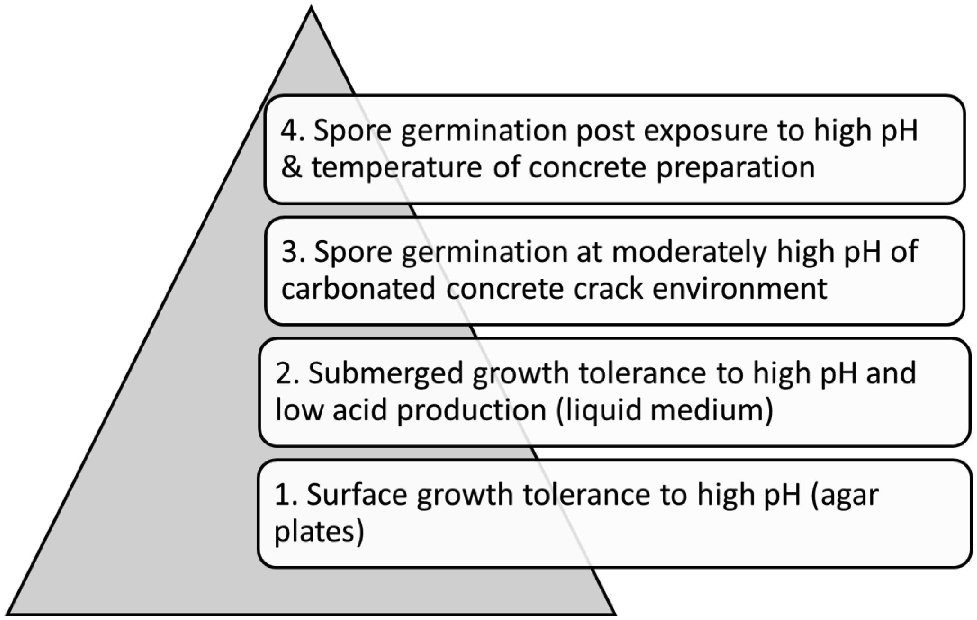
This work addresses the problem of having no existent protocol that closely simulates the harsh concrete environment, namely, high pH (about 13) and temperature (up to above 50 °C during cement hydration), for selecting microorganisms to use in microbe-effected self-healing concrete (MESHC). This problem critically impedes the progress of MESHC development, which has tremendous significance because concrete cracking reduces service life while making new concrete consumes substantial energy and emits a large amount of CO2. The objective of this work was to develop an effective and innovative screening protocol that mimics not only the concrete-relevant pH and temperature but also the essential process/scenario of MESHC, that is, microbial spores remain dormant through the harsh conditions during concrete mixing and curing and germinate later at the less harsh condition (pH 9–11, ambient temperature) in concrete cracks. The protocol includes 4 steps with increasing complexity to reduce strains for time-consuming steps. The first 2 steps screen for strains capable of vegetative growth on agar and liquid media with initial pH ≥ 10. Step 3 evaluates the spore germinability in medium with pH gradually decreasing from 12. In Step 4, spores were subjected to both high pH (12, 12.9) and temperature (45, 55 °C) for 2 h before being stored in the high-pH medium at room temperature for up to 21 days. Spores were finally collected and resuspended in pH 9.5 medium to observe germinability. 18 alkalotolerant strains were screened. Scopulariopsis brevicaulis (S. brevicaulis) is identified as the best candidate. Results also indicate the importance of incorporating designs to locally lower pH near spores, at least slightly (from 12.9 to 12), and to limit temperature to below 53 °C. Survivability of S. brevicaulis spores in contact with mortar undergoing cement hydration was also monitored. The work identifies promising fungi and indicates critical directions for developing spore-protection technologies for MESHC.
Citation: Ahsanul Kabir Sumon, Krutika Invally, Lu-Kwang Ju. Screening fungi for use as biocatalysts in self-healing concrete with protocol simulating concrete environment[J]. AIMS Bioengineering, 2024, 11(3): 323-342. doi: 10.3934/bioeng.2024017
This work addresses the problem of having no existent protocol that closely simulates the harsh concrete environment, namely, high pH (about 13) and temperature (up to above 50 °C during cement hydration), for selecting microorganisms to use in microbe-effected self-healing concrete (MESHC). This problem critically impedes the progress of MESHC development, which has tremendous significance because concrete cracking reduces service life while making new concrete consumes substantial energy and emits a large amount of CO2. The objective of this work was to develop an effective and innovative screening protocol that mimics not only the concrete-relevant pH and temperature but also the essential process/scenario of MESHC, that is, microbial spores remain dormant through the harsh conditions during concrete mixing and curing and germinate later at the less harsh condition (pH 9–11, ambient temperature) in concrete cracks. The protocol includes 4 steps with increasing complexity to reduce strains for time-consuming steps. The first 2 steps screen for strains capable of vegetative growth on agar and liquid media with initial pH ≥ 10. Step 3 evaluates the spore germinability in medium with pH gradually decreasing from 12. In Step 4, spores were subjected to both high pH (12, 12.9) and temperature (45, 55 °C) for 2 h before being stored in the high-pH medium at room temperature for up to 21 days. Spores were finally collected and resuspended in pH 9.5 medium to observe germinability. 18 alkalotolerant strains were screened. Scopulariopsis brevicaulis (S. brevicaulis) is identified as the best candidate. Results also indicate the importance of incorporating designs to locally lower pH near spores, at least slightly (from 12.9 to 12), and to limit temperature to below 53 °C. Survivability of S. brevicaulis spores in contact with mortar undergoing cement hydration was also monitored. The work identifies promising fungi and indicates critical directions for developing spore-protection technologies for MESHC.

| [1] | Mamlouk MS, Zaniewski JP (2011) Materials for Civil and Construction Engineers. New York: Prentice Hall. |
| [2] |
Pacheco J, Polder R (2012) Corrosion initiation and propagation in cracked concrete–a literature review. Advances in Modeling Concrete Service Life . Dordrecht: Springer 85-93. 
|
| [3] |
Silva FB, Boon N, De Belie N, et al. (2015) Industrial application of biological self-healing concrete: Challenges and economical feasibility. J Commer Biotechnol 21: 31-38. https://doi.org/10.5912/jcb662
|
| [4] |
Amran M, Onaizi AM, Fediuk R, et al. (2022) Self-healing concrete as a prospective construction material: A review. Materials 15: 1-46. https://doi.org/10.3390/ma15093214
|
| [5] |
Williamson RB (1968) Constitutional supersaturation in Portland cement solidified by hydration. J Cryst Growth 3: 787-794. https://doi.org/10.1016/0022-0248(68)90267-4
|
| [6] |
Behnood A, Van Tittelboom K, De Belie N (2016) Methods for measuring pH in concrete: A review. Constr Build Mater 105: 176-188. https://doi.org/10.1016/j.conbuildmat.2015.12.032
|
| [7] |
Zou X, Chao A, Tian Y, et al. (2012) An experimental study on the concrete hydration process using Fabry-Perot fiber optic temperature sensors. Measurement 45: 1077-1082. https://doi.org/10.1016/j.measurement.2012.01.034
|
| [8] |
Siang GC (2017) Determination of temperature rise and temperature differentials of CEMII/B-V cement for 20 MPa mass concrete using adiabatic temperature rise data. IOP Conf Ser Mater Sci Eng 217: 012008. https://doi.org/10.1088/1757-899X/217/1/012008
|
| [9] |
Matthieu B, Farid B, Jean-Michel T, et al. (2012) Analysis of semi-adiabatic tests for the prediction of early-age behavior of massive concrete structures. Cem Concr Compos 34: 634-641. https://doi.org/https://doi.org/10.1016/j.cemconcomp.2011.09.001
|
| [10] |
Kuriakose B, Rao BN, Dodagoudar GR (2016) Early-age temperature distribution in a massive concrete foundation. Procedia Technol 25: 107-114. https://doi.org/10.1016/j.protcy.2016.08.087
|
| [11] | Straube J (2000) Moisture properties of plaster and stucco for strawbale buildings. CMHC . https://www.ecobuildnetwork.org/ |
| [12] | Possan E, Thomaz WA, Aleandri GA, et al. (2017) CO2 uptake potential due to concrete carbonation: A case study. Case Stud Constr Mat 6: 147-161. https://doi.org/10.1016/j.cscm.2017.01.007 |
| [13] |
Ji Y, Yuan Y, Shen J, et al. (2010) Comparison of concrete carbonation process under natural condition and high CO2 concentration environments. J Wuhan Univ Technol 25: 515-522. https://doi.org/10.1007/s11595-030-0034-y
|
| [14] |
Van Wylick A, Rahier H, De Laet L, et al. (2024) Conditions for CaCO3 biomineralization by Trichoderma reesei with the perspective of developing fungi-mediated self-healing concrete. Glob Chall 8: 2300160. https://doi.org/10.1002/gch2.202300160
|
| [15] | Jonkers HM (2011) Bacteria-based self-healing concrete. Heron 56: 1-12. |
| [16] |
Jonkers HM, Thijssen A, Muyzer G, et al. (2010) Application of bacteria as self-healing agent for the development of sustainable concrete. Ecol Eng 36: 230-235. https://doi.org/10.1016/j.ecoleng.2008.12.036
|
| [17] |
Gao M, Guo J, Cao H, et al. (2020) Immobilized bacteria with pH-response hydrogel for self-healing of concrete. J Environ Manage 261: 110225. https://doi.org/10.1016/j.jenvman.2020.110225
|
| [18] |
Khushnood RA, Ali AM, Faraz Bhatti M, et al. (2022) Self-healing fungi concrete using potential strains Rhizopus oryzae and Trichoderma longibrachiatum. J Build Eng 50: 104155. https://doi.org/10.1016/j.jobe.2022.104155
|
| [19] |
Houshmand Khaneghahi M, Rahmaninezhad SA, Kamireddi D, et al. (2024) Carbonate biomineralization potential of endospore-laden polymeric fibers (BioFibers) for bio-self-healing applications. Develop Built Environment 17: 100351. https://doi.org/10.1016/j.dibe.2024.100351
|
| [20] |
Van Wylick A, Brouwers E, Rahier H, et al. (2024) Encapsulation of Trichoderma reesei and Neurospora crassa in calcium alginate capsules for fungi-mediated self-healing concrete: An explorative study on the protocol, survival of the organisms and CaCO3 biomineralization. Develop Built Environment 18: 100445. https://doi.org/10.1016/j.dibe.2024.100445
|
| [21] |
Zhang JL, Wu RS, Li YM, et al. (2016) Screening of bacteria for self-healing of concrete cracks and optimization of the microbial calcium precipitation process. Appl Microbiol Biotechnol 100: 6661-6670. https://doi.org/10.1007/s00253-016-7382-2
|
| [22] |
Martuscelli C, Soares C, Camões A, et al. (2020) Potential of fungi for concrete repair. Procedia Manuf 46: 180-185. https://doi.org/10.1016/j.promfg.2020.03.027
|
| [23] |
Luo J, Chen X, Crump J, et al. (2018) Interactions of fungi with concrete: Significant importance for bio-based self-healing concrete. Constr Build Mater 164: 275-285. https://doi.org/10.1016/j.conbuildmat.2017.12.233
|
| [24] |
Menon RR, Luo J, Chen X, et al. (2019) Screening of fungi for potential application of self-healing concrete. Sci Rep 9: 2075. https://doi.org/10.1038/s41598-019-39156-8
|
| [25] |
Zhao J, Csetenyi L, Gadd GM (2022) Fungal-induced CaCO3 and SrCO3 precipitation: A potential strategy for bioprotection of concrete. Sci Total Environ 816: 151501. https://doi.org/10.1016/j.scitotenv.2021.151501
|
| [26] | Kladwang W, Hywel-Jones N, Bhumirattana A (2003) Alkaline-tolerant fungi from Thailand. Fungal Divers 13: 69-84. |
| [27] |
Grum-Grzhimaylo AA, Georgieva ML, Debets AJM, et al. (2013) Are alkalitolerant fungi of the Emericellopsis lineage (Bionectriaceae) of marine origin?. IMA Fungus 4: 213-228. https://doi.org/10.5598/imafungus.2013.04.02.07
|
| [28] |
Nagai K, Sakai T, Rantiatmodjo RM, et al. (1995) Studies on the distribution of alkalophilic and alkali-tolerant soil fungi I. Mycoscience 36: 247-256. https://doi.org/10.1007/BF02268598
|
| [29] |
Grum-Grzhimaylo AA, Georgieva ML, Bondarenko SA, et al. (2016) On the diversity of fungi from soda soils. Fungal Divers 76: 27-74. https://doi.org/10.1007/s13225-015-0320-2
|
| [30] | Anke T, Schüffler A (2018) Physiology and Genetics: Selected Basic and Applied Aspects. Berlin: Springer. |
| [31] |
Nagai K (1998) Studies on the distribution of alkalophilic and alkali-tolerant soil fungi II: Fungal flora in two limestone caves in Japan. Mycoscience 39: 293-298. https://doi.org/10.1007/bf02464011
|
| [32] |
Kaur G, Siddique R, Rajor A (2013) Micro-structural and metal leachate analysis of concrete made with fungal treated waste foundry sand. Constr Build Mater 38: 94-100. https://doi.org/10.1016/j.conbuildmat.2012.07.112
|


Figures(4) / Tables(5)

Ahsanul Kabir Sumon, Krutika Invally, Lu-Kwang Ju. Screening fungi for use as biocatalysts in self-healing concrete with protocol simulating concrete environment[J]. AIMS Bioengineering, 2024, 11(3): 323-342. doi: 10.3934/bioeng.2024017


 DownLoad: