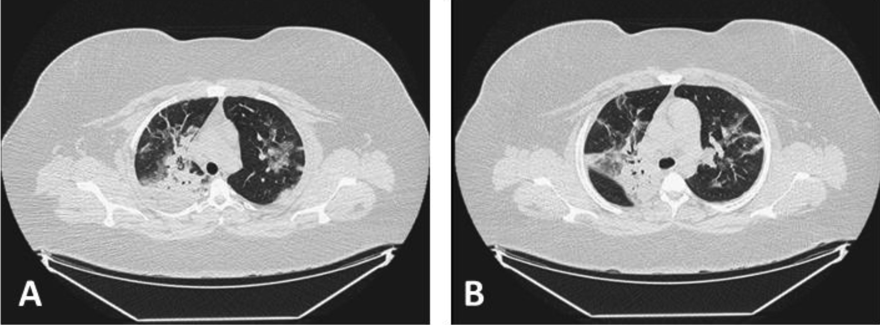
Rhabdomyolysis is an acute condition with skeletal muscle damage and release of toxins produced by myocytes with variable clinical presentation. Laboratory diagnosis is based on an increase in serum creatine phosphokinase (CPK), which can trigger irreversible renal impairment. The aim was to describe the association of COVID-19 with rhabdomyolysis as a serious complication, in order to promote early diagnosis and treatment. Case description: Female patient, 18 years old, grade 3 obesity, complaining of dry cough for 7 days, associated with continuous fever for 4 days, myalgia of the lower limbs and moderate dyspnea for 2 days, was admitted to the emergency room and sent to the Intensive Care Unit (ICU), with positive PCR-RT. After 12 hours of admission, the patient developed severe hypoxemic acute respiratory failure. On the second day of hospitalization, an exponential increase in CPK was found and measures of volume optimization were initiated. On the seventh day of hospitalization, associated with increased CPK, a decline in renal function was observed, evolving to the need for renal replacement therapy. On the ninth day of hospitalization, she presented with multiple organ dysfunction and death. In conclusion, COVID-19 can generate rhabdomyolysis by direct myocyte injury by SARS-CoV-2, an exacerbated immune response to the virus resulting in a cytokine storm with concomitant muscle damage and through direct injury by circulating viral toxins. However, as it is an uncommon manifestation and presents a nonspecific clinical condition, early diagnosis is not always performed. This conclusion was based on the case report presented and not only on the results of the literature review, since the clinical case is in accordance with the literature and can contribute to the recognition of these conditions, leading to better management in the treatment.
Citation: Uri Adrian Prync Flato, Karina Vilariço Ferreira, Piero Biteli, Daniela Ortega Balbo Rodrigues Reina, Fábio Tadeu Rodrigues Reina, Fausto Tucunduva Vernaschi, Gabriela Achete de Souza, Gyovanna Sorrentino dos Santos Campanari, Júlia Novaes Matias, Vinícius Marinho Lima, Tereza Lais Menegucci Zutin, Rogério Leone Buchaim, Daniela Vieira Buchaim, Sandra Maria Barbalho. Rabdomyolysis as a serious complication of COVID-19[J]. AIMS Bioengineering, 2021, 8(2): 165-172. doi: 10.3934/bioeng.2021015
Rhabdomyolysis is an acute condition with skeletal muscle damage and release of toxins produced by myocytes with variable clinical presentation. Laboratory diagnosis is based on an increase in serum creatine phosphokinase (CPK), which can trigger irreversible renal impairment. The aim was to describe the association of COVID-19 with rhabdomyolysis as a serious complication, in order to promote early diagnosis and treatment. Case description: Female patient, 18 years old, grade 3 obesity, complaining of dry cough for 7 days, associated with continuous fever for 4 days, myalgia of the lower limbs and moderate dyspnea for 2 days, was admitted to the emergency room and sent to the Intensive Care Unit (ICU), with positive PCR-RT. After 12 hours of admission, the patient developed severe hypoxemic acute respiratory failure. On the second day of hospitalization, an exponential increase in CPK was found and measures of volume optimization were initiated. On the seventh day of hospitalization, associated with increased CPK, a decline in renal function was observed, evolving to the need for renal replacement therapy. On the ninth day of hospitalization, she presented with multiple organ dysfunction and death. In conclusion, COVID-19 can generate rhabdomyolysis by direct myocyte injury by SARS-CoV-2, an exacerbated immune response to the virus resulting in a cytokine storm with concomitant muscle damage and through direct injury by circulating viral toxins. However, as it is an uncommon manifestation and presents a nonspecific clinical condition, early diagnosis is not always performed. This conclusion was based on the case report presented and not only on the results of the literature review, since the clinical case is in accordance with the literature and can contribute to the recognition of these conditions, leading to better management in the treatment.

| [1] |
Matias JN, dos Santos Campanari GS, de Souza GA, et al. (2020) Metabolic syndrome and COVID-19. AIMS Bioengineering 7: 242-253. doi: 10.3934/bioeng.2020021

|
| [2] |
Guan WJ, Ni ZY, Hu Y, et al. (2020) Clinical characteristics of coronavirus disease 2019 in China. N Engl J Med 382: 1708-1720. doi: 10.1056/NEJMoa2002032
|
| [3] |
Zhai P, Ding Y, Wu X, et al. (2020) The epidemiology, diagnosis and treatment of COVID-19. Int J Antimicrob Agents 55: 105955. doi: 10.1016/j.ijantimicag.2020.105955
|
| [4] |
Tian S, Hu N, Lou J, et al. (2020) Characteristics of COVID-19 infection in Beijing. J Infect 80: 401-406. doi: 10.1016/j.jinf.2020.02.018
|
| [5] |
Ahmad I, Rathore FA (2020) Neurological manifestations and complications of COVID-19: A literature review. J Clin Neurosci 77: 8-12. doi: 10.1016/j.jocn.2020.05.017
|
| [6] |
Miesbach W, Makris M (2020) COVID-19: Coagulopathy, risk of thrombosis, and the rationale for anticoagulation. Clin Appl Thromb Hemost 26: 1076029620938149. doi: 10.1177/1076029620938149
|
| [7] |
Shao M, Li X, Liu F, et al. (2020) Acute kidney injury is associated with severe infection and fatality in patients with COVID-19: A systematic review and meta-analysis of 40 studies and 24,527 patients. Pharmacol Res 161: 105107. doi: 10.1016/j.phrs.2020.105107
|
| [8] |
Wang L, Wang Y, Ye D, et al. (2020) Review of the 2019 novel coronavirus (SARS-CoV-2) based on current evidence. Int J Antimicrob Agents 55: 105948. doi: 10.1016/j.ijantimicag.2020.105948
|
| [9] | Kannan S, Shaik Syed Ali P, Sheeza A, et al. (2020) COVID-19 (Novel Coronavirus 2019)-recent trends. Eur Rev Med Pharmacol Sci 24: 2006-2011. |
| [10] |
Shi Y, Wang G, Cai XP, et al. (2020) An overview of COVID-19. J Zhejiang Univ Sci B 21: 343-360. doi: 10.1631/jzus.B2000083
|
| [11] |
Ge H, Wang X, Yuan X, et al. (2020) The epidemiology and clinical information about COVID-19. Eur J Clin Microbiol Infect Dis 39: 1011-1019. doi: 10.1007/s10096-020-03874-z
|
| [12] |
Singhal T (2020) A review of coronavirus disease-2019 (COVID-19). Indian J Pediatr 87: 281-286. doi: 10.1007/s12098-020-03263-6
|
| [13] |
Sohrabi C, Alsafi Z, O'Neill N, et al. (2020) World Health Organization declares global emergency: A review of the 2019 novel coronavirus (COVID-19). Int J Surg 76: 71-76. doi: 10.1016/j.ijsu.2020.02.034
|
| [14] |
Harapan H, Itoh N, Yufika A, et al. (2020) Coronavirus disease 2019 (COVID-19): A literature review. J Infect Public Health 13: 667-673. doi: 10.1016/j.jiph.2020.03.019
|
| [15] |
Madabhavi I, Sarkar M, Kadakol N COVID-19: a review (2020) . doi: 10.4081/monaldi.2020.1298
|
| [16] |
Tufan A, Avanoğlu Güler A, Matucci-Cerinic M (2020) COVID-19, immune system response, hyperinflammation and repurposing antirheumatic drugs. Turk J Med Sci 50: 620-632. doi: 10.3906/sag-2004-168
|
| [17] |
Li T, Lu H, Zhang W (2020) Clinical observation and management of COVID-19 patients. Emerg Microbes Infect 9: 687-690. doi: 10.1080/22221751.2020.1741327
|
| [18] |
Ahn DG, Shin HJ, Kim MH, et al. (2020) Current status of epidemiology, diagnosis, therapeutics, and vaccines for novel coronavirus disease 2019 (COVID-19). J Microbiol Biotechnol 30: 313-324. doi: 10.4014/jmb.2003.03011
|
| [19] |
Velavan TP, Meyer CG (2020) The COVID-19 epidemic. Trop Med Int Health 25: 278-280. doi: 10.1111/tmi.13383
|
| [20] |
Samies NL, Pinninti S, James SH (2020) Rhabdomyolysis and acute renal failure in an adolescent with COVID-19. J Pediatric Infect Dis Soc 9: 507-509. doi: 10.1093/jpids/piaa083
|
| [21] | Suwanwongse K, Shabarek NJC (2020) Rhabdomyolysis as a presentation of 2019 novel coronavirus disease. Cureus 12: e7561. |
| [22] |
Fadila MF, Wool KJ (2015) Rhabdomyolysis secondary to influenza a infection: a case report and review of the literature. N Am J Med Sci 7: 122-124. doi: 10.4103/1947-2714.153926
|
| [23] |
Chedid NR, Udit S, Solhjou Z, et al. (2020) COVID-19 and rhabdomyolysis. J Gen Intern Med 35: 3087-3090. doi: 10.1007/s11606-020-06039-y
|
| [24] |
Jin M, Tong Q (2020) Rhabdomyolysis as potential late complication associated with COVID-19. Emerg Infect Dis 26: 1618-1620. doi: 10.3201/eid2607.200445
|
| [25] |
Gefen AM, Palumbo N, Nathan SK, et al. (2020) Pediatric COVID-19-associated rhabdomyolysis: a case report. Pediatr Nephrol 35: 1517-1520. doi: 10.1007/s00467-020-04617-0
|
Figures(1) / Tables(1)

Uri Adrian Prync Flato, Karina Vilariço Ferreira, Piero Biteli, Daniela Ortega Balbo Rodrigues Reina, Fábio Tadeu Rodrigues Reina, Fausto Tucunduva Vernaschi, Gabriela Achete de Souza, Gyovanna Sorrentino dos Santos Campanari, Júlia Novaes Matias, Vinícius Marinho Lima, Tereza Lais Menegucci Zutin, Rogério Leone Buchaim, Daniela Vieira Buchaim, Sandra Maria Barbalho. Rabdomyolysis as a serious complication of COVID-19[J]. AIMS Bioengineering, 2021, 8(2): 165-172. doi: 10.3934/bioeng.2021015


 DownLoad: