Exposure to vanadium has been known to lead to a progressive neurodegenerative disorder like Parkinson's disease. Naringin is a known flavonoid glycoside that is mostly seen in the flesh of grapefruit and orange and is believed to have protective effects for the treatment of neurodegenerative disorders. This study sought to investigate the role of Naringin in the treatment of vanadium-induced neurotoxicity. Vanadium (10 mg/kg BW) was injected intraperitoneally to induce motor dysfunction, followed by treatment with Naringin (30 mg/kg BW) intraperitoneally for 14 days. Oxidative stress imbalance was monitored by checking Glutathione Peroxidase (GPX) and Catalase levels. Histological and immunohistochemical alterations were observed using RBFOX3 polyclonal antibody to determine neuronal cell distribution and NLRP3 inflammasome antibody as a marker of inflammation. Exposure to vanadium induces neurotoxicity by significantly increasing the Catalase and Glutathione Peroxidase (GPX) levels. Vanadium administration also led to increased inflammatory cells and a significant reduction of the viable neuronal cells in the SNc and CPu. Treatment with Naringin showed a neuroprotective role by dependently restoring the Catalase and Glutathione Peroxidase (GPX) levels, inflammasome activation, and neuronal damage in the SNc and CPu. Naringin demonstrated anti-oxidative, and anti-inflammatory responses by inhibiting oxidative stress, and inflammation and exerts neuroprotective effects by inhibiting apoptosis following vanadium-induced neurotoxicity in adult Wistar rats.
Citation: Adeshina O. Adekeye, Adedamola A. Fafure, Ayoola E. Ogunsemowo, Linus A. Enye, Olusola S. Saka, Oluwatosin O. Ogedengbe. Naringin ameliorates motor dysfunction and exerts neuroprotective role against vanadium-induced neurotoxicity[J]. AIMS Neuroscience, 2022, 9(4): 536-550. doi: 10.3934/Neuroscience.2022031
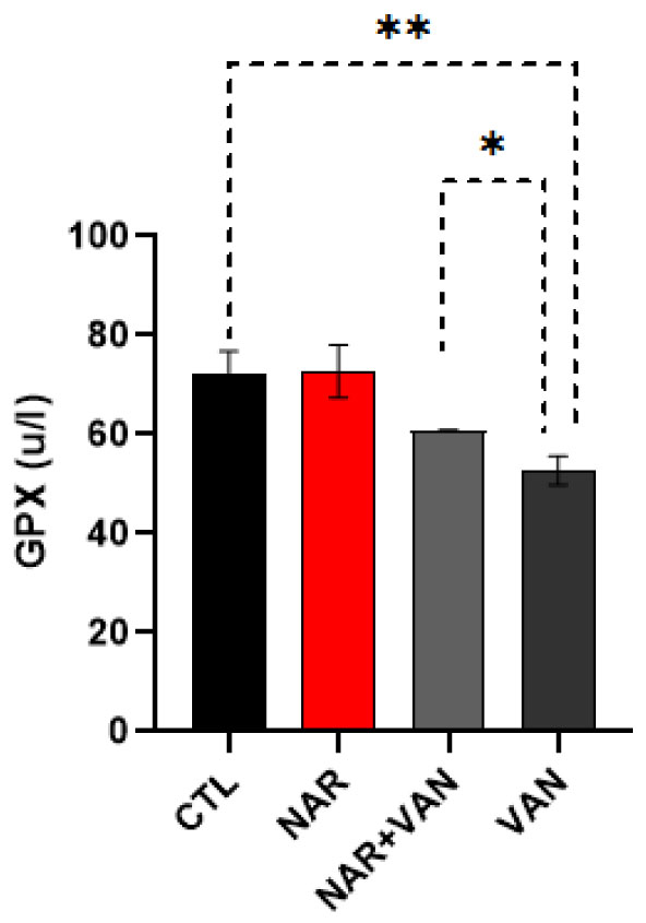
Exposure to vanadium has been known to lead to a progressive neurodegenerative disorder like Parkinson's disease. Naringin is a known flavonoid glycoside that is mostly seen in the flesh of grapefruit and orange and is believed to have protective effects for the treatment of neurodegenerative disorders. This study sought to investigate the role of Naringin in the treatment of vanadium-induced neurotoxicity. Vanadium (10 mg/kg BW) was injected intraperitoneally to induce motor dysfunction, followed by treatment with Naringin (30 mg/kg BW) intraperitoneally for 14 days. Oxidative stress imbalance was monitored by checking Glutathione Peroxidase (GPX) and Catalase levels. Histological and immunohistochemical alterations were observed using RBFOX3 polyclonal antibody to determine neuronal cell distribution and NLRP3 inflammasome antibody as a marker of inflammation. Exposure to vanadium induces neurotoxicity by significantly increasing the Catalase and Glutathione Peroxidase (GPX) levels. Vanadium administration also led to increased inflammatory cells and a significant reduction of the viable neuronal cells in the SNc and CPu. Treatment with Naringin showed a neuroprotective role by dependently restoring the Catalase and Glutathione Peroxidase (GPX) levels, inflammasome activation, and neuronal damage in the SNc and CPu. Naringin demonstrated anti-oxidative, and anti-inflammatory responses by inhibiting oxidative stress, and inflammation and exerts neuroprotective effects by inhibiting apoptosis following vanadium-induced neurotoxicity in adult Wistar rats.
Glutathione Peroxidase
Endoplasmic Reticulum
Vanadium
Naringin
Substantia nigra pars compacta
Caudate putamen
Glial cell line-derived neurotrophic factor
Tumour Necrosis Factor

| [1] |
Ohiomokhare S, Olaolorun F, Ladagu A, et al. (2020) The Pathopharmacological Interplay between Vanadium and Iron in Parkinson's Disease Models. Int J Mol Sci 21: 6719. https://doi.org/10.3390/ijms21186719 
|
| [2] |
Fatola OI, Olaolorun FA, Olopade FE, et al. (2019) Trends in vanadium neurotoxicity. Brain Res Bull 145: 75-80. https://doi.org/10.1016/j.brainresbull.2018.03.010
|
| [3] |
Onojake MC, Osuji LC, Abrakasa S (2015) Source, depositional environment and maturity levels of some crude oils in southwest Niger Delta, Nigeria. Chinese J Geochem 34: 224-232. https://doi.org/10.1007/s11631-015-0035-9
|
| [4] |
Lee AY, Lee MH, Lee S, et al. (2018) Neuroprotective effect of alpha-linolenic acid against Aβ-mediated inflammatory responses in C6 glial cell. J Agr Food Chem 66: 4853-4861. https://doi.org/10.1021/acs.jafc.8b00836
|
| [5] |
Ahmad S, Khan A, Ali W, et al. (2021) Fisetin rescues the mice brains against D-galactose-induced oxidative stress, neuroinflammation and memory impairment. Front Pharmacol 12: 612078. https://doi.org/10.3389/fphar.2021.612078
|
| [6] |
Zaidun NH, Thent ZC, Abd Latiff A (2018) Combating oxidative stress disorders with citrus flavonoid: Naringenin. Life Sci 208: 111-122. https://doi.org/10.1016/j.lfs.2018.07.017
|
| [7] |
Yang Y, Wu Y, Zou J, et al. (2021) Naringenin attenuates non-alcoholic fatty liver disease by enhancing energy expenditure and regulating autophagy via AMPK. Front Pharmacol 12: 687095. https://doi.org/10.3389/fphar.2021.687095
|
| [8] |
Adekeye AO, Irawo GJ, Fafure AA (2020) Ficus exasperata Vahl leaves extract attenuates motor deficit in vanadium-induced parkinsonism mice. Anat Cell Biol 53: 183-193. https://doi.org/10.5115/acb.19.205
|
| [9] |
Sayre LM, Perry G, Smith MA (2008) Oxidative stress and neurotoxicity. Chem Res Toxicol 21: 172-188. https://doi.org/10.1021/tx700210j
|
| [10] |
Mir NT, Saleem U, Anwar F, et al. (2019) Lawsonia Inermis markedly improves cognitive functions in animal models and modulate oxidative stress markers in the brain. Medicina 55: 192. https://doi.org/10.3390/medicina55050192
|
| [11] |
Garabadu D, Agrawal N (2020) Naringin exhibits neuroprotection against rotenone-induced neurotoxicity in experimental rodents. NeuroMolecular Medcine 22: 314-330. https://doi.org/10.1007/s12017-019-08590-2
|
| [12] |
Adebiyi OE, Olayemi FO, Olopade JO, et al. (2019) Βeta-sitosterol enhances motor coordination, attenuates memory loss and demyelination in a vanadium-induced model of experimental neurotoxicity. Pathophysiology 26: 21-29. https://doi.org/10.1016/j.pathophys.2018.12.002
|
| [13] |
Sugumar M, Sevanan M, Sekar S (2019) Neuroprotective effect of naringenin against MPTP-induced oxidative stress. Int J Neurosci 129: 534-539. https://doi.org/10.1080/00207454.2018.1545772
|
| [14] |
Folarin OR, Adaramoye OA, Akanni OO, et al. (2018) Changes in the brain antioxidant profile after chronic vanadium administration in mice. Metabolic Brain Disease 33: 377-385. https://doi.org/10.1007/s11011-017-0070-9
|
| [15] | Uddin MS, Mamun AA, Rahman MM, et al. (2021) Natural products for neurodegeneration: regulating neurotrophic signals. Oxid Med Longev 2021. https://doi.org/10.1155/2021/8820406 |
| [16] |
Nnama AU, Ekeh FN, Aguzie IO, et al. (2022) Vanadium pentoxide induces hematological, oxidative stress and histological changes in Oryctolagus cuniculus. J Hazard Mater Adv 5: 100048. https://doi.org/10.1016/j.hazadv.2022.100048
|
| [17] |
Cannon JR, Greenamyre JT (2009) NeuN is not a reliable marker of dopamine neurons in rat substantia nigra. Neurosci Lett 464: 14-17. https://doi.org/10.1016/j.neulet.2009.08.023
|
| [18] |
Zou L, Ning M, Wang W, et al. (2020) Naringenin Prevents Propofol Induced Neurodegeneration in Neonatal Mice Brain and Long-Term Neurocognitive Impacts on Adults. Drug Des Dev Ther 14: 5469. https://doi.org/10.2147/DDDT.S280443
|
| [19] |
Paldino E, D'Angelo V, Sancesario G, et al. (2020) Pyroptotic cell death in the R6/2 mouse model of Huntington's disease: new insight on the inflammasome. Cell Death Discov 6: 1-12. https://doi.org/10.1038/s41420-020-00293-z
|
| [20] | Im H, Kim E, Kwon HJ, et al. (2022) Therapeutic effect of silibinin on vanadium-induced chronic lung injury in mice. PREPRINT (Version 1) available at Research Square . https://doi.org/10.21203/rs.3.rs-1548568/v1 |
| [21] |
Cao H, Liu J, Shen P, et al. (2018) Protective effect of naringin on DSS-induced ulcerative colitis in mice. J Agr Food Chem 66: 13133-13140. https://doi.org/10.1021/acs.jafc.8b03942
|
| [22] |
Calabrese V, Mancuso C, Calvani M, et al. (2007) Nitric oxide in the central nervous system: neuroprotection versus neurotoxicity. Nat Rev Neurosci 8: 766-775. https://doi.org/10.1038/nrn2214
|


Figures(8)

Adeshina O. Adekeye, Adedamola A. Fafure, Ayoola E. Ogunsemowo, Linus A. Enye, Olusola S. Saka, Oluwatosin O. Ogedengbe. Naringin ameliorates motor dysfunction and exerts neuroprotective role against vanadium-induced neurotoxicity[J]. AIMS Neuroscience, 2022, 9(4): 536-550. doi: 10.3934/Neuroscience.2022031


 DownLoad: