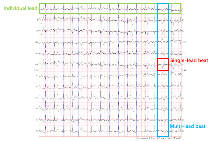
Automatic arrhythmia detection is very important for cardiovascular health. It is generally performed by measuring the electrocardiogram (ECG) signals of standard multiple leads. However, the correlations of multiple leads are often ignored. In addition, an extensive and complex feature extraction process is usually needed in most existing studies. Therefore, these challenges will not only lead to the loss of overall lead information, but also cause the detection performance to depend on the quality of features. To solve these challenges, a novel multi-lead arrhythmia detection model based on a heterogeneous graph attention network is proposed in this paper. We have modeled the multi-lead data as a heterogeneous graph to integrate diverse information and construct intra-lead and inter-lead correlations in multi-lead data, providing a reasonable and effective the data model. A heterogeneous graph network with a dual-level attention strategy has been utilized to capture the interactions among diverse information and information types. At the same time, our model does not require any feature extraction process for the ECG signals, which avoids out complex feature engineering. Extensive experimental results show that multi-lead information and complex correlations can be well captured, thus confirming that the proposed model results in significant improvements in multi-lead arrhythmia detection.
Citation: MingHao Zhong, Fenghuan Li, Weihong Chen. Automatic arrhythmia detection with multi-lead ECG signals based on heterogeneous graph attention networks[J]. Mathematical Biosciences and Engineering, 2022, 19(12): 12448-12471. doi: 10.3934/mbe.2022581
Automatic arrhythmia detection is very important for cardiovascular health. It is generally performed by measuring the electrocardiogram (ECG) signals of standard multiple leads. However, the correlations of multiple leads are often ignored. In addition, an extensive and complex feature extraction process is usually needed in most existing studies. Therefore, these challenges will not only lead to the loss of overall lead information, but also cause the detection performance to depend on the quality of features. To solve these challenges, a novel multi-lead arrhythmia detection model based on a heterogeneous graph attention network is proposed in this paper. We have modeled the multi-lead data as a heterogeneous graph to integrate diverse information and construct intra-lead and inter-lead correlations in multi-lead data, providing a reasonable and effective the data model. A heterogeneous graph network with a dual-level attention strategy has been utilized to capture the interactions among diverse information and information types. At the same time, our model does not require any feature extraction process for the ECG signals, which avoids out complex feature engineering. Extensive experimental results show that multi-lead information and complex correlations can be well captured, thus confirming that the proposed model results in significant improvements in multi-lead arrhythmia detection.

| [1] |
X. Liu, H. Wang, Z. Li, L. Qin, Deep learning in ecg diagnosis: A review, Knowl. Based Syst., 227 (2021), 107187. https://doi.org/10.1016/j.knosys.2021.107187 doi: 10.1016/j.knosys.2021.107187

|
| [2] |
H. Hao, M. Liu, P. Xiong, H. Du, H. Zhang, F. Lin, et al., Multi-lead model-based ecg signal denoising by guided filter, Eng. Appl. Artif. Intell., 79 (2019), 34–44. https://doi.org/10.1016/j.engappai.2018.12.004 doi: 10.1016/j.engappai.2018.12.004
|
| [3] |
F. M. Dias, H. L. Monteiro, T. W. Cabral, R. Naji, M. Kuehni, E. J. da S. Luz, Arrhythmia classification from single-lead ecg signals using the inter-patient paradigm, Comput. Methods Prog. Biomed., 202 (2021), 105948. https://doi.org/10.1016/j.cmpb.2021.105948 doi: 10.1016/j.cmpb.2021.105948
|
| [4] |
V. Singh, U. S. Reddy, G. M. Bhargavia, A generic and robust system for automated detection of different classes of arrhythmia, Proc. Comput. Sci., 167 (2020), 1801–1810. https://doi.org/10.1016/j.procs.2020.03.199 doi: 10.1016/j.procs.2020.03.199
|
| [5] |
H. M. Rai, K. Chatterjee, A novel adaptive feature extraction for detection of cardiac arrhythmias using hybrid technique mrdwt & mpnn classifier from ecg big data, Big Data Res., 12 (2018), 13–22. https://doi.org/10.1016/j.bdr.2018.02.003 doi: 10.1016/j.bdr.2018.02.003
|
| [6] |
J. Heo, J. J. Lee, S. Kwon, B. Kim, S. O. Hwang, Y. R. Yoon, A novel method for detecting st segment elevation myocardial infarction on a 12-lead electrocardiogram with a three-dimensional display, Biomed. Signal Process. Control, 56 (2020), 101700. https://doi.org/10.1016/j.bspc.2019.101700 doi: 10.1016/j.bspc.2019.101700
|
| [7] |
R. S. Singh, B. S. Saini, R. K. Sunkaria, Arrhythmia detection based on time-frequency features of heart rate variability and back-propagation neural network, Iran J. Comput. Sci., 2 (2019), 245–257. https://doi.org/10.1007/s42044-019-00042-1 doi: 10.1007/s42044-019-00042-1
|
| [8] |
G. Sannino, G. De Pietro, A deep learning approach for ecg-based heartbeat classification for arrhythmia detection, Future Gener. Comput. Syst., 86 (2018), 446–455. https://doi.org/10.1016/j.future.2018.03.057 doi: 10.1016/j.future.2018.03.057
|
| [9] |
E. Ramirez, P. Melin, G. Prado-Arechiga, Hybrid model based on neural networks, type-1 and type-2 fuzzy systems for 2-lead cardiac arrhythmia classification, Expert Syst. Appl., 126 (2019), 295–307. https://doi.org/10.1016/j.eswa.2019.02.035 doi: 10.1016/j.eswa.2019.02.035
|
| [10] |
M. Sharma, R. S. Tan, U. R. Acharya, Automated heartbeat classification and detection of arrhythmia using optimal orthogonal wavelet filters, Inf. Med. Unlocked, 16 (2019), 100221. https://doi.org/10.1016/j.imu.2019.100221 doi: 10.1016/j.imu.2019.100221
|
| [11] |
Z. Ebrahimi, M. Loni, M. Daneshtalab, A. Gharehbaghi, A review on deep learning methods for ecg arrhythmia classification, Expert Syst. Appl., 7 (2020), 100033. https://doi.org/10.1016/j.eswax.2020.100033 doi: 10.1016/j.eswax.2020.100033
|
| [12] |
S. Parvaneh, J. Rubin, S. Babaeizadeh, M. Xu-Wilson, Cardiac arrhythmia detection using deep learning: A review, J. Electrocardiol., 57 (2019), S70–S74. https://doi.org/10.1016/j.jelectrocard.2019.08.004 doi: 10.1016/j.jelectrocard.2019.08.004
|
| [13] |
R. Jothiramalingam, A. Jude, R. Patan, M. Ramachandran, J. H. Duraisamy, A. H. Gandomi, Machine learning-based left ventricular hypertrophy detection using multi-lead ecg signal, Neural Comput. Appl., 33 (2021), 4445–4455. https://doi.org/10.1007/s00521-020-05238-2 doi: 10.1007/s00521-020-05238-2
|
| [14] |
Z. Golrizkhatami, A. Acan, Ecg classification using three-level fusion of different feature descriptors, Expert Syst. Appl., 114 (2018), 54–64. https://doi.org/10.1016/j.eswa.2018.07.030 doi: 10.1016/j.eswa.2018.07.030
|
| [15] |
H. Martin, W. Izquierdo, M. Cabrerizo, A. Cabrera, M. Adjouadi, Near real-time single-beat myocardial infarction detection from single-lead electrocardiogram using long short-term memory neural network, Biomed. Signal Process. Control, 68 (2021), 102683. https://doi.org/10.1016/j.bspc.2021.102683 doi: 10.1016/j.bspc.2021.102683
|
| [16] |
K. Sugimoto, Y. Kon, S. Lee, Y. Okada, Detection and localization of myocardial infarction based on a convolutional autoencoder, Knowl. Based Syst., 178 (2019), 123–131. https://doi.org/10.1016/j.knosys.2019.04.023 doi: 10.1016/j.knosys.2019.04.023
|
| [17] |
K. Liu, S. Xu, N. Feng, A radial basis probabilistic process neural network model and corresponding classification algorithm, Appl. Intell., 49 (2019), 2256–2265. https://doi.org/10.1007/s10489-018-1369-x doi: 10.1007/s10489-018-1369-x
|
| [18] |
H. Fujita, D. Cimr, Decision support system for arrhythmia prediction using convolutional neural network structure without preprocessing, Appl. Intell., 49 (2019), 3383–3391. https://doi.org/10.1007/s10489-019-01461-0 doi: 10.1007/s10489-019-01461-0
|
| [19] |
M. Srinivasulu, Multi-lead ecg signal analysis using rbfnn-mso algorithm, Int. J. Speech Technol., 24 (2021), 341–350. https://doi.org/10.1007/s10772-021-09799-y doi: 10.1007/s10772-021-09799-y
|
| [20] |
G. Garcia, G. Moreira, D. Menotti, E. Luz, Inter-patient ecg heartbeat classification with temporal vcg optimized by pso, Sci. Rep., 7 (2017), 10543. https://doi.org/10.1038/s41598-017-09837-3 doi: 10.1038/s41598-017-09837-3
|
| [21] |
A. Chen, F. Wang, W. Liu, S. Chang, H. Wang, J. He, et al., Multi-information fusion neural networks for arrhythmia automatic detection, Comput. Methods Prog. Biomed., 193 (2020), 105479. https://doi.org/10.1016/j.cmpb.2020.105479 doi: 10.1016/j.cmpb.2020.105479
|
| [22] |
R. Mahajan, R. Kamaleswaran, O. Akbilgic, Comparative analysis between convolutional neural network learned and engineered features: A case study on cardiac arrhythmia detection, Cardiovass. Digital Health J., 1 (2020), 37–44. https://doi.org/10.1016/j.cvdhj.2020.04.001 doi: 10.1016/j.cvdhj.2020.04.001
|
| [23] | P. Lu, S. Guo, Y. Wang, L. Qi, X. Han, Y. Wang, Ecg classification based on long short-term memory networks, in Proceedings of the 2nd International Conference on Healthcare Science and Engineering, (2018), 129–140. |
| [24] |
J. Liao, D. Liu, G. Su, L. Liu, Recognizing diseases with multivariate physiological signals by a deepcnn-lstm network, Appl. Intell., 51 (2021), 7933–7945. https://doi.org/10.1007/s10489-021-02309-2 doi: 10.1007/s10489-021-02309-2
|
| [25] |
J. Zhang, A. Liu, M. Gao, X. Chen, X. Zhang, X. Chen, Ecg-based multi-class arrhythmia detection using spatio-temporal attention-based convolutional recurrent neural network, Artif. Intell. Med., 106 (2020), 101856. https://doi.org/10.1016/j.artmed.2020.101856 doi: 10.1016/j.artmed.2020.101856
|
| [26] |
Q. Yao, R. Wang, X. Fan, J. Liu, Y. Li, Multi-class arrhythmia detection from 12-lead varied-length ecg using attention-based time-incremental convolutional neural network, Inf. Fusion, 53 (2020), 174–182. https://doi.org/10.1016/j.inffus.2019.06.024 doi: 10.1016/j.inffus.2019.06.024
|
| [27] |
C. Che, P. Zhang, M. Zhu, Y. Qu, B. Jin, Constrained transformer network for ecg signal processing and arrhythmia classification, BMC Med. Inf. Decis. Making, 21 (2021), 184. https://doi.org/10.1186/s12911-021-01546-2 doi: 10.1186/s12911-021-01546-2
|
| [28] |
L. Wu, Y. Wang, S. Xu, K. Liu X. Li, An rbf-lvqpnn model and its application to time-varying signal classification, Appl. Intell., 51 (2021), 4548–4560. https://doi.org/10.1007/s10489-020-02094-4 doi: 10.1007/s10489-020-02094-4
|
| [29] |
P. Hao, X. Gao, Z. Li, J. Zhang, F. Wu, C. Bai, Multi-branch fusion network for myocardial infarction screening from 12-lead ecg images, Comput. Methods Prog. Biomed., 184 (2020), 105286. https://doi.org/10.1016/j.cmpb.2019.105286 doi: 10.1016/j.cmpb.2019.105286
|
| [30] |
A. K. Dohare, V. Kumar, R. Kumar, Detection of myocardial infarction in 12 lead ecg using support vector machine, Appl. Soft Comput., 64 (2018), 138–147. https://doi.org/10.1016/j.asoc.2017.12.001 doi: 10.1016/j.asoc.2017.12.001
|
| [31] |
P. Barmpoutis, K. Dimitropoulos, A. Apostolidis, N. Grammalidis, Multi-lead ecg signal analysis for myocardial infarction detection and localization through the mapping of grassmannian and euclidean features into a common hilbert space, Biomed. Signal Process. Control, 52 (2019), 111–119. https://doi.org/10.1016/j.bspc.2019.04.003 doi: 10.1016/j.bspc.2019.04.003
|
| [32] |
P. Xiong, Y. Xue, J. Zhang, M. Liu, H. Du, H. Zhang, et al., Localization of myocardial infarction with multi-lead ecg based on densenet, Comput. Methods Prog. Biomed., 203 (2021), 106024. https://doi.org/10.1016/j.cmpb.2021.106024 doi: 10.1016/j.cmpb.2021.106024
|
| [33] |
H. He, Y. Tan, J. Xing, Unsupervised classification of 12-lead ecg signals using wavelet tensor decomposition and two-dimensional gaussian spectral clustering, Knowl. Based Syst., 163 (2019), 392–403. https://doi.org/10.1016/j.knosys.2018.09.001 doi: 10.1016/j.knosys.2018.09.001
|
| [34] |
C. Han, L. Shi, Ml–resnet: A novel network to detect and locate myocardial infarction using 12 leads ecg, Comput. Methods Prog. Biomed., 185 (2020), 105138. https://doi.org/10.1016/j.cmpb.2019.105138 doi: 10.1016/j.cmpb.2019.105138
|
| [35] |
M. Sepahvand, F. Abdali-Mohammadi, A novel multi-lead ecg personal recognition based on signals functional and structural dependencies using time-frequency representation and evolutionary morphological cnn, Biomed. Signal Process. Control, 68 (2021), 102766. https://doi.org/10.1016/j.bspc.2021.102766 doi: 10.1016/j.bspc.2021.102766
|
| [36] | R. Li, S. Wang, F. Zhu, J. Huang, Adaptive graph convolutional neural networks, in Proceedings of the AAAI Conference on Artificial Intelligence, (2018), 3546–3553. https://doi.org/10.1609/aaai.v32i1.11691 |
| [37] | P. Velickovic, G. Cucurull, A. Casanova, A. Romero, P. Lio, Y. Bengio, Graph attention networks, in Proceedings of International Conference on Learning Representations(ICLR), (2018), 1–12. |
| [38] | S. Guo, Y. Lin, N. Feng, C. Song, H. Wan, Attention based spatial-temporal graph convolutional networks for traffic flow forecasting, in Proceedings of the AAAI Conference on Artificial Intelligence, (2019), 922–929. https://doi.org/10.1609/aaai.v33i01.3301922 |
| [39] | J. Justin, G. Agrim, F. F. Li, Image generation from scene graphs, in Proceedings of IEEE/CVF Conference on Computer Vision and Pattern Recognition, (2018), 1219–1228. |
| [40] | A. Fout, J. Byrd, B. Shariat, A. Ben-Hur, Protein interface prediction using graph convolutional networks, in Proceedings of the 31st International Conference on Neural Information Processing Systems, (2017), 6533–6542. |
| [41] |
C. Gunavathi, K. Sivasubramanian, P. Keerthika, C. Paramasivam, A review on convolutional neural network based deep learning methods in gene expression data for disease diagnosis, Mater. Today Proc., 45 (2021), 2282–2285. https://doi.org/10.1016/j.matpr.2020.10.263 doi: 10.1016/j.matpr.2020.10.263
|
| [42] |
A. Bessadok, M. A. Mahjoub, I. Rekik, Brain multigraph prediction using topology-aware adversarial graph neural network, Med. Image Anal., 72 (2021), 102090. https://doi.org/10.1016/j.media.2021.102090 doi: 10.1016/j.media.2021.102090
|
| [43] | B. Yu, H. Yin, Z. Zhu, Spatio-temporal graph convolutional networks: A deep learning framework for traffic forecasting, in Proceedings of Twenty-Seventh International Joint Conference on Artificial Intelligence IJCAI-18, (2018), 3634–3640. |
| [44] |
X. Yu, S. Lu, L. Guo, S. H. Wang, Y. D. Zhang, Resgnet-c: A graph convolutional neural network for detection of covid-19, Neurocomputing, 452 (2021), 592–605. https://doi.org/10.1016/j.neucom.2020.07.144 doi: 10.1016/j.neucom.2020.07.144
|
| [45] | C. Zhang, D. Song, C. Huang, Heterogeneous graph neural network, in Proceedings of the 25th ACM SIGKDD International Conference on Knowledge Discovery and Data Mining, (2019), 793–803. https://doi.org/10.1145/3292500.3330961 |
| [46] | X. Wang, H. Ji, C. Shi, B. Wang, P. Cui, P. Yu, et al., Heterogeneous graph attention network, in Proceedings of The World Wide Web Conference, (2019), 2022–2032. https://doi.org/10.1145/3308558.3313562 |
| [47] |
Y. Ding, L. P. Tian, X. Lei, B. Liao, F. X. Wu, Variational graph auto-encoders for mirna-disease association prediction, Methods, 192 (2021), 25–34. https://doi.org/10.1016/j.ymeth.2020.08.004 doi: 10.1016/j.ymeth.2020.08.004
|
| [48] |
T. Yang, L. Hu, C. Shi, H. Ji, X. Li, L. Nie, Hgat: Heterogeneous graph attention networks for semi-supervised short text classification, ACM Trans. Inf. Syst., 39 (2021), 1–29. https://doi.org/10.1145/3450352 doi: 10.1145/3450352
|
| [49] | P. PhysioBank, Physionet: components of a new research resource for complex physiologic signals, Circulation, 101 (2000), e215–e220. |
| [50] |
W. Yang, Y. Si, D. Wang, G. Zhang, A novel approach for multi-lead ecg classification using dl-ccanet and tl-ccanet, Sensors, 19 (2019), 3214. https://doi.org/10.3390/s19143214 doi: 10.3390/s19143214
|
| [51] |
J. N. Lee, Y. H. Byeon, S. B. Pan, K. C. Kwak, An eigenecg network approach based on pcanet for personal identification from ecg signal, Sensors, 18 (2018), 4024. https://doi.org/10.3390/s18114024 doi: 10.3390/s18114024
|
| [52] |
I. C. Tanoh, P. Napoletano, A novel 1-d ccanet for ecg classification, Appl. Sci., 11 (2021), 2758. https://doi.org/10.3390/app11062758 doi: 10.3390/app11062758
|


Figures(5) / Tables(13)

MingHao Zhong, Fenghuan Li, Weihong Chen. Automatic arrhythmia detection with multi-lead ECG signals based on heterogeneous graph attention networks[J]. Mathematical Biosciences and Engineering, 2022, 19(12): 12448-12471. doi: 10.3934/mbe.2022581


 DownLoad: