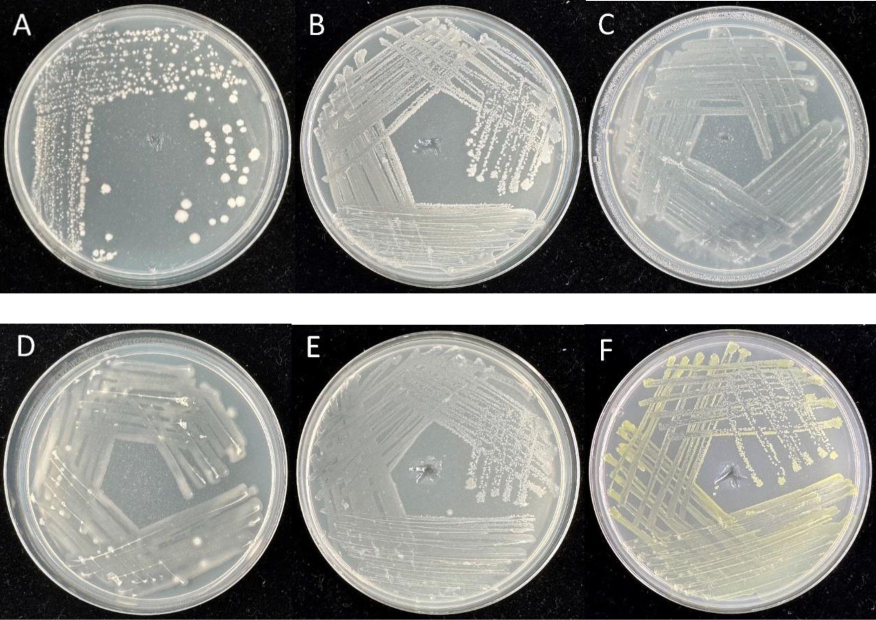
In this study, we used 16S rRNA gene sequence analysis to describe the diversity of cultivable endophytic bacteria associated with fennel (Foeniculum vulgare Mill.) and determined their plant-beneficial traits. The bacterial isolates from the roots of fennel belonged to four phyla: Firmicutes (BRN1 and BRN3), Proteobacteria (BRN5, BRN6, and BRN7), Gammaproteobacteria (BRN2), and Actinobacteria (BRN4). The bacterial isolates from the shoot of fennel represented the phyla Proteobacteria (BSN1, BSN2, BSN3, BSN5, BSN6, BSN7, and BSN8), Firmicutes (BSN4, BRN1, and BRN3), and Actinobacteria (BRN4). The bacterial species Bacillus megaterium, Bacillus aryabhattai, and Brevibacterium frigoritolerans were found both in the roots and shoots of fennel. The bacterial isolates were found to produce siderophores, HCN, and indole-3-acetic acid (IAA), as well as hydrolytic enzymes such as chitinase, protease, glucanase, and lipase. Seven bacterial isolates showed antagonistic activity against Fusarium culmorum, Fusarium solani, and Rhizoctonia. solani. Our findings show that medicinal plants with antibacterial activity may serve as a source for the selection of microorganisms that exhibit antagonistic activity against plant fungal infections and may be considered as a viable option for the management of fungal diseases. They can also serve as an active part of biopreparation, improving plant growth.
Citation: Vyacheslav Shurigin, Li Li, Burak Alaylar, Dilfuza Egamberdieva, Yong-Hong Liu, Wen-Jun Li. Plant beneficial traits of endophytic bacteria associated with fennel (Foeniculum vulgare Mill.)[J]. AIMS Microbiology, 2024, 10(2): 449-467. doi: 10.3934/microbiol.2024022
In this study, we used 16S rRNA gene sequence analysis to describe the diversity of cultivable endophytic bacteria associated with fennel (Foeniculum vulgare Mill.) and determined their plant-beneficial traits. The bacterial isolates from the roots of fennel belonged to four phyla: Firmicutes (BRN1 and BRN3), Proteobacteria (BRN5, BRN6, and BRN7), Gammaproteobacteria (BRN2), and Actinobacteria (BRN4). The bacterial isolates from the shoot of fennel represented the phyla Proteobacteria (BSN1, BSN2, BSN3, BSN5, BSN6, BSN7, and BSN8), Firmicutes (BSN4, BRN1, and BRN3), and Actinobacteria (BRN4). The bacterial species Bacillus megaterium, Bacillus aryabhattai, and Brevibacterium frigoritolerans were found both in the roots and shoots of fennel. The bacterial isolates were found to produce siderophores, HCN, and indole-3-acetic acid (IAA), as well as hydrolytic enzymes such as chitinase, protease, glucanase, and lipase. Seven bacterial isolates showed antagonistic activity against Fusarium culmorum, Fusarium solani, and Rhizoctonia. solani. Our findings show that medicinal plants with antibacterial activity may serve as a source for the selection of microorganisms that exhibit antagonistic activity against plant fungal infections and may be considered as a viable option for the management of fungal diseases. They can also serve as an active part of biopreparation, improving plant growth.

| [1] | Hui Xiang (2005) Flora of China. Tropicos Flora of China Checklist project 14: 134. |
| [2] |
Ozbek H, Ugras S, Dulger H, et al. (2003) Hepatoprotective effect of Foeniculum vulgare essential oil. Fitoterapia 74: 317-319. https://doi.org/10.1016/s0367-326x(03)00028-5 
|
| [3] |
Faudale M, Viladomat F, Bastida J, et al. (2008) Antioxidant activity and phenolic composition of wild, edible, and medicinal fennel from different mediterranean countries. J Agric Food Chem 56: 1912-1920. https://doi.org/10.1021/jf073083c
|
| [4] |
Mohsenzadeh M (2007) Evaluation of antibacterial activity of selected Iranian essential oils against Staphylococcus aureus and Escherichia coli in nutrient broth medium. Pak J Biol Sci 10: 3693-3697. https://doi.org/10.3923/pjbs.2007.3693.3697
|
| [5] |
Kaur GJ, Arora DS (2008) In-vitro antibacterial activity of three plants belonging to the family Umbelliferae. Int J Antimicrob Agents 31: 393-395. https://doi.org/10.1016/j.ijantimicag.2007.11.007
|
| [6] | Abed KF (2007) Antimicrobial activity of essential oils of some medicinal plants from Saudi Arabia. Saudi J Biol Sci 14: 53-60. |
| [7] |
Choi EM, Hwang JK (2004) Anti-inflammatory, analgesic and antioxidant activities of the fruit of Foeniculum vulgare. Fitoterapia 75: 557-565. https://doi.org/10.1016/j.fitote.2004.05.005
|
| [8] |
Tognolini M, Ballabeni V, Bertoni S, et al. (2007) Protective effect of Foeniculum vulgare essential oil and anethole in an experimental model of thrombosis. Pharmacol Res 56: 254-260. https://doi.org/10.1016/j.phrs.2007.07.002
|
| [9] | El-Soud NA, El-Laithy N, El-Saeed G, et al. (2011) Antidiabetic activities of Foeniculum vulgare Mill. essential oil in streptozotocin induced diabetic rats. Macedonian J Med Sci 173: 1857-5773. https://doi.org/10.3889/MJMS.1857-5773.2011.0173 |
| [10] | Pradhan M, Sribhuwaneswari S, Karthikeyan D, et al. (2008) In-vitro cytoprotection activity of Foeniculum vulgare and Helicteres isora in cultured human blood lymphocytes and antitumour activity against B16F10 melanoma cell line. Res J Pharm Technol 1: 450-452. |
| [11] | Reynolds JEF (1980) Essential oils and aromatic carminatives, Martindale-The Extra. Pharmacopeia, Royal Pharmaceutical Society, London . |
| [12] | Shaker GA, Alhamadany HS (2015) Isolation and identification of fungi which infect fennel Foeniculum vulgare Mill. and its impact as antifungal agent. Bulletin of the Iraq Natural History Museum 13: 31-38. |
| [13] |
Cacciola SO, Pane A, Cooke DEL, et al. (2006) First report of brown rot and wilt of fennel caused by Phytophthora megasperma in Italy. Plant Dis 90: 110. https://doi.org/10.1094/PD-90-0110A
|
| [14] |
Choi IY, Kim JH, Kim BS, et al. (2016) First report of Sclerotinia stem rot of fennel caused by Sclerotinia sclerotiorum in Korea. Plant Dis 100: 223. https://doi.org/10.1094/PDIS-05-15-0512-PDN
|
| [15] |
D'Amico M, Frisullo S, Cirulli M (2008) Endophytic fungi occurring in fennel, lettuce, chicory, and celery-commercial crops in southern Italy. Mycol Res 112: 100-107. https://doi.org/10.1016/j.mycres.2007.11.007
|
| [16] | Egamberdieva D, Wirth S, Behrendt U, et al. (2017a) Antimicrobial activity of medicinal plants correlates with the proportion of antagonistic endophytes. Front Microbiol 8: 199. https://doi.org/10.3389/fmicb.2017.00199 |
| [17] |
Egamberdieva D, Wirth S, Alqarawi AA, et al. (2017b) Phytohormones and beneficial microbes: Essential components for plants to balance stress and fitness. Front Microbiol 8: 2104. https://doi.org/10.3389/fmicb.2017.02104
|
| [18] |
Rezaei-Chiyaneh E, Battaglia ML, Sadeghpour A, et al. (2021) Optimizing intercropping systems of black cumin (Nigella sativa L.) and fenugreek (Trigonella foenum-graecum L.) through inoculation with bacteria and mycorrhizal fungi. Adv Sustainable Syst 5: 2000269. https://doi.org/10.1002/adsu.202000269
|
| [19] |
Pawlik M, Cania B, Thijs S, et al. (2017) Hydrocarbon degradation potential and plant growth-promoting activity of culturable endophytic bacteria of Lotus corniculatus and Oenothera biennis from a long-term polluted site. Environ Sci Pollut Res 24: 19640-19652. https://doi.org/10.1007/s11356-017-9496-1
|
| [20] |
Egamberdieva D, Shurigin V, Alaylar B, et al. (2020a) Bacterial endophytes from horseradish (Armoracia rusticana G. Gaertn., B. Mey. & Scherb.) with antimicrobial efficacy against pathogens. Plant Soil Environ 66: 309-316. https://doi.org/10.17221/137/2020-PSE
|
| [21] |
Egamberdieva D, Shurigin V, Alaylar B, et al. (2020b) The effect of biochars and endophytic bacteria on growth and root rot disease incidence of Fusarium infested narrow-leafed lupin (Lupinus angustifolius L.). Microorganisms 8: 496. https://doi.org/10.3390/microorganisms8040496
|
| [22] |
Nejatzadeh-Barandozi F (2013) Antibacterial activities and antioxidant capacity of Aloe vera. Bioorganic Med Chem Lett 3: 1-8. https://doi.org/10.1186/2191-2858-3-5
|
| [23] |
Bafana A, Lohiya R (2013) Diversity and metabolic potential of culturable root-associated bacteria from Origanum vulgare in sub-Himalayan region. World J Microbiol Biotechnol 29: 63-74. https://doi.org/10.1007/s11274-012-1158-3
|
| [24] |
Phetcharat P, Duangpaeng A (2012) Screening of endophytic bacteria from organic rice tissue for indole acetic acid production. Procedia Eng 32: 177-183. https://doi.org/10.1016/j.proeng.2012.01.1254
|
| [25] |
Shurigin V, Egamberdieva D, Samadiy S, et al. (2020) Endophytes from medicinal plants as biocontrol agents against Fusarium caused diseases. Mikrobiolohichnyi Zh 82: 41-52. https://doi.org/10.15407/microbiolj82.04.041
|
| [26] | Shurigin V, Alikulov B, Davranov K, et al. (2022) Bacterial endophytes from halophyte black saxaul (Haloxylon aphyllum Minkw.) and their plant growth-promoting properties. J Appl Biol Biotech 10: 45-53. https://doi.org/10.7324/JABB.2021.100106 |
| [27] |
Koberl M, Ramadan EM, Adam M, et al. (2013) Bacillus and Streptomyces were selected as broad-spectrum antagonists against soilborne pathogens from arid areas in Egypt. FEMS Microbiol Lett 342: 168-178. https://doi.org/10.1111/1574-6968.12089
|
| [28] |
Katoch M, Pull S (2017) Endophytic fungi associated with Monarda citriodora, an aromatic and medicinal plant and their biocontrol potential. Pharm Biol 55: 1528-1535. https://doi.org/10.1080/13880209.2017.1309054
|
| [29] | Tamilarasi S, Nanthakumar K, Karthikeyan K, et al. (2008) Diversity of root associated microorganisms of selected medicinal plants and influence of rhizomicroorganisms on the antimicrobial property of Coriandrum sativum. J Environ Biol 29: 127-134. |
| [30] | Salam N, Khieu TN, Liu MJ, et al. (2017) Endophytic actinobacteria associated with Dracaena cochinchinensis Lour.: isolation, diversity, and their cytotoxic activities. Biomed Res Int 1308563. https://doi.org/10.1155/2017/1308563 |
| [31] |
Rustamova N, Wubulikasimu A, Mukhamedov N, et al. (2020) Endophytic bacteria associated with medicinal plant Baccharoides anthelmintica diversity and characterization. Curr Microbiol 77: 1457-1465. https://doi.org/10.1007/s00284-020-01924-5
|
| [32] |
Shurigin V, Alaylar B, Davranov K, et al. (2021) Diversity and biological activity of culturable endophytic bacteria associated with marigold (Calendula officinalis L.). AIMS Microbiol 7: 336-353. https://doi.org/10.3934/microbiol.2021021
|
| [33] |
Mora-Ruiz MDR, Font-Verdera F, Díaz-Gil C, et al. (2015) Moderate halophilic bacteria colonizing the phylloplane of halophytes of the subfamily Salicornioideae (Amaranthaceae). Syst Appl Microbiol 38: 406-416. https://doi.org/10.1016/j.syapm.2015.05.004
|
| [34] | Dashti AA, Jadaon MM, Abdulsamad AM, et al. (2009) Heat treatment of bacteria: a simple method of DNA extraction for molecular techniques. Kuwait Med J 41: 117-122. |
| [35] | Lane DJ (1991) 16S/23S rRNA Sequencing. Nucleic acid techniques in bacterial systematic . New York: John Wiley and Sons 115-175. |
| [36] | Jinneman KC, Wetherington JH, Adams AM, et al. (1996) Differentiation of Cyclospora sp. and Eimeria spp. by using the polymerase chain reaction amplification products and restriction fragment length polymorphisms. Food and Drug Administration Laboratory Information Bulletin LIB no 4044 . |
| [37] |
Tamura K, Nei M, Kumar S (2004) Prospects for inferring very large phylogenies by using the neighbor-joining method. Proc Natl Acad Sci USA 101: 11030-11035. https://doi.org/10.1073/pnas.0404206101
|
| [38] |
Kumar S, Stecher G, Li M, et al. (2018) MEGA X: Molecular Evolutionary Genetics Analysis across computing platforms. Mol Biol Evol 35: 1547-1549. https://doi.org/10.1093/molbev/msy096
|
| [39] |
Egamberdieva D, Wirth SJ, Shurigin VV, et al. (2017d) Endophytic bacteria improve plant growth, symbiotic performance of chickpea (Cicer arietinum L.) and induce suppression of root rot caused by Fusarium solani under salt stress. Front Microbiol 8: 1887. https://doi.org/10.3389/fmicb.2017.01887
|
| [40] |
Castric PA (1975) Hydrogen cyanide, a secondary metabolite of Pseudomonas aeruginosa. Can J Microbiol 21: 613-618. https://doi.org/10.1139/m75-088
|
| [41] |
Schwyn B, Neilands JB (1987) Universal chemical assay for the detection and determination of siderophores. Anal Biochem 160: 47-56. https://doi.org/10.1016/0003-2697(87)90612-9
|
| [42] |
Brown MRW, Foster JHS (1970) A simple diagnostic milk medium for Pseudomonas aeruginosa. J Clin Pathol 23: 172-177. https://doi.org/10.1136/jcp.23.2.172
|
| [43] |
Walsh GA, Murphy RA, Killeen GF, et al. (1995) Technical note: Detection and quantification of supplemental fungal b-glucanase activity in animal feed. J Anim Sci 73: 1074-1076. https://doi.org/10.2527/1995.7341074x
|
| [44] | Malleswari D, Bagyanarayan G (2017) In vitro screening of rhizobacteria isolated from the rhizosphere of medicinal and aromatic plants for multiple plant growth promoting activities. J Microbiol Biotechnol Res 3: 84-91. |
| [45] |
Howe TG, Ward JM (1976) The utilization of tween 80 as carbon source by Pseudomonas. J Gen Microbiol 92: 234-235. https://doi.org/10.1099/00221287-92-1-234
|
| [46] |
Bano N, Musarrat J (2003) Characterization of a new Pseudomonas aeruginosa strain NJ-15 as a potential biocontrol agent. Curr Microbiol 46: 324-328. https://doi.org/10.1007/s00284-002-3857-8
|
| [47] |
Egamberdieva D, Kucharova Z (2009) Selection for root colonising bacteria stimulating wheat growth in saline soils. Biol Fertil Soils 45: 561-573. https://doi.org/10.1007/s00374-009-0366-y
|
| [48] | Chen Q, Liu S, Bai Y, et al. (2014) Screening and identification of phosphate-solubilizing bacteria from reclaimed soil in Shanxi mining area. Plant Nutr Fertilizer Sci 20: 1505-1516. |
| [49] |
Egamberdieva D, Wirth S, Li L, et al. (2017c) Microbial cooperation in the rhizosphere improves liquorice growth under salt stress. Bioengineered 8: 433-438. https://doi.org/10.1080/21655979.2016.1250983
|
| [50] |
Ali S, Duan J, Charles TC, et al. (2014) A bioinformatics approach to the determination of genes involved in endophytic behavior in Burkholderia spp. J Theor Biol 343: 193-198. https://doi.org/10.1016/j.jtbi.2013.10.007
|
| [51] |
Cho ST, Chang HH, Egamberdieva D, et al. (2015) Genome analysis of Pseudomonas fluorescens PCL1751: a rhizobacterium that controls root diseases and alleviates salt stress for its plant host. PLoS ONE 10: e0140231. https://doi.org/10.1371/journal.pone.0140231
|
| [52] |
Zhao L, Xu Y, Lai XH, et al. (2015) Screening and characterization of endophytic Bacillus and Paenibacillus strains from medicinal plant Lonicera japonica for use as potential plant growth promoters. Braz J Microbiol 46: 977-989. https://doi.org/10.1590/S1517-838246420140024
|
| [53] |
Preveena J, Bhore SJ (2013) Identification of bacterial endophytes associated with traditional medicinal plant Tridax procumbens Linn. Anc Sci Life 32: 173-177. https://doi.org/10.4103/0257-7941.123002
|
| [54] |
Webster G, Mullins AJ, Cunningham-Oakes E, et al. (2020) Culturable diversity of bacterial endophytes associated with medicinal plants of the Western Ghats, India. FEMS Microbiol Ecol 96: fiaa147. https://doi.org/10.1093/femsec/fiaa147
|
| [55] |
Liu YH, Guo JW, Salam N, et al. (2016) Culturable endophytic bacteria associated with medicinal plant Ferula songorica: molecular phylogeny, distribution and screening for industrially important traits. 3 Biotech 6: 209. https://doi.org/10.1007/s13205-016-0522-7
|
| [56] |
Chi F, Shen S, Cheng H, et al. (2005) Ascending migration of endophytic rhizobia, from roots to leaves, inside rice plants and assessment of benefits to rice growth physiology. Appl Environ Microbiol 71: 7271-7278. https://doi.org/10.1128/AEM.71.11.7271-7278.2005
|
| [57] |
Shi W, Su G, Li M, et al. (2021) Distribution of bacterial endophytes in the non-lesion tissues of potato and their response to potato common scab. Front Microbiol 12: 616013. https://doi.org/10.3389/fmicb.2021.616013
|
| [58] | Goryluk A, Rekosz-Burlaga H, Blaszczyk M (2009) Isolation and characterization of bacterial endophytes of Chelidonium majus L. Pol J Microbiol 58: 355-361. |
| [59] |
Mehanni MM, Safwat MS (2010) Endophytes of medicinal plants. Acta Hortic 854: 31-40. https://doi.org/10.17660/ActaHortic.2010.854.3
|
| [60] |
Egamberdieva D, Kucharova Z, Davranov K, et al. (2011) Bacteria able to control foot and root rot and to promote growth of cucumber in salinated soils. Biol Fertil Soils 47: 197-205. https://doi.org/10.1007/s00374-010-0523-3
|
| [61] |
Nongkhlaw FMW, Joshi SR (2014) Epiphytic and endophytic bacteria that promote growth of ethnomedicinal plants in the subtropical forests of Meghalaya, India. Rev Biol Trop 62: 1295-1308. https://doi.org/10.15517/rbt.v62i4.12138
|
| [62] |
Liu Y, Mohamad OAA, Salam N, et al. (2019) Diversity, community distribution and growth promotion activities of endophytes associated with halophyte Lycium ruthenicum Murr. 3 Biotech 9: 144. https://doi.org/10.1007/s13205-019-1678-8
|
| [63] |
Ferchichi N, Toukabri W, Boularess M, et al. (2019) Isolation, identification and plant growth promotion ability of endophytic bacteria associated with lupine root nodule grown in Tunisian soil. Arch Microbiol 201: 1333-1349. https://doi.org/10.1007/s00203-019-01702-3
|
| [64] | Fernando TC, Cruz JA (2019) Profiling and biochemical identification of potential plant growth-promoting endophytic bacteria from Nypa fruticans. Philipp J Crop Sci 44: 77-85. https://doi.org/10.13140/RG.2.2.15641.98408 |
| [65] | Rana KL, Kour D, Yadav AH (2019) Endophytic microbiomes: Biodiversity, ecological significance and biotechnological applications. Res J Biotechnol 14: 142-162. |
| [66] | Siddiqui ZA (2005) PGPR: prospective biocontrol agents of plant pathogens. PGPR: biocontrol and biofertilization . Dordrecht: Springer 111-142. https://doi.org/10.1007/1-4020-4152-7_4 |
| [67] |
Michelsen CF, Stougaard P (2012) Hydrogen cyanide synthesis and antifungal activity of the biocontrol strain Pseudomonas fluorescens In5 from Greenland is highly dependent on growth medium. Can J Microbiol 58: 381-390. https://doi.org/10.1139/w2012-004
|
| [68] |
Ahmed EA, Hassan EA, El Tobgy KMK, et al. (2014) Evaluation of rhizobacteria of some medicinal plants for plant growth promotion and biological control. Ann Agric Sci 59: 273-280. https://doi.org/10.1016/j.aoas.2014.11.016
|
| [69] |
Arun B, Gopinath B, Sharma S (2012) Plant growth promoting potential of bacteria isolated on N free media from rhizosphere of Cassia occidentalis. World J Microbiol Biotechnol 28: 2849-2857. https://doi.org/10.1007/s11274-012-1095-1
|
| [70] |
Ray S, Singh S, Sarma BK, et al. (2016) Endophytic alcaligenes isolated from horticultural and medicinal crops promotes growth in Okra (Abelmoschus esculentus). J Plant Growth Regul 35: 401-412. https://doi.org/10.1007/s00344-015-9548-z
|
| [71] |
Chowdhury EK, Jeon J, Rim SK, et al. (2017) Composition, diversity and bioactivity of culturable bacterial endophytes in mountain-cultivated ginseng in Korea. Sci Rep 7: 1-10. https://doi.org/10.1038/s41598-017-10280-7
|
| [72] |
Wozniak M, Gałazka A, Tyskiewicz R, et al. (2019) Endophytic bacteria potentially promote plant growth by synthesizing different metabolites and their phenotypic/physiological profiles in the Biolog GEN III MicroPlateTM Test. Int J Mol Sci 20: 1-24. https://doi.org/10.3390/ijms20215283
|
| [73] |
Musa Z, Ma J, Egamberdieva D, et al. (2020) Diversity and antimicrobial potential of cultivable endophytic actinobacteria associated with medicinal plant Thymus roseus. Front Microbiol 11: 191. https://doi.org/10.3389/fmicb.2020.00191
|
| [74] |
Glick BR (2014) Bacteria with ACC deaminase can promote plant growth and help to feed the world. Microbiol Res 169: 30-39. https://doi.org/10.1016/j.micres.2013.09.009
|
| [75] |
Leong J (1986) Siderophores: their biochemistry and possible role in the biocontrol of plant pathogens. Annu Rev Phytopathol 24: 187-209. https://doi.org/10.1146/annurev.py.24.090186.001155
|
| [76] |
Neilands JB, Leong SA (1986) Siderophores in relation to plant growth and disease. Annu Rev Plant Physiol 37: 187-208. https://doi.org/10.1146/annurev.pp.37.060186.001155
|
| [77] |
Goldstein AH (1986) Bacterial solubilization of mineral phosphates: Historical perspective and future prospects. Amer J Alternat Agric 1: 51-57. https://doi.org/10.1017/S0889189300000886
|
| [78] |
Sudarshna, Sharma N (2024) Endophytic bacteria associated with critically endangered medicinal plant Trillium govanianum (Wall ex. Royle) and their potential in soil nutrition alleviation. Plant Stress 11: 100349. https://doi.org/10.1016/j.stress.2024.100349
|
| [79] |
Deepa N, Chauhan Sh, Singh A (2024) Unraveling the functional characteristics of endophytic bacterial diversity for plant growth promotion and enhanced secondary metabolite production in Pelargonium graveolens. Microbiol Res 283: 127673. https://doi.org/10.1016/j.micres.2024.127673
|


Figures(3) / Tables(5)

Vyacheslav Shurigin, Li Li, Burak Alaylar, Dilfuza Egamberdieva, Yong-Hong Liu, Wen-Jun Li. Plant beneficial traits of endophytic bacteria associated with fennel (Foeniculum vulgare Mill.)[J]. AIMS Microbiology, 2024, 10(2): 449-467. doi: 10.3934/microbiol.2024022


 DownLoad: