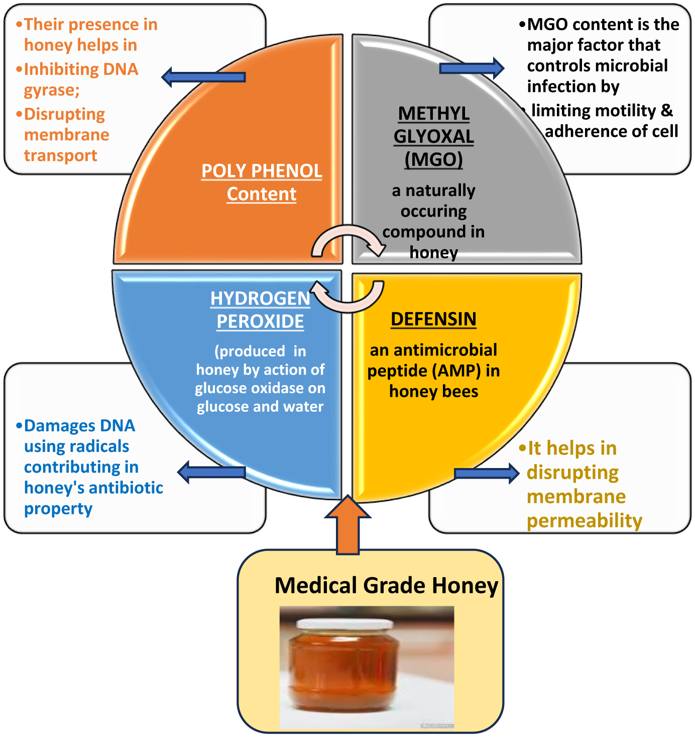
Presently, most of the reported infections are of a bacterial origin; however, this leads to a limit within the literature and research around infections caused by fungal pathogens, which are now developing resistance to antibiotic medicines. Of the natural antimicrobial agents, honey has been observed with demonstrable and highly exploitable antimicrobial and infection control related to wound healing properties; therefore, it has been incorporated into many standard pharmaceutical formulations. Generally, these products utilize a pure sample of honey as a bioactive ingredient in a product which has been purposely designed for the convenience of application. This article aims to review information available from published reports on various bioactivities of a variety of medical-grade honey products, including manuka and other conventional non-manuka types sourced from different floral types and geographical regions. Additionally, this review highlights the antibiotic activities of various types of honey products tested against pathogenic strains of bacteria, yeast and fungi, and their applications in the formulation of healthcare products.
Citation: Divakar Dahiya, Caoimhin Mackin, Poonam Singh Nigam. Studies on bioactivities of Manuka and regional varieties of honey for their potential use as natural antibiotic agents for infection control related to wound healing and in pharmaceutical formulations[J]. AIMS Microbiology, 2024, 10(2): 288-310. doi: 10.3934/microbiol.2024015
Presently, most of the reported infections are of a bacterial origin; however, this leads to a limit within the literature and research around infections caused by fungal pathogens, which are now developing resistance to antibiotic medicines. Of the natural antimicrobial agents, honey has been observed with demonstrable and highly exploitable antimicrobial and infection control related to wound healing properties; therefore, it has been incorporated into many standard pharmaceutical formulations. Generally, these products utilize a pure sample of honey as a bioactive ingredient in a product which has been purposely designed for the convenience of application. This article aims to review information available from published reports on various bioactivities of a variety of medical-grade honey products, including manuka and other conventional non-manuka types sourced from different floral types and geographical regions. Additionally, this review highlights the antibiotic activities of various types of honey products tested against pathogenic strains of bacteria, yeast and fungi, and their applications in the formulation of healthcare products.

| [1] |
. |
| [2] |
Salmanov A, Shchehlov D, Svyrydiuk O, et al. (2023) Epidemiology of healthcare-associated infections and mechanisms of antimicrobial resistance of responsible pathogens in ukraine: A multicentre study. J Hosp Infect 131: 129-138. https://doi.org/10.1016/j.jhin.2022.10.007 
|
| [3] |
Abhishek C, Prakash B, Abraham BK, et al. (2021) Incidence and impact of healthcare-associated infections on patients primarily admitted with sepsis and non-sepsis diagnoses. Indian J Crit Care Med 25: 292-295. https://doi.org/10.5005/jp-journals-10071-23760
|
| [4] | European Centre for Disease Prevention and ControlEuropean centre for disease prevention and control. assessing the health burden of infections with antibiotic-resistant bacteria in the EU/EEA, 2016-2020 (2022). Retrieved 09/08/2023, Available from: https://www.ecdc.europa.eu/en/publications-data/health-burden-infections-antibiotic-resistant-bacteria-2016-2020 |
| [5] |
Zay Ya K, Win PTN, Bielicki J, et al. (2023) Association between antimicrobial stewardship programs and antibiotic use globally: A systematic review and meta-analysis. JAMA Network Open 6: e2253806. https://doi.org/10.1001/jamanetworkopen.2022.53806
|
| [6] |
Seidelman JL, Mantyh CR, Anderson DJ (2023) Surgical site infection prevention: A review. JAMA 29: 244-252. https://doi.org/10.1001/jama.2022.24075
|
| [7] |
Sen CK (2021) Human wound and its burden: Updated 2020 compendium of estimates. Adv Wound Care 10: 281-292. https://doi.org/10.1089/wound.2021.0026
|
| [8] |
Özker E, Erkin A, Aslan HM, et al. (2021) Wound treatment strategies during COVID-19 pandemic: An expert opinion. Turkish J Vasc Surg 30: 167-173. https://doi.org/10.9739/tjvs.2020.761
|
| [9] |
Short B, Bakri A, Baz A, et al. (2023) There is more to wounds than bacteria: Fungal biofilms in chronic wounds. Curr Clin Microbiol Rep 10: 9-16. https://doi.org/10.1007/s40588-022-00187-x
|
| [10] |
Dowd SE, Delton Hanson J, Rees E, et al. (2011) Survey of fungi and yeast in polymicrobial infections in chronic wounds. J Wound Care 20: 40-47. https://doi.org/10.12968/jowc.2011.20.1.40
|
| [11] |
Bansal E, Garg A, Bhatia S, et al. (2008) Spectrum of microbial flora in diabetic foot ulcers. Indian J Pathol Microbiol 51: 204-208. https://doi.org/10.4103/0377-4929.41685
|
| [12] |
Ge Y, Wang Q (2023) Current research on fungi in chronic wounds. Front Mol Biosci 9: 1057766. https://doi.org/10.3389/fmolb.2022.1057766
|
| [13] |
Diban F, Di Lodovico S, Di Fermo P, et al. (2023) Biofilms in chronic wound infections: Innovative antimicrobial approaches using the in vitro lubbock chronic wound biofilm model. Int J Mol Sci 24: 1004. https://doi.org/10.3390/ijms24021004
|
| [14] |
Vitiello A, Ferrara F, Boccellino M, et al. (2023) Antifungal drug resistance: An emergent health threat. Biomedicines 11: 1063. https://doi.org/10.3390/biomedicines11041063
|
| [15] |
Abdelmohsen UR, Balasubramanian S, Oelschlaeger TA, et al. (2017) Potential of marine natural products against drug-resistant fungal, viral, and parasitic infections. Lancet Infect Dis 17: e30-e41. https://doi.org/10.1016/S1473-3099(16)30323-1
|
| [16] |
Sarmiento-Vizcaíno A, García LA, Blanco G (2023) Streptomyces isolated from bird feathers as a potential source for novel antibiotics. Arch Microbiol 205: 81. https://doi.org/10.1007/s00203-023-03422-1
|
| [17] |
Saadoun JH, Sogari G, Bernini V, et al. (2022) A critical review of intrinsic and extrinsic antimicrobial properties of insects. Trends Food Sci Technol 122: 40-48. https://doi.org/10.1016/j.tifs.2022.02.018
|
| [18] | Ahmed SH, Ali WK (2023) Antimicrobial activities of thirteen insect species crude body extract against some microbial pathogens. Zanco J Pure Appl Sci 35: 153-159. https://doi.org/10.21271/zjpas |
| [19] |
Abreu AC, McBain AJ, Simões M (2012) Plants as sources of new antimicrobials and resistance-modifying agents. Nat Prod Rep 29: 1007-1121. https://doi.org/10.1039/C2NP20035J
|
| [20] |
Wang JC, Fort CL, Matl CM, et al. (2023) Effects of essential oils on scars and wound healing: A systematic review. Facial Plast Surg 39: 173-179. https://doi.org/10.1055/a-1938-0343
|
| [21] |
Zhang F, Chen Z, Su F, et al. (2021) Comparison of topical honey and povidone iodine-based dressings for wound healing: A systematic review and meta-analysis. J Wound Care 30: S28-S36. https://doi.org/10.12968/jowc.2021.30.Sup4.S28
|
| [22] |
Almasaudi S (2021) The antibacterial activities of honey. Saudi J Biol Sci 28: 2188-2196. https://doi.org/10.1016/j.sjbs.2020.10.017
|
| [23] |
Hegazi NM, Elghani GEA, Farag MA (2022) The super-food manuka honey, a comprehensive review of its analysis and authenticity approaches. J Food Sci Technol 59: 2527-2534. https://doi.org/10.1007/s13197-021-05181-7
|
| [24] | Walsey E, Webster TJ (2023) Surface colonization when exposed to medical grade (manuka) honey alone or in combination with other disinfectants. Res J Med Health Sci 4. https://doi.org/10.58256/rjmhs.v4i1.1008 |
| [25] |
Lu J, Cokcetin NN, Burke CM, et al. (2019) Honey can inhibit and eliminate biofilms produced by pseudomonas aeruginosa. Sci Rep 9: 18160-13. https://doi.org/10.1038/s41598-019-54576-2
|
| [26] |
Jenkins R, Burton N, Cooper R (2011) Manuka honey inhibits cell division in methicillin-resistant staphylococcus aureus. J Antimicrob Chemother 66: 2536-2542. https://doi.org/10.1093/jac/dkr340
|
| [27] |
Bouacha M, Besnaci S, Boudiar I (2023) Comparative study of the antibacterial activity of algerian honeys and manuka honey toward pathogenic bacteria from burn wound infections. Mikrobiolohichnyĭ Zhurnal 85: 26-36. https://doi.org/10.15407/microbiolj85.02.026
|
| [28] |
Albaridi NA (2019) Antibacterial potency of honey. Int J Microbiol 2019: 2464507-10. https://doi.org/10.1155/2019/2464507
|
| [29] |
Combarros-Fuertes P, Fresno JM, Estevinho MM, et al. (2020) Honey: Another alternative in the fight against antibiotic-resistant bacteria?. Antibiotics (Basel) 9: 774. https://doi.org/10.3390/antibiotics9110774
|
| [30] |
Johnston M, McBride M, Dahiya D, et al. (2018) Antibacterial activity of manuka honey and its components: An overview. AIMS Microbiol 4: 655-664. https://doi.org/10.3934/microbiol.2018.4.655
|
| [31] |
Hulea A, Obiştioiu D, Cocan I, et al. (2022) Diversity of monofloral honey based on the antimicrobial and antioxidant potential. Antibiotics (Basel) 11: 595. https://doi.org/10.3390/antibiotics11050595
|
| [32] |
Lin T, Huang L, Cheng N, et al. (2022) The in vitro and in vivo antibacterial activities of uniflorous honey from a medicinal plant, scrophularia ningpoensis hemsl., and characterization of its chemical profile with UPLC-MS/MS. J Ethnopharmacol 296: 115499. https://doi.org/10.1016/j.jep.2022.115499
|
| [33] |
Gkoutzouvelidou M, Panos G, Xanthou MN, et al. (2021) Comparing the antimicrobial actions of greek honeys from the island of lemnos and manuka honey from new zealand against clinically important bacteria. Foods 10: 1402. https://doi.org/10.3390/foods10061402
|
| [34] |
Zhang Y, Si J, Li S, et al. (2021) Chemical analyses and antimicrobial activity of nine kinds of unifloral chinese honeys compared to manuka honey (12+ and 20+). Molecules (Basel, Switzerland) 26: 2778. https://doi.org/10.3390/molecules26092778
|
| [35] | Lu J, Turnbull L, Burke CM, et al. (2014) Manuka-type honeys can eradicate biofilms produced by staphylococcus aureus strains with different biofilm-forming abilities. PeerJ (San Francisco, CA) 2: e326. https://doi.org/10.7717/peerj.326 |
| [36] |
Portokalakis I, Yusof H, Ghanotakis D, et al. (2016) Manuka honey-induced cytotoxicity against MCF7 breast cancer cells is correlated to total phenol content and antioxidant power. J Adv Biol Biotechnol 8: 1-10. https://doi.org/10.9734/JABB/2016/27899
|
| [37] |
Henderson K, Aldhirgham T, Nigam P, et al. (2016) Evaluation of manuka honey estrogen activity using the MCF-7 cell proliferation assay. J Adv Biol Biotechnol 10: 1-11. https://doi.org/10.9734/JABB/2016/29887
|
| [38] |
Bazaid AS, Alamri A, Almashjary MN, et al. (2022) Antioxidant, anticancer, antibacterial, antibiofilm properties and gas chromatography and mass spectrometry analysis of manuka honey: A nature's bioactive honey. Appl Sci 12: 9928. https://doi.org/10.3390/app12199928
|
| [39] |
Kirkpatrick G, Nigam P, Owusu-Apenten R (2017) Total phenols, antioxidant capacity and antibacterial activity of manuka honey chemical constituents. J Adva Biol Biotechnol 15: 1-7. https://doi.org/10.9734/JABB/2017/37242
|
| [40] |
Chau T, Owusu-Apenten R, Nigam P (2017) Total phenols, antioxidant capacity and antibacterial activity of manuka honey extract. J Adv Biol Biotechnol 15: 1-6. https://doi.org/10.9734/JABB/2017/37101
|
| [41] |
Yusof H, Owusu-Apenten R, Nigam P (2018) Determination of iron (III) reducing antioxidant capacity for manuka honey and comparison with ABTS and other methods. J Adv Biol Biotechnol 18: 1-9. https://doi.org/10.9734/JABB/2018/42202
|
| [42] |
Hayashi K, Fukushima A, Hayashi-Nishino M, et al. (2014) Effect of methylglyoxal on multidrug-resistant pseudomonas aeruginosa. Front Microbiol 5: 180. https://doi.org/10.3389/fmicb.2014.00180
|
| [43] |
Tonks AJ, Dudley E, Porter NG, et al. (2007) A 5.8-kDa component of manuka honey stimulates immune cells via TLR4. J Leukocyte Biol 82: 1147-1155. https://doi.org/10.1189/jlb.1106683
|
| [44] |
Majtan J (2014) Honey: An immunomodulator in wound healing. Wound Repair Regener 22: 187-192. https://doi.org/10.1111/wrr.12117
|
| [45] |
Green KJ, Lawag I, Locher C, et al. (2022) Correlation of the antibacterial activity of commercial manuka and leptospermum honeys from australia and new zealand with methylglyoxal content and other physicochemical characteristics. PloS One 17: e0272376. https://doi.org/10.1371/journal.pone.0272376
|
| [46] |
Irish J, Carter DA, Shokohi T, et al. (2006) Honey has an antifungal effect against candida species. Med Mycol 44: 289-291. https://doi.org/10.1080/13693780500417037
|
| [47] |
Kunat-Budzyńska M, Rysiak A, Wiater A, et al. (2023) Chemical composition and antimicrobial activity of new honey varietals. Int J Environ Res Public Health 20: 2458. https://doi.org/10.3390/ijerph20032458
|
| [48] |
Anyanwu CU (2012) Investigation of in vitro antifungal activity of honey. J Med Plant Res 6. https://doi.org/10.5897/JMPR12.577
|
| [49] |
Oliveira AMP, Devesa JSP, Hill PB (2018) In vitro efficacy of a honey-based gel against canine clinical isolates of staphylococcus pseudintermedius and malassezia pachydermatis. Vet Dermatol 29: 180-e65. https://doi.org/10.1111/vde.12533
|
| [50] |
de Groot T, Janssen T, Faro D, et al. (2021) Antifungal activity of a medical-grade honey formulation against candida auris. J Fungi (Basel) 7: 50. https://doi.org/10.3390/jof7010050
|
| [51] |
Hau-Yama NE, Magaña-Ortiz D, Oliva AI, et al. (2020) Antifungal activity of honey from stingless bee melipona beecheii against candida albicans. J Apic Res 59: 12-18. https://doi.org/10.1080/00218839.2019.1665247
|
| [52] |
Fernandes L, Ribeiro H, Oliveira A, et al. (2021) Portuguese honeys as antimicrobial agents against candida species. J Trad Complement Med 11: 130-136. https://doi.org/10.1016/j.jtcme.2020.02.007
|
| [53] |
Hamid Z, Mohamad I, Harun A, et al. (2018) Antifungal effect of three local malaysian honeys on selected pathogenic fungi of otomycosis: An in vitro evaluation. J Young Pharm 10: 414-417. https://doi.org/10.5530/jyp.2018.10.91
|
| [54] |
Vică ML, Glevitzky M, Dumitrel G, et al. (2022) Qualitative characterization and antifungal activity of romanian honey and propolis. Antibiotics (Basel) 11: 1552. https://doi.org/10.3390/antibiotics11111552
|
| [55] |
François EA, Bertin G, Armand P, et al. (2018a) Polyphenolic profile, and antioxidant and antifungal activities of honey products in benin. Afr J Microbiol Res 12: 9-18. https://doi.org/10.5897/AJMR2017.8749
|
| [56] | Marimuthu MP, Makhtar A, Rashid NA, et al. Potential of malaysian stingless bee kelulut in wound healing (2023). https://doi.org/10.48047/ecb/2023.12.si4.461 |
| [57] |
Skadiņš I, Labsvārds KD, Grava A, et al. (2023) Antimicrobial and antibiofilm properties of latvian honey against causative agents of wound infections. Antibiotics (Basel) 12: 816. https://doi.org/10.3390/antibiotics12050816
|
| [58] |
Yamani HA, Fakiha K (2023) Evaluation the effect of formulation made of talh honey, whey protein and collagen on acute excisional skin wound healing in wistar male rat. J Contemp Med Sci 9. https://doi.org/10.22317/jcms.v9i2.1328
|
| [59] |
Tsavea E, Vardaka F, Savvidaki E, et al. (2022) Physicochemical characterization and biological properties of pine honey produced across greece. Foods 11: 943. https://doi.org/10.3390/foods11070943
|
| [60] |
Bucekova M, Buriova M, Pekarik L, et al. (2018) Phytochemicals-mediated production of hydrogen peroxide is crucial for high antibacterial activity of honeydew honey. Sci Rep 8: 9061-9. https://doi.org/10.1038/s41598-018-27449-3
|
| [61] |
Alvarez-Suarez JM, Giampieri F, Cordero M, et al. (2016) Activation of AMPK/Nrf2 signalling by manuka honey protects human dermal fibroblasts against oxidative damage by improving antioxidant response and mitochondrial function promoting wound healing. J Funct Foods 25: 38-49. https://doi.org/10.1016/j.jff.2016.05.008
|
| [62] |
Del Rio D, Rodriguez-Mateos A, Spencer JPE, et al. (2013) Dietary (poly)phenolics in human health: Structures, bioavailability, and evidence of protective effects against chronic diseases. Antioxid Redox Signaling 18: 1818-1892. https://doi.org/10.1089/ars.2012.4581
|
| [63] | Sell SA, Wolfe PS, Spence AJ, et al. (2012) A preliminary study on the potential of manuka honey and platelet-rich plasma in wound healing. Int J Biomater 2012: 313781-14. https://doi.org/10.1155/2012/313781 |
| [64] | Heal CF, Banks JL, Lepper PD, et al. (2016) Topical antibiotics for preventing surgical site infection in wounds healing by primary intention. Cochrane Database Syst Rev 2016: CD011426. https://doi.org/10.1002/14651858.CD011426.pub2 |
| [65] |
Hossain ML, Lim LY, Hammer K, et al. (2023) Determination of antioxidant and antibacterial activities of honey-loaded topical formulations: A focus on western australian honeys. Appl Sci 13: 7440. https://doi.org/10.3390/app13137440
|
| [66] |
de la Torre Bolanos, Amparo Angelica S, Henderson T, et al. (2015a) A universally calibrated microplate ferric reducing antioxidant power (FRAP) assay for foods and applications to manuka honey. Food Chem 174: 119-123. https://doi.org/10.1016/j.foodchem.2014.11.009
|
| [67] |
Yasir M, Sultana B, Nigam PS, et al. (2016) Antioxidant and genoprotective activity of selected cucurbitaceae seed extracts and LC–ESIMS/MS identification of phenolic components. Food Chem 199: 307-313. https://doi.org/10.1016/j.foodchem.2015.11.138
|
| [68] |
Kwok T, Kirkpatrick G, Yusof H, et al. (2016) Rapid colorimetric determination of methylglyoxal equivalents for manuka honey. J Adv Biol Biotechnol 7: 1-6. https://doi.org/10.9734/JABB/2016/26592
|
| [69] |
Váczi P, Čonková E, Malinovská Z (2023) Efficacy of manuka honey with conventional antifungals on malassezia pachydermatis. Polish J Vet Sci 26: 257-263. https://doi.org/10.24425/pjvs.2023.145037
|
| [70] |
Harrison F, Blower A, de Wolf C, et al. (2023) Sweet and sour synergy: Exploring the antibacterial and antibiofilm activity of acetic acid and vinegar combined with medical-grade honeys. Microbiology (Reading) 169. https://doi.org/10.1099/mic.0.001351
|
| [71] |
Sparacino A, Merlino VM, Blanc S, et al. (2022) A choice experiment model for honey attributes: Italian consumer preferences and socio-demographic profiles. Nutrients 14: 4797. https://doi.org/10.3390/nu14224797
|
| [72] |
Farkasovska J, Bugarova V, Godocikova J, et al. (2019) The role of hydrogen peroxide in the antibacterial activity of different floral honeys. Euro Food Res Technol 245: 2739-2744. https://doi.org/10.1007/s00217-019-03393-y
|
| [73] | (2023) Medical DressingsMedihoney manuka honey barrier cream (50g). Medical Dressings. (Retrieved 15/08/2023) Available from: https://medicaldressings.co.uk/medihoney-manuka-honey-barrier-cream-50g/ |
| [74] | Manuka IG dressings IMS euro ltdIMS Euro (2014). (Retrieved 15/08/2023) Available from: https://www.imseuro.co.uk/manuka-ig-dressings# |
| [75] | Actilite manuka honey dressings. Advancis Medical. (Retrieved 15/08/2023). Available from: https://uk.advancismedical.com/products/actilite |
| [76] | Melmax. Principelle. (Retrieved 15/08/23) Available from: https://www.principelle.com/project/melmax-honey-wound-dressing/ |
| [77] | HoneySoft® medical grade honey. Taureon. (Retrieved 15/08/2023) Available from: https://www.ortobrand.ro/wp-content/uploads/2012/08/HONEY-SOFT-Engelse-flyer-SRC.pdf |
| [78] | Nature's Gold. TGA listed skin cream with certified manuka honey for face and body-natural moisturizer soothes and heals dry, itchy and irritated skin. Amazon. (Retrieved 18/02/2024) Available from: https://www.amazon.com.au/Therapeutic-Certified-Medicinal-Natures-Gold/dp/B016NBXLP0 |
| [79] | L-mesitran soft. hrhealthcare. (Retrieved 15/08/2023) Available from: https://www.hrhealthcare.co.uk/portfolio/l-mesitran-ointment/ |
| [80] | Activon-tube. Advancis Medical. (Retrieved 17/02/2024) Available from: https://uk.advancismedical.com/products/activon-tube |
| [81] | Comvita-medihoney-antibacterial-wound-gel. Comvita. (Retrieved 17/02.2024) Available from: https://www.comvita.co.uk/medihoney/comvita-medihoney-antibacterial-wound-gel |
| [82] | Algivon plus manuka honey alginate square wound dressing. Amazon. (Retrieved 17/02/2024) Available from: https://www.amazon.co.uk/s?k=ALGIVON |
| [83] | REVAMIL® hydrophilic wound gel. Revamil. (Retrieved 18/02/2024) Available from: https://bbuy.nl/collections/revamil/products/revamil%C2%AE-hydrofiele-wondgel |
| [84] | Melladerm® plus, bacteriostatic wound gel. Sanomed Manufacturing. (Retrieved 18/02/2024) Available from: https://www.sanomed-manufacturing.eu/en/activity/wound-care/assortment/melladerm-plus |
| [85] | L-mesitran ointment. L-Meistran. (Retrieved 18/02/2024) Available from: https://mesitran.com/products/ |
| [86] |
Smaropoulos E, Cremers NA (2020) Medical grade honey for the treatment of paediatric abdominal wounds: A case series. J Wound Care 29: 94-99. https://doi.org/10.12968/jowc.2020.29.2.94
|
| [87] |
Holubová A, Chlupáčová L, Krocová J, et al. (2023) The use of medical grade honey on infected chronic diabetic foot Ulcers—A prospective case-control study. Antibiotics (Basel) 12: 1364. https://doi.org/10.3390/antibiotics12091364
|
| [88] |
Mackin C, Dahiya D, Nigam PS (2023) Honey as a natural nutraceutical: Its combinational therapeutic strategies applicable to blood Infections—Septicemia, HIV, SARS-CoV-2, malaria. Pharmaceuticals (Basel, Switzerland) 16: 1154. https://doi.org/10.3390/ph16081154
|
| [89] |
Zhu G, Wang Q, Lu S, et al. (2017) Hydrogen peroxide: A potential wound therapeutic target. Med Princ Pract 26: 301-308. https://doi.org/10.1159/000475501
|
| [90] | Dogan-Guner EM, Mohamed H, Orbey N, et al. (2019) Stabilization and controlled release of micro-encapsulated hydrogen peroxide for wound treatment applications. J Appl Microbiol 126: 965-972. https://doi.org/10.1111/jam.14177 |
| [91] |
Münstedt K, Momm F, Hübner J (2019) Honey in the management of side effects of radiotherapy- or radio/chemotherapy-induced oral mucositis. A systematic review. Complement Therapies Clin Pract 34: 145-152. https://doi.org/10.1016/j.ctcp.2018.11.016
|
Figures(2) / Tables(3)

Divakar Dahiya, Caoimhin Mackin, Poonam Singh Nigam. Studies on bioactivities of Manuka and regional varieties of honey for their potential use as natural antibiotic agents for infection control related to wound healing and in pharmaceutical formulations[J]. AIMS Microbiology, 2024, 10(2): 288-310. doi: 10.3934/microbiol.2024015


 DownLoad: