The açaizeiro is a palm tree present on a large scale in the northern region of Brazil and in others countries, such as Colombia and Peru, its fruit constitutes one of the main forest products of great economic potential to exportation. However, a generation of large amounts of waste during its processing represents a serious environmental problem, since about 365 tons of açaí stone are discarded in landfills daily in Brazil. The objective of this work was to evaluate the potential of using açaí stone as a substitute of 25% in mass natural sand with filler function, in the development of structural mortars with reference mixture of ratio 1:2:0.45 (cement:sand:water) and waste mixture with1:1.5:0.5:0.45 (cement:sand:açai stone:water), that both mixtures are relationship adopted for structural mortar in the literature. Three different types of mortar were made for evaluation, the reference (without adding stones) and with the addition of natural stone (without treatment and treated with NaOH). After the incorporation of the stones, consistency, water retention, incorporated air content and density in fresh mortars were analyzed, in the fresh state. For evaluation in a hardened state, cylindrical specimens (50 mm × 100 mm) were molded, for compression strength and density tests. The optimum composition was also analyzed with confocal microscopy. It can be seen that in the compression strength tests, mortars with the addition of natural and treated stone showed a reduction, decreasing from 6.25 MPa (reference), to 5.55 MPa (natural stone) and 1.89 MPa (treated stone), this showed that the natural stone mortar was above the minimum of 5.00 MPa reported in the literature. As for density, the evaluations demonstrate a beneficial effect to the incorporation of the stone, which formed lighter mortars, decreasing from 2.12 to 1.79 g/cm3 in the natural composition and 1.85 g/cm3 in the treated composition, in both situations with additions, the values were within the maximum limit of 2 g/cm3 that the literature suggests for structural mortars. In addition, the results of water retention showed an increase in the treated composition (97.28%) in relation to the reference (95.84%), an increase characterized by the treatment in NaOH that reduced the hygroscopic characteristics of the stones, the mixture mortar with treated seed, as well as the reference, presented values above 95%, which is recommended by other studies. The evaluations show that there is a potential for specific structural applications of these mortars, since in all tests the composition with natural stone is within the ideal parameters by Brazilian standard, in addition to helping to solve the environmental impacts caused by discard this waste. As it is a mortar with low compressive strength, its application is suggested for the purpose of repairing specific structural defects that arise in the molding stage of beams, slabs and columns, located in regions with low load demand.
Citation: Gabriel Pereira Monteiro, Afonso Rangel Garcez de Azevedo, Markssuel Teixeira Marvila. Effect of the addition of the natural and treated açaí stone in structural mortars[J]. AIMS Materials Science, 2021, 8(4): 608-621. doi: 10.3934/matersci.2021037
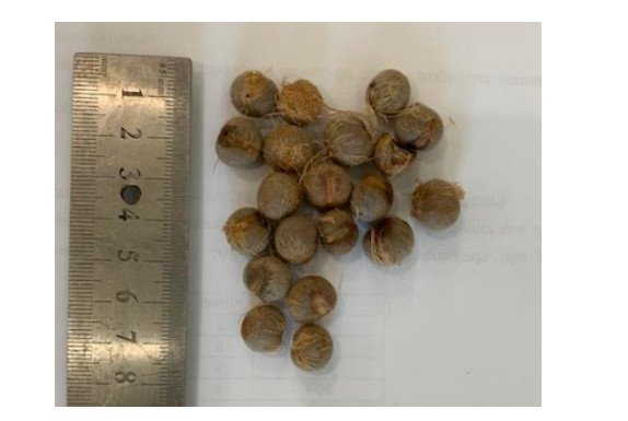
The açaizeiro is a palm tree present on a large scale in the northern region of Brazil and in others countries, such as Colombia and Peru, its fruit constitutes one of the main forest products of great economic potential to exportation. However, a generation of large amounts of waste during its processing represents a serious environmental problem, since about 365 tons of açaí stone are discarded in landfills daily in Brazil. The objective of this work was to evaluate the potential of using açaí stone as a substitute of 25% in mass natural sand with filler function, in the development of structural mortars with reference mixture of ratio 1:2:0.45 (cement:sand:water) and waste mixture with1:1.5:0.5:0.45 (cement:sand:açai stone:water), that both mixtures are relationship adopted for structural mortar in the literature. Three different types of mortar were made for evaluation, the reference (without adding stones) and with the addition of natural stone (without treatment and treated with NaOH). After the incorporation of the stones, consistency, water retention, incorporated air content and density in fresh mortars were analyzed, in the fresh state. For evaluation in a hardened state, cylindrical specimens (50 mm × 100 mm) were molded, for compression strength and density tests. The optimum composition was also analyzed with confocal microscopy. It can be seen that in the compression strength tests, mortars with the addition of natural and treated stone showed a reduction, decreasing from 6.25 MPa (reference), to 5.55 MPa (natural stone) and 1.89 MPa (treated stone), this showed that the natural stone mortar was above the minimum of 5.00 MPa reported in the literature. As for density, the evaluations demonstrate a beneficial effect to the incorporation of the stone, which formed lighter mortars, decreasing from 2.12 to 1.79 g/cm3 in the natural composition and 1.85 g/cm3 in the treated composition, in both situations with additions, the values were within the maximum limit of 2 g/cm3 that the literature suggests for structural mortars. In addition, the results of water retention showed an increase in the treated composition (97.28%) in relation to the reference (95.84%), an increase characterized by the treatment in NaOH that reduced the hygroscopic characteristics of the stones, the mixture mortar with treated seed, as well as the reference, presented values above 95%, which is recommended by other studies. The evaluations show that there is a potential for specific structural applications of these mortars, since in all tests the composition with natural stone is within the ideal parameters by Brazilian standard, in addition to helping to solve the environmental impacts caused by discard this waste. As it is a mortar with low compressive strength, its application is suggested for the purpose of repairing specific structural defects that arise in the molding stage of beams, slabs and columns, located in regions with low load demand.

| [1] | Lavorato VN, de Miranda DC, Isoldi MC, et al. (2021) Effects of aerobic exercise training and açai supplementation on cardiac structure and function in rats submitted to a high-fat diet. Food Res Int 141: 110168. |
| [2] |
Sato MK, de Lima HV, Noronha Costa A, et al. (2020) Biochar as a sustainable alternative to açaí waste disposal in Amazon, Brazil. Process Saf Environ 139: 36-46. doi: 10.1016/j.psep.2020.04.001

|
| [3] |
Song H, Shen X, Deng R, et al. (2021) Dietary anthocyanin-rich extract of açai protects from diet-induced obesity, liver steatosis, and insulin resistance with modulation of gut microbiota in mice. Nutrition 86: 111176. doi: 10.1016/j.nut.2021.111176
|
| [4] |
Melo PS, Selani MM, Gonçalves RH, et al. (2021) Açaí seeds: An unexplored agro-industrial residue as a potential source of lipids, fibers, and antioxidant phenolic compounds. Ind Crop Prod 161: 113204. doi: 10.1016/j.indcrop.2020.113204
|
| [5] |
Lee J (2019) Anthocyanins of açai products in the United States. NFS J 14-15: 14-21. doi: 10.1016/j.nfs.2019.05.001
|
| [6] |
de Azevedo AR, Marvila MT, Tayeh BA, et al. (2021) Technological performance of açaí natural fibre reinforced cement-based mortars. J Build Eng 33: 101675. doi: 10.1016/j.jobe.2020.101675
|
| [7] |
Marques EDS, Froder JG, Oliveira PRD, et al. (2019) Cytotoxic effects of Euterpe oleraceae fruit oil (açaí) in rat liver and thyroid tissues. Rev Bras Farmacogn 29: 54-61. doi: 10.1016/j.bjp.2018.12.001
|
| [8] |
Marvila MT, Azevedo ARG, Alexandre J, et al. (2020) Circular economy in cementitious ceramics: Replacement of hydrated lime with a stoichiometric balanced combination of clay and marble waste. Int J Appl Ceram Tec 18: 192-202. doi: 10.1111/ijac.13634
|
| [9] |
França BR, Azevedo ARG, Monteiro SN, et al. (2018) Durability of soil-cement blocks with the incorporation of limestone residues from the processing of marble. Mater Res 21: 1-6. doi: 10.1590/1980-5373-mr-2017-0837
|
| [10] | Deus DWP, Franco RS, Correia LS, et al. (2019) Concrete comparison analysis using acid resistance. Technical Scientific Congress of Engineering and Agronomy CONTECC. (In Portuguese) |
| [11] | Marvila MT, Azevedo ARG, Alexandre J, et al. (2020) Study of the compressive strength of mortars as a function of material composition, workability, and specimen geometry. Model Simul Eng 2020: 167610. |
| [12] |
Marvila MT, Azevedo ARG, Monteiro SN (2020) Verification of the application potential of the mathematical models of lyse, abrams and molinari in mortars based on cement and lime. J Mater Res Technol 9: 7327-7334. doi: 10.1016/j.jmrt.2020.04.077
|
| [13] | Marvila MT, Azevedo ARG, Cecchin D, et al. (2020) Durability of coating mortars containing açaí fibers. Case Stud Constr Mat 13: e00406. |
| [14] | Zhang K, Pan L, Li J, et al. (2021) What is the mechanism of the fiber effect on the rheological behavior of cement paste with polycarboxylate superplasticizer? Constr Build Mater 281: 122542. |
| [15] |
Barrios AM, Vega DF, Martínez PS, et al. (2021) Study of the properties of lime and cement mortars made from recycled ceramic aggregate and reinforced with fibers. J Build Eng 35: 102097. doi: 10.1016/j.jobe.2020.102097
|
| [16] |
Tripathi P, Gupta VK, Dixit A, et al. (2018) Development and characterization of low cost jute, bagasse and glass fiber reinforced advanced hybrid epoxy composites. AIMS Mater Sci 5: 320-337. doi: 10.3934/matersci.2018.2.320
|
| [17] | Barbosa AM, Rebelo VSM, Martorano LG, et al. (2019) Characterization of acai waste particles for civil construction use. Rev Mater 24. |
| [18] | Brazilian Association of Technical Standards (2016) Mortar for laying and coating walls and ceilings—Determination of consistency index, NBR 13276. (In Portuguese) |
| [19] | Brazilian Association of Technical Standards (2005) Mortar for laying and coating walls and ceilings—Determination of water retention, NBR 13277. (In Portuguese) |
| [20] | Brazilian Association of Technical Standards (2005) Mortar for laying and coating walls and ceilings—Determination of mass density and incorporated air content, NBR 13278. (In Portuguese) |
| [21] | Brazilian Association of Technical Standards (2018) Concrete—Compression test of cylindrical specimens, NBR 5739. (In Portuguese) |
| [22] | Brazilian Association of Technical Standards (2005) Mortar for laying and coating walls and ceilings—Determination of bulk density in hardened state, NBR 13280. (In Portuguese) |
| [23] |
Thomas C, de Brito J, Cimentada A, et al. (2020) Macro- and micro- properties of multi-recycled aggregate concrete. J Clean Prod 245: 118843. doi: 10.1016/j.jclepro.2019.118843
|
| [24] |
Hamdani, Rizal S, Riza M, et al. (2018) Mechanical properties of concrete containing beeswax/dammar gum as phase change material for thermal energy storage. AIMS Energy 6: 521-529. doi: 10.3934/energy.2018.3.521
|
| [25] |
Marvila MT, Alexandre J, Azevedo ARG, et al. (2019) Study on the replacement of the hydrated lime by kaolinitic clay in mortars. Adv Appl Ceram 118: 373-380. doi: 10.1080/17436753.2019.1595266
|
| [26] | Cheng W, Elliott JR, Hover KC (2019) High-volume carbon sequestration for controlled low-strength materials. ACI Mater J 116: 235-244. |
| [27] |
Azevedo AR, Marvila MT, Zanelato EB, et al. (2020) Development of mortar for laying and coating with pineapple fiber. Rev Bras Eng Agr Amb 24: 187-193. doi: 10.1590/1807-1929/agriambi.v24n3p187-193
|
| [28] |
Lertwattanaruk P, Suntijitto A (2015) Properties of natural fiber cement materials containing coconut coir and oil palm fibers for residential building applications. Constr Build Mater 94: 664-669. doi: 10.1016/j.conbuildmat.2015.07.154
|
| [29] |
Fediuk R, Mochalov A, Timokhin R (2018) Review of methods for activation of binder and concrete mixes. AIMS Mater Sci 5: 916-931. doi: 10.3934/matersci.2018.5.916
|
| [30] | Azevedo ARG, Alexandre J, Marvila MT, et al. (2019) Development of methodology for the characterization and incorporation of waste from the paper industry in cementitious materials, In: Li B, Li J, Ikhmayies S, et al., Characterization of Minerals, Metals, and Materials 2019, Springer, Cham, 2019: 583-590. |
| [31] | Zanelato E, Alexandre J, Azevedo A, et al. (2020) Evaluation of the incorporation of marble and granite residue in coating mortars, In: Li B, Baker SP, Zhai HZ, et al., Advances in Powder and Ceramic Materials Science, Springer, Cham, 101-108. |
| [32] |
Marvila MT, Alexandre J, de Azevedo AR, et al. (2019) Evaluation of the use of marble waste in hydrated lime cement mortar based. J Mater Cycles Waste 21: 1250-1261. doi: 10.1007/s10163-019-00878-6
|
| [33] |
Marvila MT, Azevedo ARG, Barroso LS, et al. (2020) Gypsum plaster using rock waste: A proposal to repair the renderings of historical buildings in Brazil. Constr Build Mater 250: 118786. doi: 10.1016/j.conbuildmat.2020.118786
|
| [34] |
de Azevedo ARG, Marvila MT, da Silva Barroso L, et al. (2019) Effect of granite residue incorporation on the behavior of mortars. Materials 12: 1449. doi: 10.3390/ma12091449
|
| [35] |
Ferrara G, Pepe M, Martinelli E, et al. (2019) Influence of an impregnation treatment on the morphology and mechanical behaviour of flax yarns embedded in hydraulic lime mortar. Fibers 7: 30. doi: 10.3390/fib7040030
|
| [36] |
Korniejenko K, Frączek E, Pytlak E, et al. (2016) Mechanical properties of geopolymer composites reinforced with natural fibers. Procedia Eng 151: 388-393. doi: 10.1016/j.proeng.2016.07.395
|
| [37] |
Alonso MM, Gismera S, Blanco MT, et al. (2017) Alkali-activated mortars: Workability and rheological behaviour. Constr Build Mater 145: 576-587. doi: 10.1016/j.conbuildmat.2017.04.020
|
| [38] |
Rajaei S, Shoaei P, Shariati M, et al. (2021) Rubberized alkali-activated slag mortar reinforced with polypropylene fibres for application in lightweight thermal insulating materials. Constr Build Mater 270: 121430. doi: 10.1016/j.conbuildmat.2020.121430
|
| [39] |
Movilla-Quesada D, Vega-Zamanillo Á, Castro-Fresno D, et al. (2015) Sustainability in construction works: Reuse of sludge from tunnel boring in lime mortars. Appl Clay Sci 114: 402-406. doi: 10.1016/j.clay.2015.05.019
|
| [40] |
Marvila MT, Alexandre J, Azevedo ARG, et al. (2019) Study on the replacement of the hydrated lime by kaolinitic clay in mortars. Adv Appl Ceram 118: 373-380. doi: 10.1080/17436753.2019.1595266
|
| [41] |
Kim MO, Lee HK, Kim HK (2021) Cost and environmental effects of ocean-borne plastic flakes in cement mortar considering equivalent-strength mix design. Constr Build Mater 291: 123267. doi: 10.1016/j.conbuildmat.2021.123267
|
| [42] |
Jin L, Yu H, Wang Z, et al. (2021) Effect of crack and damaged zone on chloride penetration in recycled aggregate concrete: A seven-phase mesoscale numerical method. Constr Build Mater 291: 123383. doi: 10.1016/j.conbuildmat.2021.123383
|


Figures(8)

Gabriel Pereira Monteiro, Afonso Rangel Garcez de Azevedo, Markssuel Teixeira Marvila. Effect of the addition of the natural and treated açaí stone in structural mortars[J]. AIMS Materials Science, 2021, 8(4): 608-621. doi: 10.3934/matersci.2021037


 DownLoad: