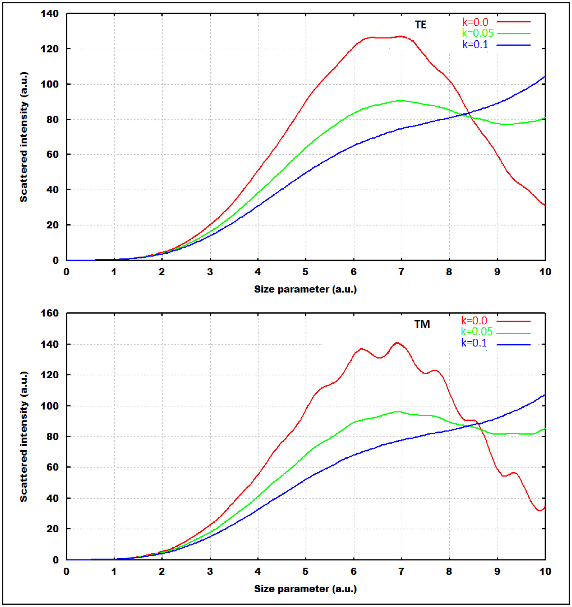
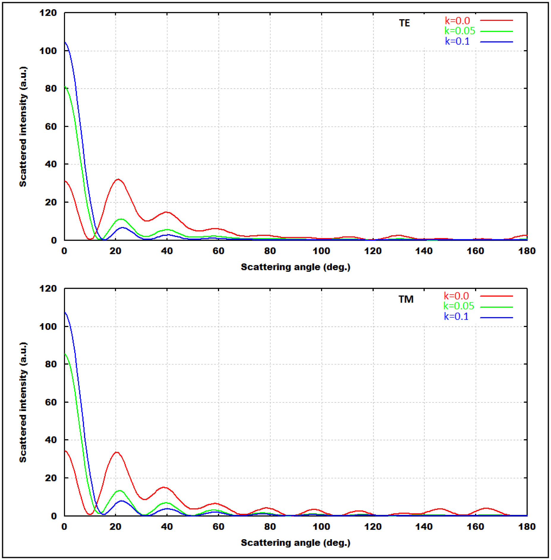
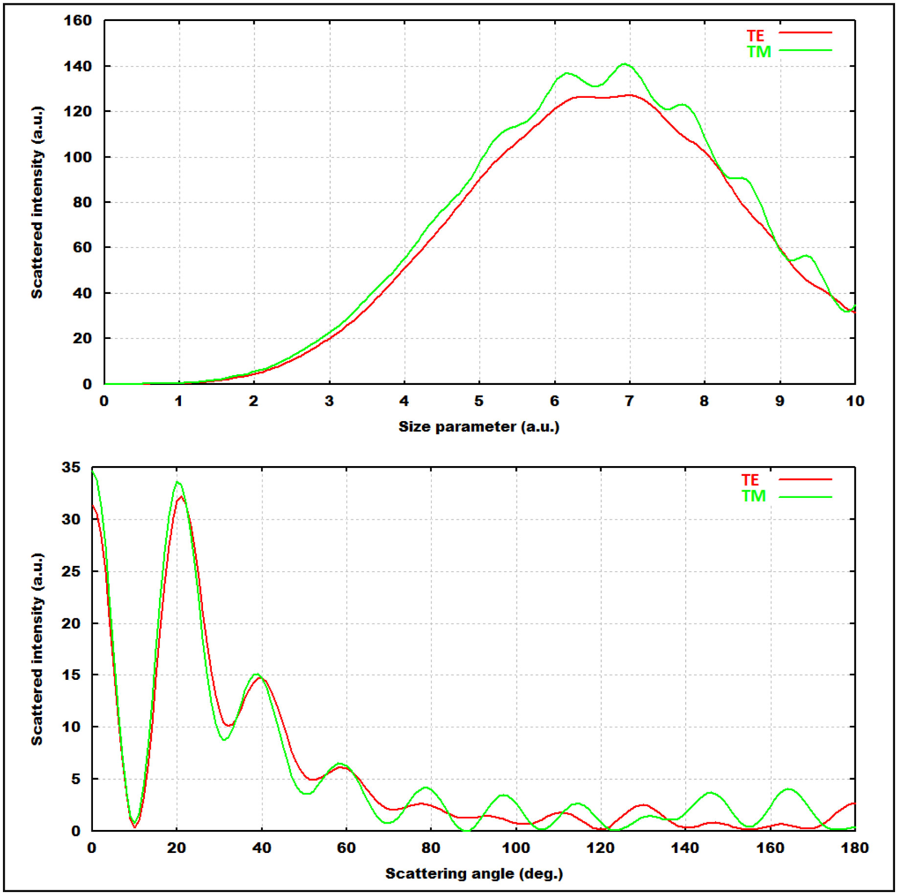
In diagnosing the urinary tract and related diseases, the problem of light scattering from human urine was examined on the basis of classical electromagnetic theory. Numerical calculations were made for the designed cylindrical model with the help of optical parameters in the literature obtained from the laboratory test results of urine samples. In the designed model, the changes of the scattered intensity of the light from the urine solution according to the size parameter of the particles and the angular distribution of the system (including forward, side and back scattering) in the equatorial plane were obtained, in both transverse electric (TE) and transverse magnetic (TM) of the polarization states of the light. It was observed that the molecular density changes caused by the materials in the urine sample changed primarily the optical parameters and indirectly the intensity distribution of the scattered light. Thus, with the contribution of standard data provided as a result of light scatter calculations from urine samples taken from people with normal and different diseases, it will be easier to diagnose diseases that will be encountered later.
Citation: Suleyman Yilmaz. Using elastic scattering to determination of diseases via urine samples[J]. AIMS Biophysics, 2021, 8(4): 307-317. doi: 10.3934/biophy.2021024
In diagnosing the urinary tract and related diseases, the problem of light scattering from human urine was examined on the basis of classical electromagnetic theory. Numerical calculations were made for the designed cylindrical model with the help of optical parameters in the literature obtained from the laboratory test results of urine samples. In the designed model, the changes of the scattered intensity of the light from the urine solution according to the size parameter of the particles and the angular distribution of the system (including forward, side and back scattering) in the equatorial plane were obtained, in both transverse electric (TE) and transverse magnetic (TM) of the polarization states of the light. It was observed that the molecular density changes caused by the materials in the urine sample changed primarily the optical parameters and indirectly the intensity distribution of the scattered light. Thus, with the contribution of standard data provided as a result of light scatter calculations from urine samples taken from people with normal and different diseases, it will be easier to diagnose diseases that will be encountered later.

| [1] |
Foxman B (2014) Urinary tract infection syndromes: occurrence, recurrence, bacteriology, risk factors, and disease burden. Infec Dis Clin N Am 28: 1-13. doi: 10.1016/j.idc.2013.09.003

|
| [2] |
Medina-Bombardó D, Jover-Palmer A (2011) Does clinical examination aid in the diagnosis of urinary tract infections in women? A systematic review and meta-analysis. BMC Fam Pract 12: 111. doi: 10.1186/1471-2296-12-111
|
| [3] |
Benramdane L, Bouatia M, Idrissi MOB, et al. (2008) Infrared analysis of urinary stones, using a single reflection accessory and a KBr pellet transmission. Spect Lett 41: 72-80. doi: 10.1080/00387010801943806
|
| [4] | Petrova EV, Gvozdev NV, Rashkovich LN (2004) Growth and dissolution of calcium oxalate monohydrate (COM) crystals. J Optoelectron Adv Mater 6: 261-268. |
| [5] |
Smith KF, Goldberg M, Rosenthal S, et al. (2014) Global rise in human infectious disease outbreaks. J R Soc Interface 11: 0950. doi: 10.1098/rsif.2014.0950
|
| [6] |
Locke A, Fitzgerald S, Mahadevan-Jansen A (2020) Advances in optical detection of human-associated pathogenic bacteria. Molecules 25: 5256. doi: 10.3390/molecules25225256
|
| [7] |
Newell DG, Koopmans M, Verhoef L, et al. (2010) Food-borne diseases—the challenges of 20 years ago still persist while new ones continue to emerge. Int J Food Microbiol 139: 3-15. doi: 10.1016/j.ijfoodmicro.2010.01.021
|
| [8] |
Bhunia AK (2014) One day to one hour: How quickly can foodborne pathogens be detected? Future Microbiol 9: 935-946. doi: 10.2217/fmb.14.61
|
| [9] |
Tuchin V (2015) Tissue optics: light scattering methods and instruments for medical diagnostics Bellingham: SPIE Press. doi: 10.1117/3.1003040
|
| [10] |
Prywer J, Kozanecki M, Mielniczek-Brzóska E, et al. (2018) Solid phases precipitating in artificial urine in the absence and presence of bacteria. Crystal 8: 164. doi: 10.3390/cryst8040164
|
| [11] |
Delanghe JR (2007) New screening diagnostic techniques in urinalysis. Acta Clin Belg 62: 155-161. doi: 10.1179/acb.2007.026
|
| [12] | Genuer V, Gal O, Méteau J, et al. (2016) Optical elastic scattering for early label-free identification of clinical pathogens, advanced biomedical and clinical diagnostic and surgical guidance systems. International Society for Optics and Photonics . |
| [13] |
Moshaver B, de Boer F, van Egmond-Kreileman H, et al. (2016) Fast and accurate prediction of positive and negative urine cultures by flow cytometry. BMC Infect Dis 16: 21. doi: 10.1186/s12879-016-1557-4
|
| [14] |
Gardiner C, Shaw M, Hole P, et al. (2014) Measurement of refractive index by nanoparticle tracking analysis reveals heterogeneity in extracellular vesicles. J Extra Vesic 3: 25361. doi: 10.3402/jev.v3.25361
|
| [15] |
Buzás EI, Gardiner C, Lee C, et al. (2016) Single particle analysis: Methods for detection of platelet extracellular vesicles in suspension (excluding flow cytometry). Platelets 28: 249-255. doi: 10.1080/09537104.2016.1260704
|
| [16] | Barber PW, Hill SC (1990) Light scattering by particles: computational methods. World Scientific Singapore: 276. |
| [17] | Sheng P (1995) Introduction to wave scattering, localization, and mesoscopic phenomena San Diego: Academic Press, 45-74. |
| [18] | Bohren CF, Huffman DR (1983) Absorption and scattering of the light by small articles. John Wiley & Sons, New York . |
| [19] | Barber PW, Huffman DR (1990) Light scattering by small particles computational methods. World Scientific Singapore: 25-77. |
| [20] |
van de Hulst HC (1957) Light scattering by small particles. Phys Today 10: 28. doi: 10.1063/1.3060205
|
| [21] |
Hammer M, Schweitzer D, Michel B, et al. (1998) Single scattering by red blood cells. Appl Opt 37: 7410-7418. doi: 10.1364/AO.37.007410
|
| [22] |
Chance B (1991) Optical method. Annu Rev Biophys Biophys Chem 20: 1-28. doi: 10.1146/annurev.bb.20.060191.000245
|
| [23] |
Yodh AG, Chance B (1995) Spectroscopy and imaging with diffusing light. Phys Today 48: 34-40. doi: 10.1063/1.881445
|
| [24] | Backman V (1998) Early diagnosis of cancer using light scattering spectroscopy. Mass Inst Technol . |
| [25] |
Yilmaz S, Kalkandelen A, Mamedova NI (2006) Comparison of the T-matrix calculations for erythrocytes with oxygenated hemoglobin and immersed in water. J Quant Spectrosc Radiat Transfer 97: 20-28. doi: 10.1016/j.jqsrt.2004.12.031
|
| [26] |
Yilmaz S, Koc H (2005) Light scattering by DNA structure due to phase transition. Mol Phys 103: 2633-2637. doi: 10.1080/00268970500229753
|
| [27] |
Mishchenko MI, Travis LD, Macke A (1996) Scattering of light by polydisperse, randomly oriented, finite circular cylinders. Appl Opt 35: 4927-4940. doi: 10.1364/AO.35.004927
|
| [28] |
Plummer DT, Leathwood PD (1967) Some properties of lactate dehydrogenase found in human urine. J Biochem 103: 172-176. doi: 10.1042/bj1030172
|
| [29] |
Wyness-Sara P, Hunsaker-Joshua JH, Snow-Taylor M, et al. (2016) Evaluation and analytical validation of a handheld digital refractometer for urine specific gravity measurement. Pract Lab Med 5: 65-74. doi: 10.1016/j.plabm.2016.06.001
|
| [30] |
Lonappan A, Hamsakkutty V, Bindu G, et al. (2004) Dielectric properties of human urine at microwave frequencies. Microw Opt Techn Lett 42: 500-503. doi: 10.1002/mop.20349
|
| [31] |
Plotnikova L, Polyanichko A, Kobeleva M, et al. (2017) The Influence of protein fractions and electrolyte imbalance on refractive index of serum in patenti with multiple myeloma. J phys conf ser 784: 012047. doi: 10.1088/1742-6596/784/1/012047
|
| [32] | Rao SV (2010) Study of multimode fiber optic sensor. PhD thesis of Jawaharlal Nehru Technological University . |
| [33] | Guo S (2004) Optical scattering for bacterial colony detection and characterization. Purdue University . |
| [34] |
Schmitt JM, Kumar G (1998) Optical scattering properties of soft tissue: a discrete particle model. Appl Opt 37: 2788-2797. doi: 10.1364/AO.37.002788
|
| [35] |
Dunn A, Kortum-Richards R (1996) Three-dimensional computation of light scattering from cells. IEEE J Sel Top Quantum Electron 2: 898-905. doi: 10.1109/2944.577313
|
| [36] |
Travis LD, Mishchenco MI (1994) Light scattering by polydispersions of randomly oriented spheroids with sizes comparable to wavelengths of observation. App Opt 33: 7206. doi: 10.1364/AO.33.007206
|
| [37] |
Videen G, Ngo D (1997) Light scattering from a cylinder near a plane interface: theory and comparison with experimental data. J Opt Soc Am A 14: 70. doi: 10.1364/JOSAA.14.000070
|
| [38] | van de Hulst HC (1981) Light scattering by small particles New York: Dover Publications, Inc. |
| [39] |
Yilmaz S (2012) Determination of the structural properties of a columnar hexagonal liquid crystal by light scattering. J Mod Opt 59: 912-916. doi: 10.1080/09500340.2012.676093
|
Figures(3)

Suleyman Yilmaz. Using elastic scattering to determination of diseases via urine samples[J]. AIMS Biophysics, 2021, 8(4): 307-317. doi: 10.3934/biophy.2021024


 DownLoad: