At present, radiotherapy (RT) is widely used in cancer treatment, but traditional RT methods using ionizing radiation cannot avoid damage to normal tissues. Therefore, the development of a more precise RT is an important research direction for relevant researchers. Concurrently, research on radiosensitizers (RSs) using nanotechnology is developing rapidly, and RSs that are selective for cancerous tissues or cancer cells may become an important part of future precision RT.
Using RSs and RT as keywords, the relevant papers in the PubMed database from 2013 to 2022 were summarized. Articles on RS with selectivity to cancer tissue were collected. Among the selected articles, RSs were classified into “active selectivity”, “passive selectivity” and “others” according to the different selectivity principles of RSs.
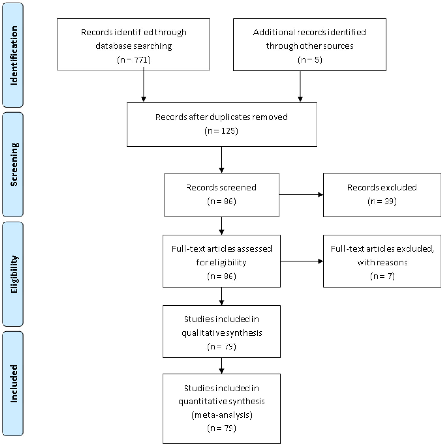
A total of 771 articles were retrieved from PubMed. After screening, the research content of the remaining 79 articles was found to be related to the selectivity of RSs to cancer tissues. Among them, 28 articles were classified as “active selectivity”, and most of the sensitizers in this category could target specific targets in cancer tissues. There were 30 papers classified as “passive selectivity” and the selectivity principles were mainly the enhanced permeability and retention (EPR) effect, aggregation caused by pH sensitivity, and aggregation in anoxic environments. There were 21 papers classified as “others”. The sensitizers in these studies showed selectivity for cancer tissue, but the mechanism was not clear. This review attempts to summarize studies on RSs that are selective for cancer tissues.
We reviewed nearly ten years of literature on selective RSs and classified the selectivity of different RSs into active and passive selectivities.
Citation: Hengmao Zhang, Haobo Zhao, Ming Chi, Kaizhen Yang, Yukang Chen, Jiahui Mao, Peilin Li, Zukang Wang, Faqiao Song, Wenxuan Guo, Miyu Sakai, Junko Takahashi. A systematic review on the development of radiosensitizers, with cancer selectivity, for radiotherapy using ionizing radiation[J]. AIMS Bioengineering, 2023, 10(2): 89-110. doi: 10.3934/bioeng.2023008
At present, radiotherapy (RT) is widely used in cancer treatment, but traditional RT methods using ionizing radiation cannot avoid damage to normal tissues. Therefore, the development of a more precise RT is an important research direction for relevant researchers. Concurrently, research on radiosensitizers (RSs) using nanotechnology is developing rapidly, and RSs that are selective for cancerous tissues or cancer cells may become an important part of future precision RT.
Using RSs and RT as keywords, the relevant papers in the PubMed database from 2013 to 2022 were summarized. Articles on RS with selectivity to cancer tissue were collected. Among the selected articles, RSs were classified into “active selectivity”, “passive selectivity” and “others” according to the different selectivity principles of RSs.
A total of 771 articles were retrieved from PubMed. After screening, the research content of the remaining 79 articles was found to be related to the selectivity of RSs to cancer tissues. Among them, 28 articles were classified as “active selectivity”, and most of the sensitizers in this category could target specific targets in cancer tissues. There were 30 papers classified as “passive selectivity” and the selectivity principles were mainly the enhanced permeability and retention (EPR) effect, aggregation caused by pH sensitivity, and aggregation in anoxic environments. There were 21 papers classified as “others”. The sensitizers in these studies showed selectivity for cancer tissue, but the mechanism was not clear. This review attempts to summarize studies on RSs that are selective for cancer tissues.
We reviewed nearly ten years of literature on selective RSs and classified the selectivity of different RSs into active and passive selectivities.

| [1] |
Baumann M, Krause M, Overgaard J, et al. (2016) Radiation oncology in the era of precision medicine. Nat Rev Cancer 16: 234-249. https://doi.org/10.1038/nrc.2016.18 
|
| [2] |
Komorowska D, Radzik T, Kalenik S, et al. (2022) Natural radiosensitizers in radiotherapy: cancer treatment by combining ionizing radiation with resveratrol. Int J Mol Sci 23: 10627. https://doi.org/10.3390/ijms231810627
|
| [3] |
Zhang J, Liu Y, Wang X, et al. (2020) Nanozyme-incorporated biodegradable bismuth mesoporous radiosensitizer for tumor microenvironment-modulated hypoxic tumor thermoradiotherapy. ACS Appl Mater Interfaces 12: 57768-57781. https://doi.org/10.1021/acsami.0c18853
|
| [4] |
Zhang H, Zhang W, Zhou Y, et al. (2017) Dual functional mesoporous silicon nanoparticles enhance the radiosensitivity of VPA in glioblastoma. Transl Oncol 10: 229-240. https://doi.org/10.1016/j.tranon.2016.12.011
|
| [5] |
Huang Y, Luo Y, Zheng W, et al. (2014) Rational design of cancer-targeted BSA protein nanoparticles as radiosensitizer to overcome cancer radioresistance. ACS Appl Mater Interfaces 6: 19217-19228. https://doi.org/10.1021/am505246w
|
| [6] |
Mirrahimi M, Hosseini V, Shakeri-Zadeh A, et al. (2019) Modulation of cancer cells' radiation response in the presence of folate conjugated Au@Fe2O3 nanocomplex as a targeted radiosensitizer. Clin Transl Oncol 21: 479-488. https://doi.org/10.1007/s12094-018-1947-8
|
| [7] |
Nosrati H, Charmi J, Salehiabar M, et al. (2019) Tumor targeted albumin coated bismuth sulfide nanoparticles (Bi2S3) as radiosensitizers and carriers of curcumin for enhanced chemoradiation therapy. ACS Biomater Sci Eng 5: 4416-4424. https://doi.org/10.1021/acsbiomaterials.9b00489
|
| [8] |
Cho WJ, Kessel D, Rakowski J, et al. (2021) Photodynamic therapy as a potent radiosensitizer in head and neck squamous cell carcinoma. Cancers 13: 1193. https://doi.org/10.3390/cancers13061193
|
| [9] |
Wang L, Zhang T, Huo M, et al. (2019) Construction of nucleus-targeting iridium nanocrystals for photonic hyperthermia-synergized cancer radiotherapy. Small 15: e1903254. https://doi.org/10.1002/smll.201903254
|
| [10] |
Enferadi M, Fu SY, Hong JH, et al. (2018) Radiosensitization of ultrasmall GNP-PEG-cRGDfK in ALTS1C1 exposed to therapeutic protons and kilovoltage and megavoltage photons. Int J Radiat Biol 94: 124-136. https://doi.org/10.1080/09553002.2018.1407462
|
| [11] |
Wang J, Pang X, Tan X, et al. (2017) A triple-synergistic strategy for combinational photo/radiotherapy and multi-modality imaging based on hyaluronic acid-hybridized polyaniline-coated WS2 nanodots. Nanoscale 9: 5551-5564. https://doi.org/10.1039/c6nr09219e
|
| [12] |
Liu J, Yang Y, Zhu W, et al. (2016) Nanoscale metal-organic frameworks for combined photodynamic & radiation therapy in cancer treatment. Biomaterials 97: 1-9. https://doi.org/10.1016/j.biomaterials.2016.04.034
|
| [13] |
Mayahi S, Neshasteh-Riz A, Pornour M, et al. (2020) Investigation of combined photodynamic and radiotherapy effects of gallium phthalocyanine chloride on MCF-7 breast cancer cells. J Biol Inorg Chem 25: 39-48. https://doi.org/10.1007/s00775-019-01730-w
|
| [14] |
Sazgarnia A, Montazerabadi AR, Bahreyni-Toosi MH, et al. (2013) In vitro survival of MCF-7 breast cancer cells following combined treatment with ionizing radiation and mitoxantrone-mediated photodynamic therapy. Photodiagnosis Photodyn Ther 10: 72-78. https://doi.org/10.1016/j.pdpdt.2012.06.001
|
| [15] |
Miyake M, Tanaka N, Hori S, et al. (2019) Dual benefit of supplementary oral 5-aminolevulinic acid to pelvic radiotherapy in a syngenic prostate cancer model. Prostate 79: 340-351. https://doi.org/10.1002/pros.23740
|
| [16] |
Takahashi J, Misawa M, Iwahashi H (2016) Combined treatment with X-ray irradiation and 5-aminolevulinic acid elicits better transcriptomic response of cell cycle-related factors than X-ray irradiation alone. Int J Radiat Biol 92: 774-789. https://doi.org/10.1080/09553002.2016.1230240
|
| [17] |
Laird JH, Lok BH, Ma J, et al. (2018) Talazoparib is a potent radiosensitizer in small cell lung cancer cell lines and xenografts. Clin Cancer Res 24: 5143-5152. https://doi.org/10.1158/1078-0432.CCR-18-0401
|
| [18] |
Li D, Zhao J, Ma J, et al. (2022) GMT8 aptamer conjugated PEGylated Ag@Au core-shell nanoparticles as a novel radiosensitizer for targeted radiotherapy of glioma. Colloids Surf B Biointerfaces 211: 112330. https://doi.org/10.1016/j.colsurfb.2022.112330
|
| [19] |
Jiao X, Yu Y, Meng J, et al. (2019) Dual-targeting and microenvironment-responsive micelles as a gene delivery system to improve the sensitivity of glioma to radiotherapy. Acta Pharm Sin B 9: 381-396. https://doi.org/10.1016/j.apsb.2018.12.001
|
| [20] |
Park M, Kwon J, Shin HJ, et al. (2020) Butyrate enhances the efficacy of radiotherapy via FOXO3A in colorectal cancer patient‑derived organoids. Int J Oncol 57: 1307-1318. https://doi.org/10.3892/ijo.2020.5132
|
| [21] |
Pan W, Gong S, Wang J, et al. (2019) A nuclear-targeted titanium dioxide radiosensitizer for cell cycle regulation and enhanced radiotherapy. Chem Commun 55: 8182-8185. https://doi.org/10.1039/c9cc01651a
|
| [22] | Ji F, Sha H, Meng F, et al. (2018) Tumor‑penetrating peptide fused EGFR single‑domain antibody enhances radiation responses following EGFR inhibition in gastric cancer. Oncol Rep 40: 1583-1591. https://doi.org/10.3892/or.2018.6532 |
| [23] |
Zeng D, Deng S, Sang C, et al. (2018) Rational design of cancer-targeted selenadiazole derivative as efficient radiosensitizer for precise cancer therapy. Bioconjug Chem 29: 2039-2049. https://doi.org/10.1021/acs.bioconjchem.8b00247
|
| [24] |
Bromma K, Chithrani DB (2020) Advances in gold nanoparticle-based combined cancer therapy. Nanomaterials 10: 1671. https://doi.org/10.3390/nano10091671
|
| [25] |
Koo T, Kim IA (2016) Brain metastasis in human epidermal growth factor receptor 2-positive breast cancer: from biology to treatment. Radiat Oncol J 34: 1-9. https://doi.org/10.3857/roj.2016.34.1.1
|
| [26] |
Nambiar DK, Rajamani P, Deep G, et al. (2015) Silibinin preferentially radiosensitizes prostate cancer by inhibiting DNA repair signaling. Mol Cancer Ther 14: 2722-2734. https://doi.org/10.1158/1535-7163.MCT-15-0348
|
| [27] |
Reda M, Ngamcherdtrakul W, Gu S, et al. (2019) PLK1 and EGFR targeted nanoparticle as a radiation sensitizer for non-small cell lung cancer. Cancer Lett 467: 9-18. https://doi.org/10.1016/j.canlet.2019.09.014
|
| [28] |
Guo XX, Guo ZH, Lu JS, et al. (2021) All-purpose nanostrategy based on dose deposition enhancement, cell cycle arrest, DNA damage, and ROS production as prostate cancer radiosensitizer for potential clinical translation. Nanoscale 13: 14525-14537. https://doi.org/10.1039/d1nr03869a
|
| [29] |
Luo D, Johnson A, Wang X, et al. (2020) Targeted radiosensitizers for MR-guided radiation therapy of prostate cancer. Nano Lett 20: 7159-7167. https://doi.org/10.1021/acs.nanolett.0c02487
|
| [30] |
Luo D, Wang X, Zeng S, et al. (2019) Targeted gold nanocluster-enhanced radiotherapy of prostate cancer. Small 15: e1900968. https://doi.org/10.1002/smll.201900968
|
| [31] |
Maeda H, Wu J, Sawa T, et al. (2000) Tumor vascular permeability and the EPR effect in macromolecular therapeutics: a review. J Control Release 65: 271-284. https://doi.org/10.1016/s0168-3659(99)00248-5
|
| [32] |
Hua S, He J, Zhang F, et al. (2021) Multistage-responsive clustered nanosystem to improve tumor accumulation and penetration for photothermal/enhanced radiation synergistic therapy. Biomaterials 268: 120590. https://doi.org/10.1016/j.biomaterials.2020.120590
|
| [33] |
Hatoyama K, Kitamura N, Takano-Kasuya M, et al. (2019) Quantitative analyses of amount and localization of radiosensitizer gold nanoparticles interacting with cancer cells to optimize radiation therapy. Biochem Biophys Res Commun 508: 1093-1100. https://doi.org/10.1016/j.bbrc.2018.12.016
|
| [34] |
Xu X, Chong Y, Liu X, et al. (2019) Multifunctional nanotheranostic gold nanocages for photoacoustic imaging guided radio/photodynamic/photothermal synergistic therapy. Acta Biomater 84: 328-338. https://doi.org/10.1016/j.actbio.2018.11.043
|
| [35] |
Cheng K, Sano M, Jenkins CH, et al. (2018) Synergistically enhancing the therapeutic effect of radiation therapy with radiation activatable and reactive oxygen species-releasing nanostructures. ACS Nano 12: 4946-4958. https://doi.org/10.1021/acsnano.8b02038
|
| [36] |
Zhang XD, Chen J, Min Y, et al. (2014) Metabolizable Bi2Se3 nanoplates: biodistribution, toxicity, and uses for cancer radiation therapy and imaging. Adv Funct Mater 24: 1718-1729. https://doi.org/10.1002/adfm.201302312
|
| [37] |
Zhang XD, Chen J, Luo Z, et al. (2014) Enhanced tumor accumulation of sub-2 nm gold nanoclusters for cancer radiation therapy. Adv Healthc Mater 3: 133-141. https://doi.org/10.1002/adhm.201300189
|
| [38] |
Feng L, Dong Z, Liang C, et al. (2018) Iridium nanocrystals encapsulated liposomes as near-infrared light controllable nanozymes for enhanced cancer radiotherapy. Biomaterials 181: 81-91. https://doi.org/10.1016/j.biomaterials.2018.07.049
|
| [39] |
Hua Y, Wang Y, Kang X, et al. (2021) A multifunctional AIE gold cluster-based theranostic system: tumor-targeted imaging and Fenton reaction-assisted enhanced radiotherapy. J Nanobiotechnol 19: 438. https://doi.org/10.1186/s12951-021-01191-x
|
| [40] |
Gao S, Zhang W, Wang R, et al. (2020) Nanoparticles encapsulating nitrosylated maytansine to enhance radiation therapy. ACS Nano 14: 1468-1481. https://doi.org/10.1021/acsnano.9b05976
|
| [41] |
Jung J, Jeong SY, Park SS, et al. (2015) A cisplatin‑incorporated liposome that targets the epidermal growth factor receptor enhances radiotherapeutic efficacy without nephrotoxicity. Int J Oncol 46: 1268-1274. https://doi.org/10.3892/ijo.2014.2806
|
| [42] |
Liu H, Xie Y, Zhang Y, et al. (2017) Development of a hypoxia-triggered and hypoxic radiosensitized liposome as a doxorubicin carrier to promote synergetic chemo-/radiotherapy for glioma. Biomaterials 121: 130-143. https://doi.org/10.1016/j.biomaterials.2017.01.001
|
| [43] |
Menon JU, Kuriakose A, Iyer R, et al. (2017) Dual-drug containing core-shell nanoparticles for lung cancer therapy. Sci Rep 7: 13249. https://doi.org/10.1038/s41598-017-13320-4
|
| [44] |
Zhang Z, Niu X, Feng X, et al. (2021) Construction of a pH/TGase “dual key”-responsive gold nano-radiosensitizer with liver tumor-targeting ability. ACS Biomater Sci Eng 7: 3434-3445. https://doi.org/10.1021/acsbiomaterials.1c00428
|
| [45] |
Lv S, Long W, Chen J, et al. (2020) Dual pH-triggered catalytic selective Mn clusters for cancer radiosensitization and radioprotection. Nanoscale 12: 548-557. https://doi.org/10.1039/c9nr08192e
|
| [46] |
Bonnet M, Hong CR, Wong WW, et al. (2018) Next-generation hypoxic cell radiosensitizers: nitroimidazole alkylsulfonamides. J Med Chem 61: 1241-1254. https://doi.org/10.1021/acs.jmedchem.7b01678
|
| [47] |
Kouhsari E, Ghadimi-Daresajini A, Abdollahi H, et al. (2018) The potential roles of bacteria to improve radiation treatment outcome. Clin Transl Oncol 20: 127-139. https://doi.org/10.1007/s12094-017-1701-7
|
| [48] |
Yang S, Han G, Chen Q, et al. (2021) Au-Pt nanoparticle formulation as a radiosensitizer for radiotherapy with dual effects. Int J Nanomedicine 16: 239-248. https://doi.org/10.2147/IJN.S287523
|
| [49] |
Chen J, Chen Q, Liang C, et al. (2017) Albumin-templated biomineralizing growth of composite nanoparticles as smart nano-theranostics for enhanced radiotherapy of tumors. Nanoscale 9: 14826-14835. https://doi.org/10.1039/c7nr05316a
|
| [50] |
Ma X, Lee C, Zhang T, et al. (2021) Image-guided selection of Gd@C-dots as sensitizers to improve radiotherapy of non-small cell lung cancer. J Nanobiotechnol 19: 284. https://doi.org/10.1186/s12951-021-01018-9
|
| [51] |
Wang H, Mu X, He H, Zhang XD (2018) Cancer Radiosensitizers. Trends Pharmacol Sci 39: 24-48. https://doi.org/10.1016/j.tips.2017.11.003
|
| [52] |
Kefayat A, Ghahremani F, Motaghi H, et al. (2019) Ultra-small but ultra-effective: Folic acid-targeted gold nanoclusters for enhancement of intracranial glioma tumors' radiation therapy efficacy. Nanomedicine 16: 173-184. https://doi.org/10.1016/j.nano.2018.12.007
|
| [53] | Dorsey JF, Sun L, Joh DY, et al. (2013) Gold nanoparticles in radiation research: potential applications for imaging and radiosensitization. Transl Cancer Res 2: 280-291. https://doi.org/10.3978/j.issn.2218-676X.2013.08.09 |
| [54] |
Besse HC, Bos C, Zandvliet MMJM, et al. (2018) Triggered radiosensitizer delivery using thermosensitive liposomes and hyperthermia improves efficacy of radiotherapy: An in vitro proof of concept study. PLoS One 13: e0204063. https://doi.org/10.1371/journal.pone.0204063
|
| [55] |
Tung FI, Zheng LJ, Hou KT, et al. (2020) One-stop radiotherapeutic targeting of primary and distant osteosarcoma to inhibit cancer progression and metastasis using 2DG-grafted graphene quantum dots. Nanoscale 12: 8809-8818. https://doi.org/10.1039/c9nr10823h
|
| [56] |
Zhao L, Qiu G, Wang K, et al. (2020) A nano-integrated diagnostic and therapeutic platform with oxidation-reduction reactions in tumor microenvironments. Nanoscale Adv 2: 2192-2202. https://doi.org/10.1039/c9na00786e
|
| [57] |
Mehdi Z, Petronek MS, Stolwijk JM, et al. (2021) Utilization of pharmacological ascorbate to enhance hydrogen peroxide-mediated radiosensitivity in cancer therapy. Int J Mol Sci 22: 10880. https://doi.org/10.3390/ijms221910880
|
| [58] |
Pan P, Dong X, Chen Y, et al. (2022) Engineered bacteria for enhanced radiotherapy against breast carcinoma. ACS Nano 16: 801-812. https://doi.org/10.1021/acsnano.1c08350
|
| [59] |
Hou X, Chang YX, Yue YX, et al. (2022) Supramolecular radiosensitizer based on hypoxia-responsive macrocycle. Adv Sci 9: e2104349. https://doi.org/10.1002/advs.202104349
|
| [60] |
Nandy P, Mukherjee A, Pradhan C, et al. (2020) Radio-sensitizing effects of CuII and ZnII complexes of ornidazole: role of nitro radical anion. ACS Omega 5: 25668-25676. https://doi.org/10.1021/acsomega.0c02811
|
| [61] |
Minea RO, Duc TC, Swenson SD, et al. (2020) Developing a clinically relevant radiosensitizer for temozolomide-resistant gliomas. PLoS One 15: e0238238. https://doi.org/10.1371/journal.pone.0238238
|
| [62] |
Chastagner P, Sudour H, Mriouah J, et al. (2015) Preclinical studies of pegylated- and non-pegylated liposomal forms of doxorubicin as radiosensitizer on orthotopic high-grade glioma xenografts. Pharm Res 32: 158-166. https://doi.org/10.1007/s11095-014-1452-x
|
| [63] |
Guo Y, Zhai J, Zhang J, et al. (2019) Improved radiotherapy sensitivity of nasopharyngeal carcinoma cells by miR-29-3p targeting COL1A1 3′-UTR. Med Sci Monit 25: 3161-3169. https://doi.org/10.12659/MSM.915624
|
| [64] |
Li H, Jiang M, Cui M, et al. (2019) MiR-365 enhances the radiosensitivity of non-small cell lung cancer cells through targeting CDC25A. Biochem Biophys Res Commun 512: 392-398. https://doi.org/10.1016/j.bbrc.2019.03.082
|
| [65] |
Wang S, Pan Y, Zhang R, et al. (2016) Hsa-miR-24-3p increases nasopharyngeal carcinoma radiosensitivity by targeting both the 3′UTR and 5′UTR of Jab1/CSN5. Oncogene 35: 6096-6108. https://doi.org/10.1038/onc.2016.147
|
| [66] |
Mekkawy MH, Fahmy HA, Nada AS, et al. (2020) Study of the radiosensitizing and radioprotective efficacy of bromelain (a pineapple extract): in vitro and in vivo. Integr Cancer Ther 19: 1534735420950468. https://doi.org/10.1177/1534735420950468
|
| [67] |
Ahire V, Kumar A, Mishra KP, et al. (2017) Ellagic acid enhances apoptotic sensitivity of breast cancer cells to γ-Radiation. Nutr Cancer 69: 904-910. https://doi.org/10.1080/01635581.2017.1339811
|
| [68] |
Yi J, Zhu J, Zhao C, et al. (2021) Potential of natural products as radioprotectors and radiosensitizers: opportunities and challenges. Food Funct 12: 5204-5218. https://doi.org/10.1039/d1fo00525a
|
| [69] |
J Jayakumar S, Patwardhan RS, Pal D, et al. (2016) Dimethoxycurcumin, a metabolically stable analogue of curcumin enhances the radiosensitivity of cancer cells: Possible involvement of ROS and thioredoxin reductase. Biochem Biophys Res Commun 478: 446-454. https://doi.org/10.1016/j.bbrc.2016.06.144
|
| [70] |
Luo H, Wang L, Schulte BA, et al. (2013) Resveratrol enhances ionizing radiation-induced premature senescence in lung cancer cells. Int J Oncol 43: 1999-2006. https://doi.org/10.3892/ijo.2013.2141
|
| [71] |
Meza-Menchaca T, Poblete-Naredo I, Albores-Medina A, et al. (2020) Ergosterol peroxide isolated from oyster medicinal mushroom, pleurotus ostreatus (agaricomycetes), potentially induces radiosensitivity in cervical cancer. Int J Med Mushrooms 22: 1109-1119. https://doi.org/10.1615/IntJMedMushrooms.2020036673
|
| [72] |
Liu X, Sun C, Liu B, et al. (2016) Genistein mediates the selective radiosensitizing effect in NSCLC A549 cells via inhibiting methylation of the keap1 gene promoter region. Oncotarget 7: 27267-27279. https://doi.org/10.18632/oncotarget.8403
|
| [73] |
Lin C, Yu Y, Zhao HG, et al. (2012) Combination of quercetin with radiotherapy enhances tumor radiosensitivity in vitro and in vivo. Radiother Oncol 104: 395-400. https://doi.org/10.1016/j.radonc.2011.10.023
|
| [74] | Si LB, Zhang MZ, Han Q, et al. (2018) Sensitization of keloid fibroblasts by quercetin through the PI3K/Akt pathway is dependent on regulation of HIF-1α. Am J Transl Res 10: 4223-4234. https://doi.org/10.4172/2155-9554.1000409 |
| [75] |
Yang YP, Chang YL, Huang PI, et al. (2012) Resveratrol suppresses tumorigenicity and enhances radiosensitivity in primary glioblastoma tumor initiating cells by inhibiting the STAT3 axis. J Cell Physiol 227: 976-993. https://doi.org/10.1002/jcp.22806
|
| [76] |
Cho HJ, Ahn KC, Choi JY, et al. (2015) Luteolin acts as a radiosensitizer in non-small cell lung cancer cells by enhancing apoptotic cell death through activation of a p38/ROS/caspase cascade. Int J Oncol 46: 1149-1158. https://doi.org/10.3892/ijo.2015.2831
|
| [77] |
Veeraraghavan J, Natarajan M, Lagisetty P, et al. (2011) Impact of curcumin, raspberry extract, and neem leaf extract on rel protein-regulated cell death/radiosensitization in pancreatic cancer cells. Pancreas 40: 1107-1119. https://doi.org/10.1097/MPA.0b013e31821f677d
|
| [78] |
Zhang G, Wang Y, Zhang Y, et al. (2012) Anti-cancer activities of tea epigallocatechin-3-gallate in breast cancer patients under radiotherapy. Curr Mol Med 12: 163-176. https://doi.org/10.2174/156652412798889063
|
| [79] |
Choi C, Cho Y, Son A, et al. (2020) Therapeutic potential of (-)-agelamide d, a diterpene alkaloid from the marine sponge Agelas sp., as a natural radiosensitizer in hepatocellular carcinoma models. Mar Drugs 18: 500. https://doi.org/10.3390/md18100500
|
| [80] |
Kang Q, Zhang X, Cao N, et al. (2019) EGCG enhances cancer cells sensitivity under 60Coγ radiation based on miR-34a/Sirt1/p53. Food Chem Toxicol 133: 110807. https://doi.org/10.1016/j.fct.2019.110807
|
| [81] |
Askar MA, Thabet NM, El-Sayyad GS, et al. (2021) Dual hyaluronic acid and folic acid targeting pH-sensitive multifunctional 2DG@DCA@MgO-nano-core-shell-radiosensitizer for breast cancer therapy. Cancers 13: 5571. https://doi.org/10.3390/cancers13215571
|
Figures(1) / Tables(3)

Hengmao Zhang, Haobo Zhao, Ming Chi, Kaizhen Yang, Yukang Chen, Jiahui Mao, Peilin Li, Zukang Wang, Faqiao Song, Wenxuan Guo, Miyu Sakai, Junko Takahashi. A systematic review on the development of radiosensitizers, with cancer selectivity, for radiotherapy using ionizing radiation[J]. AIMS Bioengineering, 2023, 10(2): 89-110. doi: 10.3934/bioeng.2023008


 DownLoad: