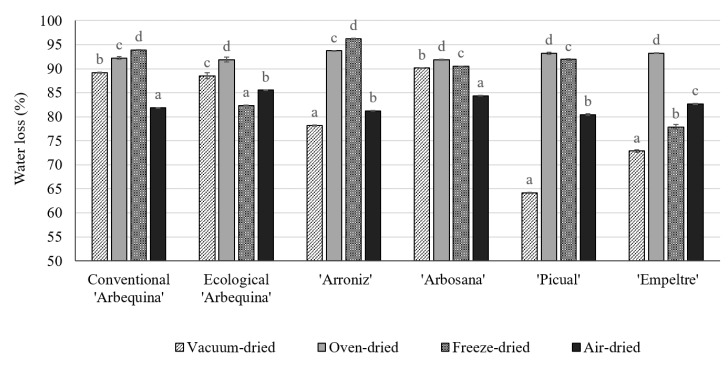
Up to 5% of the total olive weight arriving at the mill is discarded as leaves. Interest in the possible uses of these residues is growing, because they constitute a potential cheap and abundant source of compounds with high total antioxidant capacity (TAC) associated with total phenolic content (TPC) and biophenols such as hydroxytyrosol (HC) and oleuropein (OC), which could be used as nutraceuticals or as natural substitutes for synthetic antioxidants. However, studies that characterize specific cultivars, interannual variability, and different drying methods are lacking. This work investigates the TAC, TPC, HC and OC in olive (Olea europaea L.) leaves under four drying methods (vacuum-drying, oven-drying, freeze-drying and air-drying). Leaves were collected from cultivars 'Arbequina' grown under organic methods and from 'Arroniz', 'Empeltre', 'Arbosana', 'Picual' and 'Arbequina' grown under conventional systems. Among fresh samples, 'Arbosana' leaves presented the highest TPC (34.0 ± 1.1 mg gallic acid equivalents/g dry weight (DW)) and TAC (146 ± 20 μmol Trolox equivalents/g DW) and the lowest interannual variability of the TPC (3.2%). The four tested drying methods were also compared as the effect on TPC, TAC, HC and OC. Freeze-drying and air-drying best preserved TPC and TAC in olive leaves. However, air-drying maintained greater OC (14–40 mg/g DW) than freeze-drying (3–20 mg/g DW). Air-dried ecological 'Arbequina' leaves exhibited the highest TPC and TAC. Consequently, this cultivar presented more valorization opportunities as a source of nutraceuticals or natural antioxidants.
Citation: Itxaso Filgueira-Garro, Carolina González-Ferrero, Diego Mendiola, María R. Marín-Arroyo. Effect of cultivar and drying methods on phenolic compounds and antioxidant capacity in olive (Olea europaea L.) leaves[J]. AIMS Agriculture and Food, 2022, 7(2): 250-264. doi: 10.3934/agrfood.2022016
Up to 5% of the total olive weight arriving at the mill is discarded as leaves. Interest in the possible uses of these residues is growing, because they constitute a potential cheap and abundant source of compounds with high total antioxidant capacity (TAC) associated with total phenolic content (TPC) and biophenols such as hydroxytyrosol (HC) and oleuropein (OC), which could be used as nutraceuticals or as natural substitutes for synthetic antioxidants. However, studies that characterize specific cultivars, interannual variability, and different drying methods are lacking. This work investigates the TAC, TPC, HC and OC in olive (Olea europaea L.) leaves under four drying methods (vacuum-drying, oven-drying, freeze-drying and air-drying). Leaves were collected from cultivars 'Arbequina' grown under organic methods and from 'Arroniz', 'Empeltre', 'Arbosana', 'Picual' and 'Arbequina' grown under conventional systems. Among fresh samples, 'Arbosana' leaves presented the highest TPC (34.0 ± 1.1 mg gallic acid equivalents/g dry weight (DW)) and TAC (146 ± 20 μmol Trolox equivalents/g DW) and the lowest interannual variability of the TPC (3.2%). The four tested drying methods were also compared as the effect on TPC, TAC, HC and OC. Freeze-drying and air-drying best preserved TPC and TAC in olive leaves. However, air-drying maintained greater OC (14–40 mg/g DW) than freeze-drying (3–20 mg/g DW). Air-dried ecological 'Arbequina' leaves exhibited the highest TPC and TAC. Consequently, this cultivar presented more valorization opportunities as a source of nutraceuticals or natural antioxidants.

| [1] | International Olive Council (2019) International Olive Council Newsletter, The international market, 2019. Available from: https://www.internationaloliveoil.org/wp-content/uploads/2019/12/newsletter_144_english.pdf. |
| [2] |
Molina-Alcaide E, Yáñez-Ruiz DR (2008) Potential use of olive by-products in ruminant feeding: A review. Anim Feed Sci Technol 147: 247–264. https://doi.org/10.1016/j.anifeedsci.2007.09.021 doi: 10.1016/j.anifeedsci.2007.09.021

|
| [3] |
Žugčić T, Abdelkebir R, Alcantara C, et al. (2019) From extraction of valuable compounds to health promoting benefits of olive leaves through bioaccessibility, bioavailability and impact on gut microbiota. Trends Food Sci Technol 83: 63–77. https://doi.org/10.1016/j.tifs.2018.11.005 doi: 10.1016/j.tifs.2018.11.005
|
| [4] |
Nunes MA, Pimentel FB, Costa ASG, et al. (2016) Olive by-products for functional and food applications: Challenging opportunities to face environmental constraints. Innov Food Sci Emerg Technol 35: 139–148. https://doi.org/10.1016/j.ifset.2016.04.016 doi: 10.1016/j.ifset.2016.04.016
|
| [5] |
Talhaoui N, Taamalli A, Gómez-Caravaca AM, et al. (2015) Phenolic compounds in olive leaves: Analytical determination, biotic and abiotic influence, and health benefits. Food Res Int 77: 92–108. https://doi.org/10.1016/j.foodres.2015.09.011 doi: 10.1016/j.foodres.2015.09.011
|
| [6] |
Talhaoui N, Gómez-Caravaca AM, Roldán C, et al. (2015) Chemometric analysis for the evaluation of phenolic patterns in olive leaves from six cultivars at different growth stages. J Agric Food Chem 63: 1722–1729. https://doi.org/10.1021/jf5058205 doi: 10.1021/jf5058205
|
| [7] | Şahin S, Ahmed Malik NS, Perez JL, et al. (2012) Seasonal changes of individual phenolic compounds in leaves of twenty olive cultivars grown in Texas. J Agric Sci Technol 2: 242–247. |
| [8] |
Özcan MM, Fındık S, AlJuhaimi F, et al. (2019) The effect of harvest time and varieties on total phenolics, antioxidant activity and phenolic compounds of olive fruit and leaves. J Food Sci Technol 56: 2373–2385. https://doi.org/10.1007/s13197-019-03650-8 doi: 10.1007/s13197-019-03650-8
|
| [9] |
Nicolì F, Negro C, Vergine M, et al. (2019) Evaluation of phytochemical and antioxidant properties of 15 Italian Olea europaea L. cultivar leaves. Molecules 24: 1998. https://doi.org/10.3390/molecules24101998 doi: 10.3390/molecules24101998
|
| [10] |
Hülya-Orak H, Karamać M, Amarowicz R, et al. (2019) Genotype-related differences in the phenolic compound profile and antioxidant activity of extracts from olive (Olea europaea L.) leaves. Molecules 24: 1130. https://doi.org/10.3390/molecules24061130 doi: 10.3390/molecules24061130
|
| [11] |
Olmo-García L, Bajoub A, Benlamaalam S, et al. (2018) Establishing the phenolic composition of Olea europaea L. leaves from cultivars grown in Morocco as a crucial step towards their subsequent exploitation. Molecules 23: 2524. https://doi.org/10.3390/molecules23102524 doi: 10.3390/molecules23102524
|
| [12] |
Guinda Á, Castellano JM, Santos-Lozano JM, et al. (2015) Determination of major bioactive compounds from olive leaf. LWT-Food Sci Technol 64: 431–438. https://doi.org/10.1016/j.lwt.2015.05.001 doi: 10.1016/j.lwt.2015.05.001
|
| [13] |
Romero C, Medina E, Mateo MA, et al. (2017) Quantification of bioactive compounds in Picual and Arbequina olive leaves and fruit. J Sci Food Agric 97: 1725–1732. https://doi.org/10.1002/jsfa.7920 doi: 10.1002/jsfa.7920
|
| [14] |
Lama-Muñoz A, Contreras MM, Espínola F, et al. (2020) Content of phenolic compounds and mannitol in olive leaves extracts from six Spanish cultivars: Extraction with the Soxhlet method and pressurized liquids. Food Chem 320: 126626. https://doi.org/10.1016/j.foodchem.2020.126626 doi: 10.1016/j.foodchem.2020.126626
|
| [15] |
Papoti VT, Papageorgiou M, Dervisi K, et al. (2018) Screening olive leaves from unexploited traditional Greek cultivars for their phenolic antioxidant dynamic. Foods 7: 197. https://doi.org/10.3390/foods7120197 doi: 10.3390/foods7120197
|
| [16] |
De Leonardis A, Macciola V, Cuomo F, et al. (2015) Evidence of oleuropein degradation by olive leaf protein extract. Food Chem 175: 568–574. https://doi.org/10.1016/j.foodchem.2014.12.016 doi: 10.1016/j.foodchem.2014.12.016
|
| [17] |
Afaneh I, Yateem H, Al-Rimawi F (2015) Effect of olive leaves drying on the content of oleuropein. Am J Anal Chem 6: 246–252. http://dx.doi.org/10.4236/ajac.2015.63023 doi: 10.4236/ajac.2015.63023
|
| [18] |
Kamran M, Hamlin AS, Scott CJ, et al. (2015) Drying at high temperature for a short time maximizes the recovery of olive leaf biophenols. Ind Crop Prod 78: 29–38. https://doi.org/10.1016/j.indcrop.2015.10.031 doi: 10.1016/j.indcrop.2015.10.031
|
| [19] |
Attya M, Benabdelkamel H, Perri E, et al. (2010) Effects of conventional heating on the stability of major olive oil phenolic compounds by tandem mass spectrometry and isotope dilution assay. Molecules 15: 8734. https://doi.org/10.3390/molecules15128734 doi: 10.3390/molecules15128734
|
| [20] |
Şahin S, Elhussein E, Bilgin M, et al. (2018) Effect of drying method on oleuropein, total phenolic content, flavonoid content, and antioxidant activity of olive (Olea europaea) leaf. J Food Process Preserv 42: e13604. https://doi.org/10.1111/jfpp.13604 doi: 10.1111/jfpp.13604
|
| [21] |
Ahmad-Qasem MH, Ahmad-Qasem BH, Barrajón-Catalán E, et al. (2016) Drying and storage of olive leaf extracts. Influence on polyphenols stability. Ind Crop Prod 79: 232–239. http://dx.doi.org/10.1016/j.indcrop.2015.11.006 doi: 10.1016/j.indcrop.2015.11.006
|
| [22] |
Ali Elhussein EA, Şahin S (2018) Drying behaviour, effective diffusivity and energy of activation of olive leaves dried by microwave, vacuum and oven drying methods. Heat Mass Transf 54: 1901–1911. https://doi.org/10.1007/s00231-018-2278-6 doi: 10.1007/s00231-018-2278-6
|
| [23] |
Obied HK, Allen MS, Bedgood DR, et al. (2005) Investigation of Australian olive mill waste for recovery of biophenols. J Agric Food Chem 53: 9911–9920. https://doi.org/10.1021/jf0518352 doi: 10.1021/jf0518352
|
| [24] |
Brand-Williams W, Cuvelier M., Berset C (1995) Use of a free radical method to evaluate antioxidant activity. Food Sci Technol 28: 25–30. https://doi.org/10.1016/S0023-6438(95)80008-5 doi: 10.1016/S0023-6438(95)80008-5
|
| [25] |
Suárez M, Macià A, Romero MP, et al. (2008) Improved liquid chromatography tandem mass spectrometry method for the determination of phenolic compounds in virgin olive oil. J Chromatogr A 1214: 90–99. https://doi.org/10.1016/j.chroma.2008.10.098 doi: 10.1016/j.chroma.2008.10.098
|
| [26] |
Abaza L, Ben-Youssef N, Manai H, et al. (2011) Chétoui olive leaf extracts: influence of the solvent type on phenolics and antioxidant activities. Grasas y aceites 62: 96–104. https://doi.org/10.3989/gya.044710 doi: 10.3989/gya.044710
|
| [27] |
López-Yerena A, Lozano-Castellón J, Olmo-Cunillera A, et al. (2019) Effects of organic and conventional growing systems on the phenolic profile of extra-virgin olive oil. Molecules 24: 1986. https://doi.org/10.3390/molecules24101986 doi: 10.3390/molecules24101986
|
| [28] |
Taamalli A, Lozano-Sánchez J, Jebabli H, et al. (2019) Monitoring the bioactive compounds status in Olea europaea according to collecting period and drying conditions. Energies 12: 947. https://doi.org/10.3390/en12050947 doi: 10.3390/en12050947
|
| [29] |
Babu AK, Kumaresan G, Aroul-Raj VA, et al. (2018) Review of leaf drying: Mechanism and influencing parameters, drying methods, nutrient preservation, and mathematical models. Renew Sustain Energy Rev 90: 536–556. https://doi.org/10.1016/j.rser.2018.04.002 doi: 10.1016/j.rser.2018.04.002
|


Figures(4) / Tables(1)

Itxaso Filgueira-Garro, Carolina González-Ferrero, Diego Mendiola, María R. Marín-Arroyo. Effect of cultivar and drying methods on phenolic compounds and antioxidant capacity in olive (Olea europaea L.) leaves[J]. AIMS Agriculture and Food, 2022, 7(2): 250-264. doi: 10.3934/agrfood.2022016


 DownLoad: