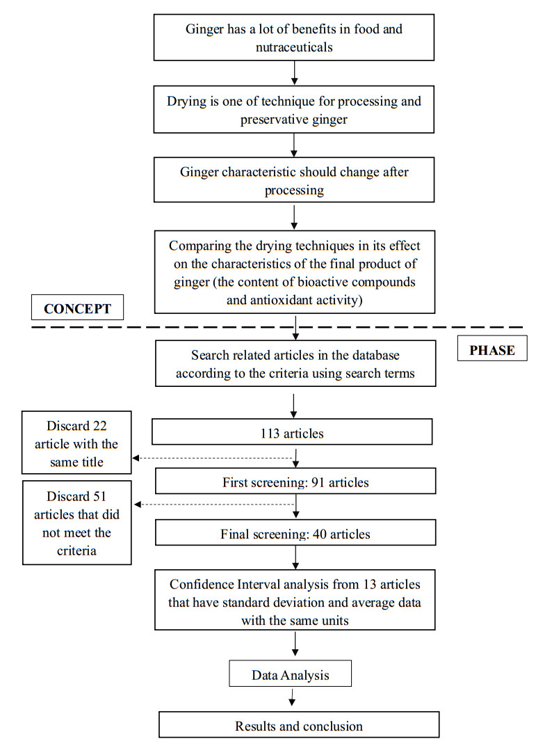
Ginger is a spice with various uses for humans, such as flavoring agents and nutraceuticals. Drying is commonly used in the processing and preserving of ginger and affects the characteristics of the final ginger product. This study aimed to review the studies that have evaluated the effects of drying techniques on the bioactivity of ginger. A meta-analysis investigation was conducted to identify a study that evaluated the effects of drying techniques on the levels of bioactivity in ginger. The database search found 113 results. There are 13 articles from 2010 to 2020 that met the inclusion criteria. The drying techniques have different effects on the optimum levels of ginger characteristics. After drying treatment there were significant different on total flavonoid and antioxidant activity and there were not significant on total phenolic content and 6-gingerol content of ginger. In conclusion, drying has different effects on ginger in terms of bioactivity. Therefore, choosing the best method must be made based on the purpose of the process and the final product criteria.
Citation: Hesti Kurniasari, Wahyudi David, Laras Cempaka, Ardiansyah. Effects of drying techniques on bioactivity of ginger (Zingiber officinale): A meta-analysis investigation[J]. AIMS Agriculture and Food, 2022, 7(2): 197-211. doi: 10.3934/agrfood.2022013
Ginger is a spice with various uses for humans, such as flavoring agents and nutraceuticals. Drying is commonly used in the processing and preserving of ginger and affects the characteristics of the final ginger product. This study aimed to review the studies that have evaluated the effects of drying techniques on the bioactivity of ginger. A meta-analysis investigation was conducted to identify a study that evaluated the effects of drying techniques on the levels of bioactivity in ginger. The database search found 113 results. There are 13 articles from 2010 to 2020 that met the inclusion criteria. The drying techniques have different effects on the optimum levels of ginger characteristics. After drying treatment there were significant different on total flavonoid and antioxidant activity and there were not significant on total phenolic content and 6-gingerol content of ginger. In conclusion, drying has different effects on ginger in terms of bioactivity. Therefore, choosing the best method must be made based on the purpose of the process and the final product criteria.

| [1] |
Bhandary U, Sharma JN, Zafar R (1997) Effect of protection action of ethanolic ginger (Zingiber officinales) extract in cholestered fed rabbits. J Ethnopharm 61: 167-175. https://doi.org/10.1016/S0378-8741(98)00026-9 doi: 10.1016/S0378-8741(98)00026-9

|
| [2] |
Phoungchandang S, Saentaweesuk S (2011) Effect of two stage, tray and heat pump assisted-dehumidified drying on drying characteristics and qualities of dried ginger. Food Bioprod Proc 89: 429-437. https://doi.org/10.1016/S0378-8741(98)00026-9 doi: 10.1016/S0378-8741(98)00026-9
|
| [3] |
Pinela J, Barros L, Carvalho AM, et al. (2011) Influence of the drying technique in the antioxidant potential and chemical composition of four shrubby flowering plants from the tribe genisteae (Fabaceae). Food Chem Toxicol 49: 2983-2989. https://doi.org/10.1016/j.fct.2011.07.054 doi: 10.1016/j.fct.2011.07.054
|
| [4] |
Chan E, Lim YY, Wong SK, et al. (2009) Effects of different drying techniques on the antioxidant properties of leaves and tea of ginger species. Food Chem 113: 166-172. https://doi.org/10.1016/j.foodchem.2008.07.090 doi: 10.1016/j.foodchem.2008.07.090
|
| [5] |
Dugasani S, Pichika MR, Nadarajah VD, et al. (2009) Comparative antioxidant and anti-inflammatory effects of 6-gingerol, 8-gingerol, 10-gingerol and 6-shogaol. J Ethnopharm 127: 515-520. https://doi.org/10.1016/j.jep.2009.10.004 doi: 10.1016/j.jep.2009.10.004
|
| [6] |
Jolad SD, Lantz RC, Chen GJ, et al. (2005) Commercially processed dry ginger (Zingiber officinale): composition and effects on LPS-stimulated PGE2 production. Phytochem 66: 1614-1635. https://doi.org/10.1016/j.phytochem.2005.05.007 doi: 10.1016/j.phytochem.2005.05.007
|
| [7] |
Park M, Bae J, Lee DS (2008) Antibacterial activity of 10-gingerol and 12-gingerol isolated from ginger rhizome against periodontal bacteria. Phytothe Res 22: 1446-1449. https://doi.org/10.1002/ptr.2473 doi: 10.1002/ptr.2473
|
| [8] | Gilani AH, Ghayur MN (2005) Ginger: from Myth to Reality. In: Gottshalk-Batsschkus CE, Green JC (Eds.), Ethnotherepies in The Cycle of Life, Ethnomed Institute fur Ethnomedzirine.V. Munich, 307-315. |
| [9] |
Ajay M, Gilanui AH, Mustafa MR (2003) Effect of flavonoids on vascular smooth muscles of the isolated rat thoracic aorta. Life Sci 74: 603-612. https://doi.org/10.1016/j.lfs.2003.06.039 doi: 10.1016/j.lfs.2003.06.039
|
| [10] |
Stoilova I, Krastanov A, Stoyanova A, et al. (2007) Antioxidant activity of a ginger extract (Zingiber officinale). Food Chem 102: 764-70. https://doi.org/10.1016/j.foodchem.2006.06.023 doi: 10.1016/j.foodchem.2006.06.023
|
| [11] | Cosmovici A, Sonia A (2017) Study on the influence of heat treatment on the antioxidant properties of ginger. J Fac Food Engin 16: 153-159. |
| [12] |
Yuxin L, Yang H, Yanquan H, et al. (2016) Chemical characterization and antioxidant activities comparison in fresh, dried, stir-frying and carbonized ginger. J Chromatogr 1011: 223-232. https://doi.org/10.1016/j.jchromb.2016.01.009 doi: 10.1016/j.jchromb.2016.01.009
|
| [13] |
Eze JI, Agbo KE (2011) Comparative studies of sun and solar drying of peeled and unpeeled ginger. American J Sci Indust Res 2: 136-143. https://doi.org/10.5251/ajsir.2011.2.2.136.143 doi: 10.5251/ajsir.2011.2.2.136.143
|
| [14] |
Gumusay OA, Borazan AA, Ercal N, et al. (2014) Drying effects on the antioxidant properties of tomatoes and ginger. Food Chem 173: 156-162. https://doi.org/10.1016/j.foodchem.2014.09.162 doi: 10.1016/j.foodchem.2014.09.162
|
| [15] |
Jelled A, Fernandes Â, Barros L, et al. (2015) Chemical and Antioxidant Parameters of Dried Forms of Ginger Rhizomes. Ind Crops Prod 77: 30-35. https://doi.org/10.1016/j.indcrop.2015.08.052 doi: 10.1016/j.indcrop.2015.08.052
|
| [16] | Dahlan MS (2012) Pengantar Meta-Analisis: Disertai Aplikasi Meta-Analisis Epiyudin. Jakarta: PT. Epidemiologi Indonesia. |
| [17] |
Keijing A, Dandan Z, Zhengfu W, et al. (2015) Comparison of different drying techniques on chinese ginger (Zingiber officinale Roscoe): Changes in volatils, chemical profile, antioxidant properties, and microstructure. Food Chem 197: 1292-1300. https://doi.org/10.1016/j.foodchem.2015.11.033 doi: 10.1016/j.foodchem.2015.11.033
|
| [18] |
Osae R, Gloria E, Raphael NA, et al. (2019) Drying of ginger slices-evaluation of quality attributes, energy consumption, and kinetics study. J Food Proc Engin 43: e1334. https://doi.org/10.1111/jfpe.13348 doi: 10.1111/jfpe.13348
|
| [19] |
Mustafa, I, Nyuk LC, Sharida F, et al. (2019) Comparison of phytochemicals, antioxidant and anti-inflammatory properties of sun-, oven- and freeze-dried ginger extracts. Foods 8: 456. https://doi.org/10.3390/foods8100456 doi: 10.3390/foods8100456
|
| [20] |
Valadez-Carmona L, Cortez-García RM, Plazola-Jacinto CP, et al. (2016) Effect of microwave drying and oven drying on the water activity, color, phenolic compounds content and antioxidant activity of coconut husk (Cocos nucifera L.). J Food Sci and Tech 53: 3495-3501. https://doi.org/10.1007/s13197-016-2324-7 doi: 10.1007/s13197-016-2324-7
|
| [21] |
Jihene L, Touil A, Chemkhi S, et al. (2013) Impact of infra-red drying temperature on total phenolic and flavonoid content, on antioxidan and antimicrobial activities of ginger. J Environ Sci Toxicol Food Technol 6: 38-46. https://doi.org/10.9790/2402-0653846 doi: 10.9790/2402-0653846
|
| [22] |
Ghasemzadeh A, Hawa ZE, Jaafar AR (2016) Variation of the phytochemical constituents and antioxidant activities of zingiber officinale var. Rubrum theilade associated with different drying techniques and polyphenol oxidase activity. Molecules 21: 780. https://doi.org/10.3390/molecules21060780 doi: 10.3390/molecules21060780
|
| [23] |
Cherrat S, Boulkebache-Makhlouf L, Iqbal J, et al. (2019) Effect of different drying temperatures on the composition and antioxidant activity of ginger powder. The Ann Univ Dunarea de Jos of Galati Fascicle VI-Food Technol 43: 125-142. https://doi.org/10.35219/foodtechnology.2019.2.09 doi: 10.35219/foodtechnology.2019.2.09
|
| [24] |
Lv W, Li S, Han Q, et al. (2016) Study of the drying process of ginger slices in microwave fluidized bed dryer. J Drying Technol 34: 1690-1699. https://doi.org/10.1080/07373937.2015.1137932 doi: 10.1080/07373937.2015.1137932
|
| [25] |
Kubra IR, Rao LJM (2012) Microwave drying of ginger (Zingiber officinale R) and its effects on polyphenolic content and antioxidant activity. Inter J Food Sci Technol 47: 2311-2317. https://doi.org/10.1111/j.1365-2621.2012.03104.x doi: 10.1111/j.1365-2621.2012.03104.x
|
| [26] |
Jung MY, Lee MK, Park HJ, et al. (2018) Heat-induced conversion of gingerols to shogaols in ginger as affected by heat type (dry or moist heat), sample type (fresh or dried), temperature and time. Food Sci Biotechnol 27: 687-693. https://doi.org/10.1007/s10068-017-0301-1 doi: 10.1007/s10068-017-0301-1
|
| [27] | Prakash A, Rigelhof F, Miller E (2001) Antioxidant activity: medallion laboratories. Analit Progr 19: 1-4. |
| [28] |
Chumreonphat T, Intha K, Luchai B (2011) Stability of phytochemicals and antioxidant properties in ginger (Zingiber officinale Roscoe) with different drying techniques. J Herbs, Spices & Medic Plants 17: 361-374. https://doi.org/10.1080/10496475.2011.629776 doi: 10.1080/10496475.2011.629776
|
| [29] | Sida S, Rajnibhas SS, Niramon U (2019) Influence of maturity and drying temperature on antioxidant activity and chemical compositions in ginger. J Curr App Sci Technol 19: 28-42. |
| [30] |
Bevilacqua A, Petruzzi L, Perricone M, et al. (2018) Nonthermal technologies for fruit and vegetale juices and beverages: overview and advances. Compre Rev Food Sci Food Safety 17: 2-62. https://doi.org/10.1111/1541-4337.12299 doi: 10.1111/1541-4337.12299
|
| [31] |
Osae R, Chunsan Z, Raphael NA, et al. (2019) Effects of Various Nonthermal pretreatments on the physicochemical properties of dried ginger (Zingiber officinale R) slices from two geographical locations. J Food Sci 84: 2847-2858. https://doi.org/10.1111/1750-3841.14790 doi: 10.1111/1750-3841.14790
|
| [32] |
Thuwapanichayanan R, Charotorn P, Donludee J, et al. (2014) Effects of pretreatments and drying temperatures on drying characteristics, antioxidant properties and color of ginger slice. Acta Univers Agric Mendelianae Brunensis 62: 1125-1134. https://doi.org/10.11118/actaun201462051125 doi: 10.11118/actaun201462051125
|
| [33] | Famurewa AV, Emuekele PO, Jaiyeoba KF (2011) Effect of drying and size reduction on the chemical and volatil oil contents of ginger (Zingiber officinale). J Medic Plants Res 5: 2941-2944. |


Figures(11)

Hesti Kurniasari, Wahyudi David, Laras Cempaka, Ardiansyah. Effects of drying techniques on bioactivity of ginger (Zingiber officinale): A meta-analysis investigation[J]. AIMS Agriculture and Food, 2022, 7(2): 197-211. doi: 10.3934/agrfood.2022013


 DownLoad: