Research article

Influence of heat shock proteins in individual sensitivity of human neutrophils to heat stress

  • Received: 26 March 2019 Accepted: 19 July 2019 Published: 01 August 2019
  • We have developed a simple and reliable method to measure the sensitivity of individuals to oxidative stress. This method utilizes luminol-amplified chemiluminescence to quantify production of reactive oxygen species (ROS) by opsonized zymosan-stimulated neutrophils that have been subjected to short-term stress via heat shock. In this study, the chemiluminescence reaction was used to monitor the dynamics of ROS production in neutrophils derived from 17 patients of different ages and genders before and after these neutrophils were subjected to heat shock. In addition, we determined expression of Toll-like receptors using fluorescent-labeled antibody. The effects of adrenaline, dexamethasone, aspirin, and indomethacin, as well as different doses of exogenous heat shock protein 70 (Hsp70), on the production of ROS by stimulated neutrophils was also investigated. Our data showed that adrenaline and exogenous Hsp70 both suppressed ROS production by stimulated neutrophils. Furthermore, TLR4 expression was upregulated upon heat stress. Thus, adrenaline, HSPs, and TLRs may all play a role in regulating stress responses in phagocytes.

    Citation: Viktor Semenkov, Anatolii Mikhalskii, Alexandr Sapoznikov. Influence of heat shock proteins in individual sensitivity of human neutrophils to heat stress[J]. AIMS Molecular Science, 2019, 6(2): 38-51. doi: 10.3934/molsci.2019.2.38

    Related Papers:

    [1] Noor Riyadh Thiab, Nicola King, Mary McMillan, Amer Almashhadany, Graham L Jones . Age-related protein and mRNA expression of glutathione peroxidases (GPx) and Hsp-70 in different regions of rat kidney with and without stressor. AIMS Molecular Science, 2016, 3(2): 125-137. doi: 10.3934/molsci.2016.2.125
    [2] Nataša Čerekovic, Palmiro Poltronieri . Plant signaling pathways activating defence response and interfering mechanisms by pathogen effectors, protein decoys and bodyguards. AIMS Molecular Science, 2017, 4(3): 370-388. doi: 10.3934/molsci.2017.3.370
    [3] Zhaoping Qin, Patrick Robichaud, Taihao Quan . Oxidative stress and CCN1 protein in human skin connective tissue aging. AIMS Molecular Science, 2016, 3(2): 269-279. doi: 10.3934/molsci.2016.2.269
    [4] Shreya Shetty, Sampat Kumar Srigiri, Karunakar Shetty . The potential role of neutrophil extracellular traps (NETS) in periodontal disease—A scanning electron microscopy (SEM) study. AIMS Molecular Science, 2023, 10(3): 205-212. doi: 10.3934/molsci.2023014
    [5] Valentina Contrò, John R. Basile, Patrizia Proia . Sex steroid hormone receptors, their ligands, and nuclear and non-nuclear pathways. AIMS Molecular Science, 2015, 2(3): 294-310. doi: 10.3934/molsci.2015.3.294
    [6] Francisco Les, Zsuzsa Iffiú-Soltész, Josep Mercarder, Christian Carpéné . Tyramine activates lipid accumulation in rat adipocytes: influences of in vitro and in vivo administration. AIMS Molecular Science, 2017, 4(3): 339-351. doi: 10.3934/molsci.2017.3.339
    [7] Ahmed Yaqinuddin, Abdul Hakim Almakadma, Junaid Kashir . Kawasaki like disease in SARS-CoV-2 infected children – a key role for neutrophil and macrophage extracellular traps. AIMS Molecular Science, 2021, 8(3): 174-183. doi: 10.3934/molsci.2021013
    [8] Giulia Ambrosi, Pamela Milani . Endoplasmic reticulum, oxidative stress and their complex crosstalk in neurodegeneration: proteostasis, signaling pathways and molecular chaperones. AIMS Molecular Science, 2017, 4(4): 424-444. doi: 10.3934/molsci.2017.4.424
    [9] Marta Monzón . Approaches to therapy against prion diseases focused on the individual defence system. AIMS Molecular Science, 2017, 4(3): 241-251. doi: 10.3934/molsci.2017.3.241
    [10] William M. Scovell, Sachindra R. Joshi . The changing paradigm: estrogen receptor α recognition on DNA and within the dynamic nature of nucleosomes. AIMS Molecular Science, 2015, 2(2): 48-63. doi: 10.3934/molsci.2015.2.48
  • We have developed a simple and reliable method to measure the sensitivity of individuals to oxidative stress. This method utilizes luminol-amplified chemiluminescence to quantify production of reactive oxygen species (ROS) by opsonized zymosan-stimulated neutrophils that have been subjected to short-term stress via heat shock. In this study, the chemiluminescence reaction was used to monitor the dynamics of ROS production in neutrophils derived from 17 patients of different ages and genders before and after these neutrophils were subjected to heat shock. In addition, we determined expression of Toll-like receptors using fluorescent-labeled antibody. The effects of adrenaline, dexamethasone, aspirin, and indomethacin, as well as different doses of exogenous heat shock protein 70 (Hsp70), on the production of ROS by stimulated neutrophils was also investigated. Our data showed that adrenaline and exogenous Hsp70 both suppressed ROS production by stimulated neutrophils. Furthermore, TLR4 expression was upregulated upon heat stress. Thus, adrenaline, HSPs, and TLRs may all play a role in regulating stress responses in phagocytes.


    Clinical and experimental data demonstrate that, to a large extent, sensitivity to external damaging stressors and development of inflammation depends on the functions of the adrenal medulla and cortex, which are under the control of the hypothalamus-hypophysis system. Inflammation at different pathologies can be attributed to excessive reactive oxygen species (ROS) production by patients' phagocytes. Increase of ROS production combines with rising pro-inflammatory cytokines TNF, IL-1, IL-6 and activation of TLR of phagocytes, especially TLR-4. TLR-4 evokes activation NF-kB and associates with LPS and some HSPs [1][3]. Extremely high ROS concentrations damage proteins, lipids, and DNA, which lead to cell aging and the so-called free radical diseases such as atherosclerosis, essential hypertension, senile dementia of Alzheimer type, Parkinson disease, cancer, and so on [4]. In turn, these diseases are stressors for the organism and amplify intercellular oxidative stress leading to further agammaavation of the pathologic process [5]. Treatment of these diseases is realized by using anti-inflammatory agents like synthetic glucocorticoid dexamethason and nonsteroid drags (aspirin, indomethacin and other). Early identification of individuals with high sensitivity to oxidative stress will allow implementation of preemptive measures to prevent or delay the onset of these diseases.

    Methodologies currently used to measure levels of psycho-emotional stress involve determination of informative physiological parameters (such as heart rate, breathing rate, systolic and diastolic blood pressure, electromyogram parameters, galvanic skin response, and so on). Some investigations are focused on stress responses of lymphocytes, which are not able to produce ROS. By contrast, neutrophils are micro-phagocytes that are able to produce ROS. In this study, we measured the sensitivity of individuals to oxidative stress by using luminol-amplified chemiluminescence to quantify ROS production by neutrophils (activated by opsonized zymosan) after heat shock. Figure 1 illustrates the action of different factors of environment on manifestation of stress reaction.

    Figure 1.  Action of different environmental factors on manifestation of stress reaction.

    A total of 17 patients aged 21–92 years (three men), registered in the Moscow Clinical Centre of Gerontology were recruited for the study. These donors suffered from co-morbidities, with coronary heart disease, cardiosclerosis, arterial hypertension, and cerebrovascular atherosclerosis diagnosed as the main pathologies. The inclusion criteria for participation were the absence of active pathologies (history of acute infection, tumors, apoplexy, or myocardial infarction) and absence of treatment with corticosteroids or high doses of non-steroidal anti-inflammatory drugs. The study was approved by Ethics Committee of the Pirigov Russian National Research Medical University. All participants gave their written informed consent prior to the study. Most of them were the elderly but several patients were young in order to comparison of ROS levels.

    Neutrophils were isolated from the peripheral blood of each patient 10 minutes after drawing the patients' blood via the ficoll-verographin double density-gradient centrifugation method as follows: Defibrinated or anticoagulant-treated blood was diluted with an equal volume of a sterilized balanced salt solution, and layered carefully over the ficoll-verographin gradient (the density of the bottom liquid was 1.119 g/cm3, and that of the top layer of liquid was 1.077 g/cm3) without intermixing, and then centrifuged for 40 min at 400 × g. A sterile micropipette was used to extract neutrophils from the interphase layer. The neutrophils were then washed twice using physiological solution and then re-suspended in colorless Hank's solution. Finally, the number of nuclear cells in the suspension was counted using a hemocytometer. The resulting neutrophil suspension had a purity of 96–98% determined by microscopy method. The luminol-enhanced chemiluminescence reaction was carried out using plastic test tubes containing 20 μL of colorless Hank's solution and 150 μL of 2.5 mg/mL luminol (5-Amino-2,3-dihydro-1,4-phthalazinedione, SERVA Electrophoresis GmbH, Heidelberg, Germany). Test tubes were placed in the chemiluminometer cylinder (Lum-5773, DISoft, Vjscow, Russia) and pre-incubated for one hour to reach a temperature of 37 °C. Then, 100 μL of neutrophil suspension (2 × 10⁵ nuclear cells) was added to the luminol solution and incubated for a further 60 min at 37 °C before stimulating the neutrophils with 100 μL of 20 mg/mL opsonized zymosan (Sigma, St. Louis, MO). The chemiluminescence that was induced and the dynamics of ROS production by neutrophils were registered over a period of 30 min by the luminometer that records the number of impulses-photons per minute. To expose neutrophils to short-term stress, the test tubes were heated in a 42 °C water bath for 30 seconds 1 or 3 minutes before carrying out the chemiluminescence reaction.

    To calculate individual sensitivity to stress, a coefficient of neutrophil chemiluminescence was calculated as the ratio between number of photon counts per minute before stress and number of photon counts per minute after stress. Reduction in coefficient of neutrophil chemiluminescence value corresponds to an increase in individual resistance to stress or in other words, an overall decrease in the individual's stress response.

    The effect of different amounts of exogenously added recombinant bovine Hsp70 (Stressgen Bioreagents Corp, Victoria, Canada) on the chemiluminescence produced by opsonized zymosan-stimulated neutrophils was investigated by adding the Hsp70 (0.5 µg or 5 µg) to the reaction mix in addition to the opsonized zymosan.

    The expression levels of TLRs was estimated using flow cytometric analysis of leukocytes labelled using standard protocols with fluorochrome-conjugated primary antibodies directed against TLR4 (HycultBiotech, Uden, The Netherlands). Flow cytometric analysis of a minimum of 10,000 events was carried out using the FACScan system (Becton Dickinson, Franklin Lakes, NJ).

    We also performed flow cytometric analyses evaluating the relative level of fluorescence after heating shock using as the baseline level the value of fluorescence before heating shock.

    The reaction was performed as described in Section 2.2 of Materials and methods, except that the standard Hank's solution contained Ca++ and Mg++, and adrenaline or dexamethasone was added to the experimental tubes at a concentration of 10−4 M or 10−5 M, respectively, followed by incubation for 30 min at 37 °C prior to the chemiluminescence reaction.

    We investigated dependence of ROS production by neutrophils on treatment by aspirin and indomethacin in different doses in patients of different age because of aspirin and indomethacin are anti-inflammatory medications.

    Statistical analysis was conducted using the computer program Biostat by applying the Mann-Whitney non-parametric test. Results were considered statistically significant if the p-value was <0.05. Mathematical comparison of neutrophil chemiluminescence before and after being subjected to heat shock for 30 sec, 1 min, or 3 min, was conducted using Binomial distribution in the form P(r,p)=(nr)pr(1p)nr, wherein n: total number of experiments, p: probability of success, r: number of experiments with success, (nr): binomial coefficient, P(r, p): probability of getting exactly r successes in n experiments if probability of success equals p. Estimate for probability of success p is obtained by maximization of probability P(r, p) on p and is given by formula ˆp=r/n. 95% confidence interval for probability of success was determined from exact formulas for Binomial distribution using modern software tools.

    The kinetics of chemiluminescence production by stimulated neutrophils in the absence of heat shock (control) and when subjected to heat shock for different durations, was monitored over time and recorded for each patient. In general, the chemiluminescence profiles for all patients showed a rapid burst in ROS production leading to a peak in photon emission rate, followed by a decay of chemiluminescence over time due to the consumption of reagents and decreases in the chemiluminescent quantum efficiency with time. Figures 24 show the ratio between initial chemiluminescence emission rate of neutrophils and chemiluminescence emission rate of neutrophils after heat shock of different durations in the neutrophils of the 17 patients in this study.

    Figure 2.  Plots showing the kinetics of ratio between initial chemiluminescence emission rate of patient-derived neutrophils, and chemiluminescence emission rate of patient-derived neutrophils after a 30-second heat shock for each of the 17 patients in the study.
    Figure 3.  Plots showing the kinetics of ratio between initial chemiluminescence emission rate of patient-derived neutrophils, and chemiluminescence emission rate of patient-derived neutrophils after a 1-min heat shock, for each of the 17 patients in the study.
    Figure 4.  Plots showing the kinetics of ratio between initial chemiluminescence emission rate of patient-derived neutrophils, and chemiluminescence emission rate of patient-derived neutrophils after a 3-min heat shock, for each of the 17 patients in the study.

    Dynamics of ROS production change may be attributed to individual sensitivity to stress, treatment, age differences and diseases. Additional investigations are necessary to determine degree of these factors influence on ROS production.

    Table 1 presents the estimation for probabilities that the ratio between initial chemiluminescence intensity and chemiluminescence intensity after heat shock reaches its maximum value within first 3 minutes calculated by averaging results of all patients. Confidence intervals are presented as well. For example, based on the chemiluminescence data for the 30-second heat shock (Figure 3), the probability of reaching a peak value within 3 minutes equals 14/17 = 0.82 with 95% confidence interval from 0.59 to 0.84.

    Table 1.  Estimation for probabilities and confidence intervals that ratio between initial chemiluminescence intensity and chemiluminescence intensity after heat shock reaches its maximum value within first 3 minutes.
    Heating duration Probability Lower 95% CI endpoint Upper 95% CI endpoint
    30 seconds 0.82 0.59 0.84
    1 minute 0.89 0.62 0.99
    3 minutes 0.71 0.45 0.88

     | Show Table
    DownLoad: CSV

    Our data showed that both the probabilities as well as the lower limits of the confidence intervals obtained for the 30-second and 1-min heat shock had higher values. Statistically significant increase of neutrophils' chemiluminescence coefficient was found during the first minute after stress for the three types of stress. Hypothesis that one minute duration of heating does not accompanied with the highest probability to observe maximum value for ratio between initial chemiluminescence intensity and chemiluminescence intensity after heat shock within first 3 minutes was rejected with p-value less than 10−5. This means that the individual sensitivity to oxidative stress is to be measured at the 1 min time-point.

    We present in Tables 25 some examples of chemiluminescence data to demonstrate the kinetics of ROS production in opsonized zymosan-stimulated neutrophils derived from 4 of the 17 patients in this study. In the tables, the numbers of photon per minute are presented for the different experimental conditions.

    Table 2.  Kinetics of ROS production of the patient M., 88 years, female with co-morbidities: Arterial hypertension, coronary heart disease, cardiosclerosis.
    Condition Initial 1 min 2 min 4 min 8 min 10 min 12 min 14 min
    no heating 2254 4010 7187 9149 9375 9306 8538 7331
    30 sec heating 1007 1613 3543 5015 5170 5210 4720 4172
    1 min heating 1176 1871 4160 5339 5685 5494 4784 4008
    3 min heating 347 429 1194 2073 2637 2454 2335 1892

     | Show Table
    DownLoad: CSV
    Table 3.  Kinetics of ROS production of the patient X., 86 years, male with co-morbidities: Arterial hypertension, coronary heart disease, cardiosclerosis, cerebrovascular atherosclerosis.
    Condition Initial 1 min 2 min 4 min 8 min 10 min 12 min 14 min
    no heating 1993 2619 8536 13526 15690 15824 15427 14253
    30 sec heating 366 488 1619 2941 3905 4666 4700 4463
    1 min heating 204 272 729 1308 1641 1828 1854 1913
    3 min heating 216 176 416 770 1036 1207 1080 1146

     | Show Table
    DownLoad: CSV
    Table 4.  Kinetics of ROS production of the patient F., 93 years, female with co-morbidities: Arterial hypertension, coronary heart disease, cardiosclerosis.
    Condition Initial 2 min 4 min 6 min 10 min 12 min
    no heating 1022 11068 14961 14825 11594 8721
    30 sec heating 316 2965 5234 5194 4562 3701
    1 min heating 259 2016 3845 4041 3946 3133
    3 min heating 131 1284 2754 2801 2603 1986

     | Show Table
    DownLoad: CSV
    Table 5.  Kinetics of ROS production of the patient V., 26 years, female without co-morbidity.
    Condition Initial 2 min 4 min 6 min 10 min 12 min
    no heating 1589 8289 18524 18271 16828 12784
    30 sec heating 343 1800 3700 3953 3883 2884
    1 min heating 588 2767 6940 6980 7063 5280
    3 min heating 787 2781 7120 7000 7400 6107

    Note: In the Tables one can see the statistically significant difference in ROS production between heating treatments and control (no heating) with p-value less than 0.001.

     | Show Table
    DownLoad: CSV

    Our results suggest that the change in the neutrophils' chemiluminescence coefficient may be attributed to the medical treatments being administered to the patients. Two examples are given below.

    Example 1. Patient R. (84 years old, female) had been diagnosed with hypertension disease of the 3rd stage, ischemic heart disease, cardiosclerosis, pneumo-sclerosis, and nephroangiosclerosis. Before hospitalization, the coefficient of her neutrophils' chemiluminescence (individual coefficient of resistance to stress) was 0.57 which corresponded to 7616 photon counts per minute initial (pre-stress) and 4338 photon counts per minute after stress. After two weeks of amlodipine medication, the coefficient of her neutrophils' chemiluminescence was 0.22, which corresponded to 406 photon counts per minute initial (pre-stress) and 89 photon counts per minute after stress.

    Example 2. Patient O. (92 years old, male) was diagnosed with a hip fracture. Before cure, his coefficient of neutrophil chemiluminescence was 0.35, which corresponded to 68,621 photon counts per minute initial (pre-stress) and 24,146 photon counts per minute after stress. After 7 sessions of low dose laser therapy, his neutrophils' coefficient of chemiluminescence decreased 1.3-fold and was 0.27, which corresponded to 6484 photon counts per minute initial (pre-stress) and 1725 photon counts per minute after stress. It therefore appeared that this patient's low dose laser therapy had reduced inflammation and ROS synthesis by his phagocytes.

    Figure 5.  Dependence of ROS production by neutrophils on aspirin doses in patients of different age. Age: x—21 years; □—29 years; ▪—32 years; o—57 years. Dose: 1—0.125 mg; 2—0.062 mg; 3—0.031 mg; 4—Control.
    Figure 6.  Dependence of ROS production by neutrophils on indomethacin doses in patients of different age. Age: □—27 years; ▪—45 years. Dose: 1—0.125 mg; 2—0.025 mg; 3—Control.

    Figures 5 and 6 demonstrate dependence between ROS production by neutrophils (number of photon counts per minute) and patients' age and doses (in mg) of aspirin and indomethacin, respectively. We observed a reduction in ROS production by neutrophils among patients of different ages that was proportional to dose of aspirin (Figure 5) or indomethacin (Figure 6). These trends are similar to those observed upon upregulation of anti-stress genes in cells, or when compounds exert anti-inflammatory or anti-oxidant effects on neutrophils. In addition, data presented in Figures 5 and 6 show individual sensitivity of ROS production by neutrophils in patients of different age and different doses of aspirin and indomethacin. These figures illustrate individual differences in ROS production when different doses of aspirin and indomethacin are used. To get statistical inference about significance of the differences one should collect more data in specific age groups which is out of the article scope.

    These examples show that hospitalization and proper medication may lead to decrease in sensitivity to oxidative stress due to reduction of (a) stressful pathological conditions, and (b) ROS production by phagocytic cells, probably through activation of anti-stress genes by the anti-inflammatory or anti-oxidant medications administered to these patients.

    Next, we evaluated if the chemiluminescence was affected by inclusion of different doses of exogenous recombinant bovine Hsp70 (Figure 7). Bovine serum albumin was used as control. The “oxidative burst” of the neutrophils (peak in chemiluminescence) was induced by opsonized zymosan inoculation at the 100-min time-point. We observed a striking decrease in ROS production with inclusion of increasing doses of Hsp70 in the reaction mix. Maximal suppression of chemiluminescence was observed at a dose 5 mg/ml of recombinant bovine Hsp70.

    We also performed flow cytometric analyses evaluating the kinetics of TLR4 induction in the stimulated neutrophils after heat shock (Table 6). We observed a more significant increase in TLR4 expression in neutrophils at 30 min after heat shock than in mononuclear cells. Statistical significance of increase in TLR4 expression in neutrophils at 30 min after heat shock is supported by p-value = 0.01, calculated under supposition of Poisson distribution.

    Figure 7.  Chemiluminescence analysis of the influence of different doses of exogenous Hsp70 on kinetics of extracellular ROS production by human peripheral blood neutrophils. Asterisks put presenting the statistical significant differences with control.
    Table 6.  Expression of TLR4 in neutrophils after heat shock.
    Conditions Neutrophils
    Mononuclear cells
    % TLR4-positive Rel. level of fluorescence % TLR4-positive Rel. level of fluorescence
    Before HS 1.8 34.9 0.4 18.9
    0 min after HS 5.2 36.4 2.1 16.2
    15 min after HS 5.5 38.4 1.4 15.1
    30 min after HS 11.3 34.1 2.1 13.6

     | Show Table
    DownLoad: CSV

    Next, we examined the effect of including different concentrations of adrenaline and dexamethasone in the chemiluminescence reaction mix. Figures 8 and 9 demonstrate influence of adrenaline and dexamethasone on ROS production by opsonized zymosan-stimulated neutrophils derived from two patients whose ages were 90 and 94 years. We observed that adrenaline suppressed ROS production in neutrophils to a greater degree than dexamethasone did.

    Figure 8.  Influence of adrenaline and dexamethasone on ROS production by stimulated neutrophils derived from a 90-year-old patient. Red: control, white: adrenaline 10−5 M, purple: adrenaline 10−4 M, light green: dexamethasone 10−5 M, light yellow: dexamethasone 10−4 M. Asterisks put presenting the statistical significant differences with control.
    Figure 9.  Influence of adrenaline and dexamethasone on ROS production by stimulated neutrophils derived from a 94-year-old patient. Red: control, white: adrenaline 10−5 M, purple: adrenaline 10−4 M, light green: dexamethasone 10−5 M, light yellow: dexamethasone 10−4 M. Asterisks put presenting the statistical significant differences with control.

    The same effect of adrenaline was observed in neutrophils derived from young patient presented in Figure 10.

    Figure 10.  Influence of adrenaline on ROS production by stimulated neutrophils derived from a 22-year-old patient. Red: control, gray: adrenaline 10−5 M, blue: adrenaline 10−4 M. Asterisks put presenting the statistical significant differences with control.

    Hsp70 can inhibit the main producer of ROS (NADPH oxidase) in human neutrophils, and thereby suppress ROS production [6]. The chemical oxidation of adrenaline can possible explain observed decrease in ROS production [7]. The negative correlation between intracellular Hsp70 levels and ROS production was discovered in the previous work [8]. In this article we demonstrate pronounced suppression of ROS production in neutrophils by exogenous recombinant Hsp70. Individual resistance to various oxidative stressors could arise due to the functions of anti-stress genes that are under the control of Hsp70, and also due to the levels of suprarenal hormones such as adrenaline. In our investigations, adrenaline at concentrations 10−4–10−5 M evoked prominent reduction in production of ROS by neutrophils.

    Also worthy of consideration is the role of phagocytosis as a stressor for a cell [9]. The process of phagocytosis is closely connected with expression of TLRs on cells and studies have shown that TLRs can specifically promote phagocytic clearance of bacteria during infection [10]. We demonstrated increased expression of TLR4 on neutrophils after heat shock. It is possible that besides adrenaline production by suprarenal hormones, expression of TLRs on phagocytes also influences individual sensitivity to stress. TLRs play important roles in neuroinflammation as well [11].

    Different types of inflammation in human beings cause ROS production by phagocytes to increase [12] and change the functions of anti-stress genes [13]. Environmental factors can influence the functions of anti-stress genes too [14]. Microenvironment can induce oxidative stress [15]. Individual sensitivity to stress may reflect immune response of the organism [16]. Traumatic tissue injuries in humans induce changes in expression of HSPs and in production of cytokines, and HSPs may represent trauma-associated immunomodulators [17].

    Mathematical analysis supports hypothesis that short-term stress reveals individual neutrophils sensitivity to stress during the first minute after affect. Long heat shock, for example more than 3 minutes, does not discover individual sensitivity of neutrophils to stress. Realization of individual sensitivity possible originates at mRNA level of anti-stress neutrophils genes.

    It is important in hospital to take into consideration individual sensitivity to stress for correction doses and prolonged treatment by anti-stress agents and anti-inflammatory medicine in patients with various diseases. Data of the present article show possibility to use exogenous Hsp70 as suppressive inflammation agent.

    Sensitivity of neutrophils derived from individual patients to heat stress was determined as the ratio between level of chemiluminescence before stress to the level of chemiluminescence after stress, which we termed “individual coefficient of resistance to stress”. Reduction in this coefficient shows a decrease in sensitivity to stress reaction for the individual patient. Our results demonstrate a possible role of adrenaline production in individual sensitivity to stress because different doses of this hormone suppressed production of ROS—a process normally controlled by anti-stress genes. Besides adrenaline, our results also highlight the need to consider the role of TLR expression in regulating stress responses in phagocytes.


    Abbreviation ROS: reactive oxygen species; TLR: Toll-like receptor; Hsp70: heat shock protein 70; LPS: lipopolysaccharide; IL-1: interleukin 1; TNF: tumor necrosis factor;

    Conflict of interest


    The authors declare no conflict of interest.

    [1] Wang J, Grishin AV, Ford HR (2016) Experimental anti-inflammatory drug semapimod inhibits Toll-like receptor signaling by targeting the TLR chaperone gp96. J Immunol 196: 5130–5137. doi: 10.4049/jimmunol.1502135
    [2] Gao H, Liu X, Sun W, et al. (2017) Total transhinones exhibits anti-inflammatory effects through bloking TLR4 dimerization via the MyD88 pathway. Cell Death Dis 8: e3004.
    [3] Speer EM, Dowling DJ, Xu J, et al. (2018) Pentoxifylline, dexamethasone and azithromycin demonstrate distinct age-dependent and synergistic inhibition of TLR- and inflammasome-mediated cytokine production in human newborn and adult blood in vitro. PLoS ONE 13: e0196352. doi: 10.1371/journal.pone.0196352
    [4] 
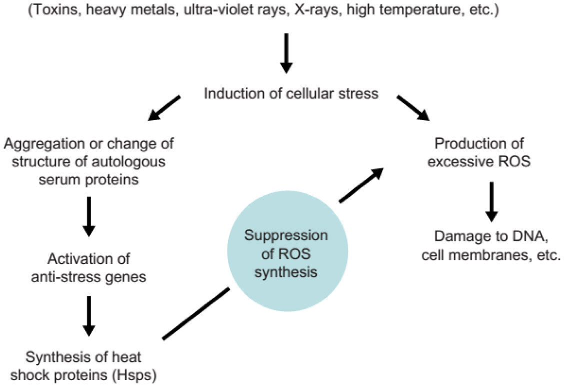
Ikwegbue PC, Masamba P, Oyinloye BE, et al. (2018) Roles of heat shock proteins in apoptosis, oxidative stress, human inflammatory diseases, and cancer. Pharmaceuticals 11: 2. doi: 10.3390/pharmaceutics11010002
    [5] Uttara B, Singh AV, Zamboni P, et al. (2009) Oxidative stress and neurodegenerative diseases: A review of upstream and downstream antioxidant therapeutic options. Curr Neuropharmacol 7: 65–74. doi: 10.2174/157015909787602823
    [6] Maridonneau-Parini I, Clerc J, Polla BS (1988) Heat shock inhibits NADPH oxidase in human neutrophils. Biochem Biophys Res Commun 154: 179–186. doi: 10.1016/0006-291X(88)90667-5
    [7] Costa VM, Silva R, Ferreira LM, et al. (2007) Oxidation process of adrenaline in freshly isolated rat cardiomyocytes: Formation of adrenochrome, quinoproteins, and GSH adduct. Chem Res Toxicol 8: 1183–1191.
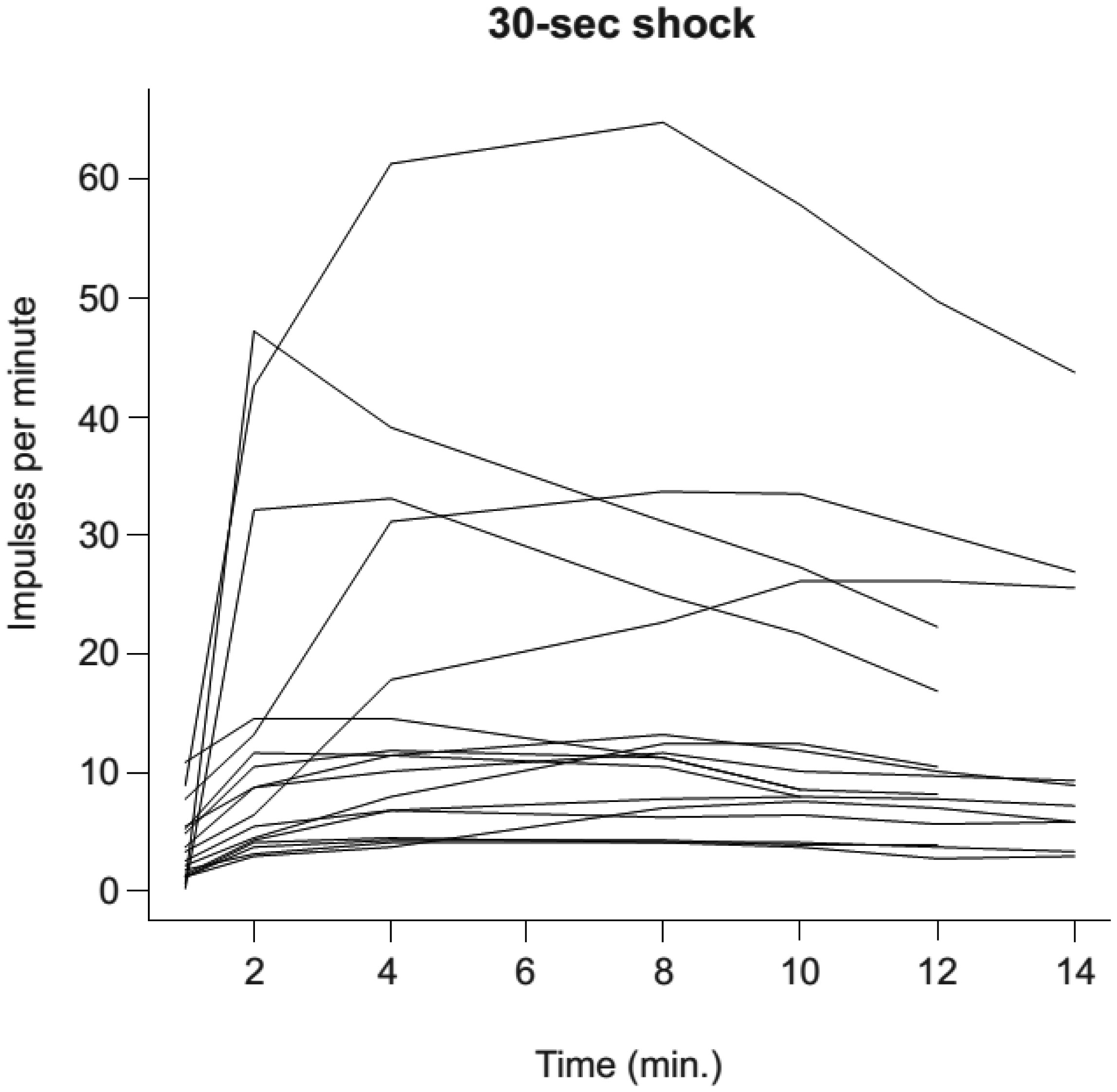
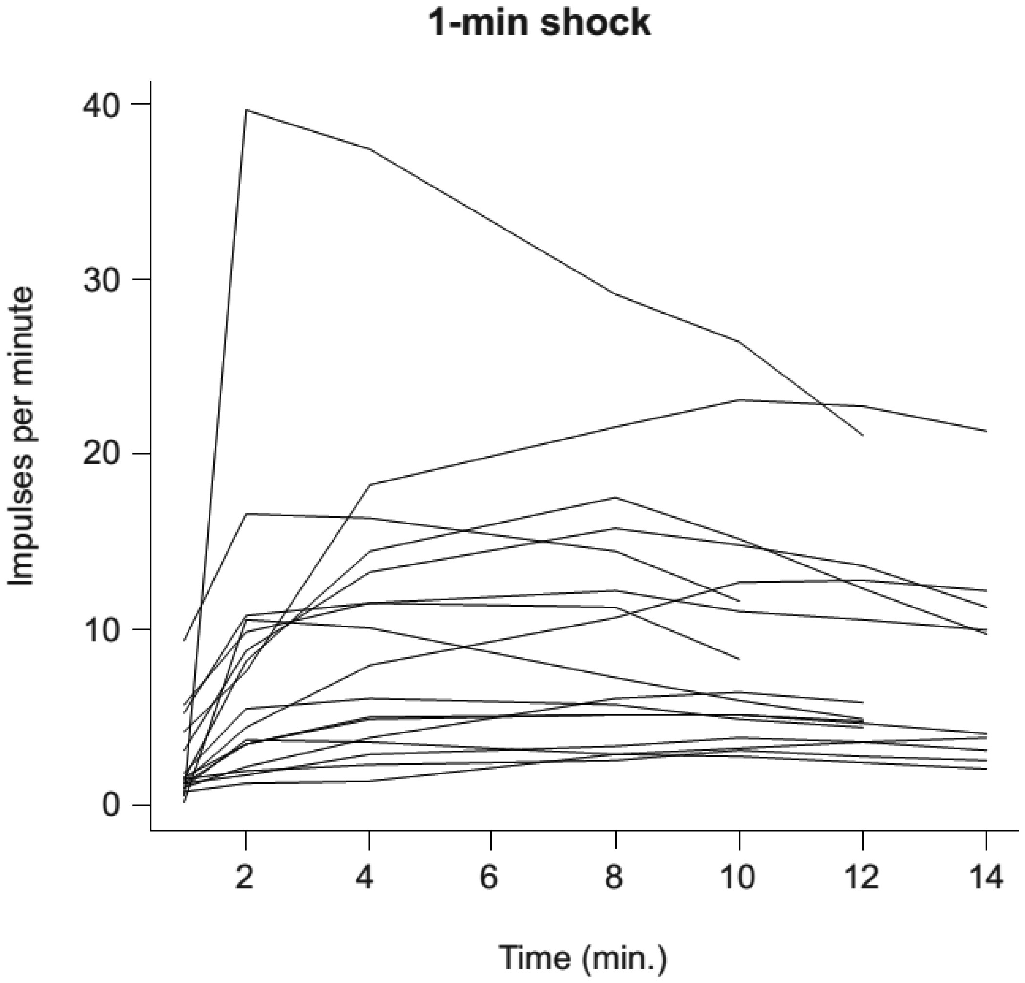
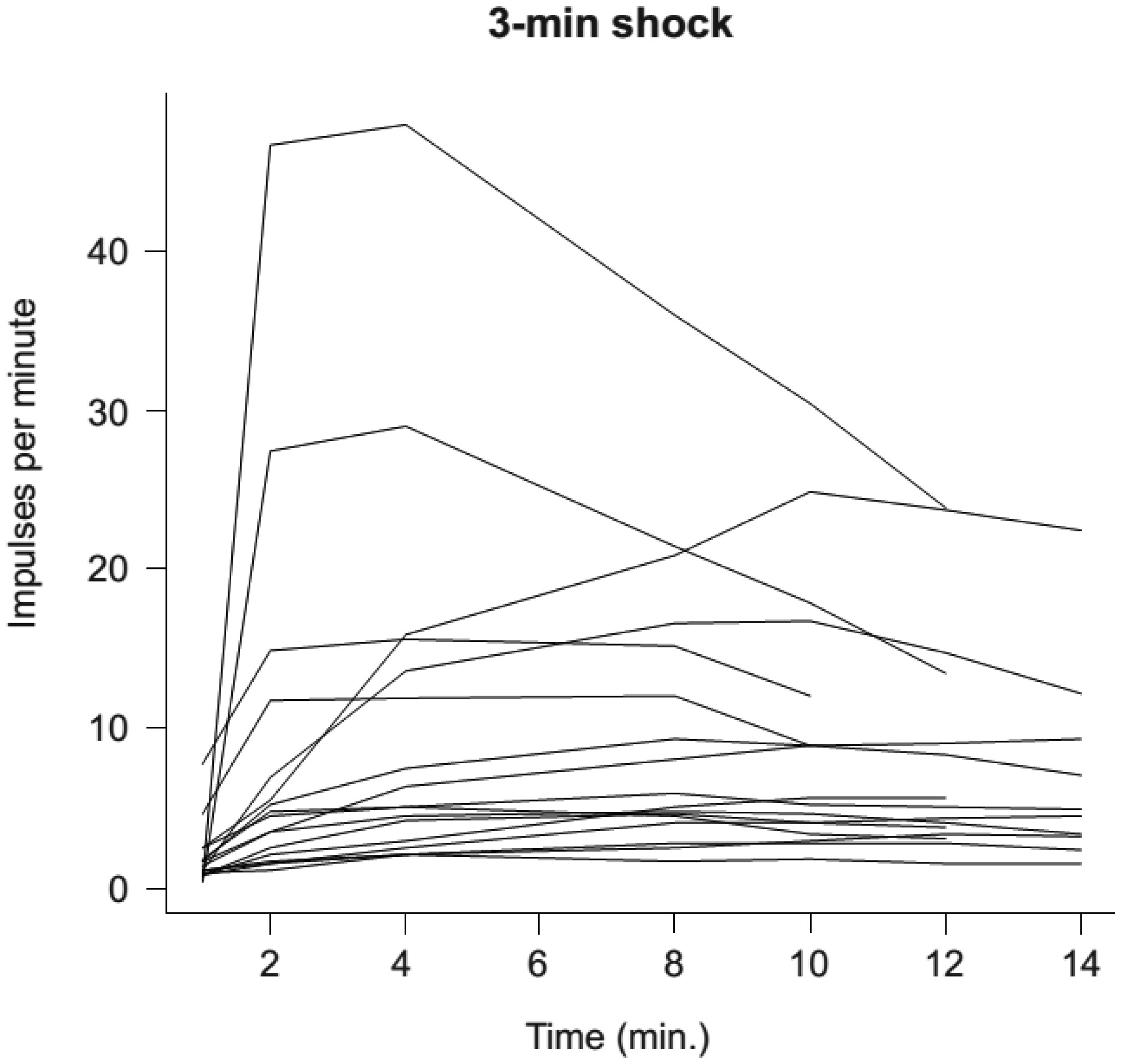
    [8] Kovalenko EI, Boyko AA, Semenkov VF, et al. (2014) ROS production, intracellular HSP70 levels and their relationship in human neutrophils: Effects of age. Oncotarget 5: 11800–11812.
    [9] Allen RC, Loose LD (1976) Phagocytic activation of a luminoldependent chemiluminescence in rabbit alveolar and peritoneal macrophages. Biochem Biophys Res Commun 69: 245–252. doi: 10.1016/S0006-291X(76)80299-9
    [10] Doyle SE, O'Connell RM, Miranda GA, et al. (2004) Toll-like receptors induce a phagocytic gene program through p38. J Exp Med 199: 81–90. doi: 10.1084/jem.20031237
    [11] Hermann JK, Lin S, Soffer A, et al. (2018) The role of Toll-like receptor 2 and 4 innate immunity pathways in intracortical microelectrode-induced neuroinflammation. Front Bioeng Biotechnol 6: 113. doi: 10.3389/fbioe.2018.00113
    [12] Ogava K, Suzuki K, Okutsu M, et al. (2008) The association of elevated reactive oxygen levels from neutrophils with low-grade inflammation in the elderly. Immun Ageing 5: 13–20. doi: 10.1186/1742-4933-5-13
    [13] Singh R, Kolvraa S, Rattan SIS (2007) Genetics of human longevity with emphasis on the relevance of HSP70 as candidate genes. Front Biosci 12: 4504–4513. doi: 10.2741/2405
    [14] Semenkov VF, Michalski AI, Sapozhnikov AM (2015) Heating and ultraviolet light activate anti-stress gene functions in humans. Front Genet 6: 245.
    [15] Chittiboyina S, Bai Y, Lelievre SA (2018) Microenvironment-cell nucleus relationship in the context of oxidative stress. Front Cell Dev Biol 6: 23. doi: 10.3389/fcell.2018.00023
    [16] Zininga T, Ramatsui L, Shonhai A (2018) Heat shock proteins as immunomodulants. Molecules 23: 2846. doi: 10.3390/molecules23112846
    [17] Guisasola MC, Alonso B, Bravo B, et al. (2017) An overview of cytokines and heat shock response in polytraumatized patients. Cell Stress Chaperones 23: 483–489.
  • This article has been cited by:

    1. Wai Myo Maung, Hidemi Nakata, Motoi Miura, Munemitsu Miyasaka, You-Kyoung Kim, Shohei Kasugai, Shinji Kuroda, Low-Intensity Pulsed Ultrasound Stimulates Osteogenic Differentiation of Periosteal Cells In Vitro, 2021, 27, 1937-3341, 63, 10.1089/ten.tea.2019.0331
  • Reader Comments
  • © 2019 the Author(s), licensee AIMS Press. This is an open access article distributed under the terms of the Creative Commons Attribution License (http://creativecommons.org/licenses/by/4.0)
通讯作者: 陈斌, bchen63@163.com
  • 1. 

    沈阳化工大学材料科学与工程学院 沈阳 110142

  1. 本站搜索
  2. 百度学术搜索
  3. 万方数据库搜索
  4. CNKI搜索

Metrics

Article views(3425) PDF downloads(576) Cited by(1)

Figures and Tables

Figures(10)  /  Tables(6)

/

DownLoad:  Full-Size Img  PowerPoint
Return
Return

Catalog