Review Special Issues

Drugs and drug delivery systems targeting amyloid-β in Alzheimer's disease

  • Received: 16 May 2015 Accepted: 20 July 2015 Published: 29 July 2015
  • Alzheimer's disease (AD) is a devastating neurodegenerative disorder with no cure and limited treatment solutions that are unable to target any of the suspected causes. Increasing evidence suggests that one of the causes of neurodegeneration is the overproduction of amyloid beta (Aβ) and the inability of Aβ peptides to be cleared from the brain, resulting in self-aggregation to form toxic oligomers, fibrils and plaques. One of the potential treatment options is to target Aβ and prevent self-aggregation to allow for a natural clearing of the brain. In this paper, we review the drugs and drug delivery systems that target Aβ in relation to Alzheimer's disease. Many attempts have been made to use anti-Aβ targeting molecules capable of targeting Aβ (with much success in vitro and in vivo animal models), but the major obstacle to this technique is the challenge posed by the blood brain barrier (BBB). This highly selective barrier protects the brain from toxic molecules and pathogens and prevents the delivery of most drugs. Therefore novel Aβ aggregation inhibitor drugs will require well thought-out drug delivery systems to deliver sufficient concentrations to the brain.

    Citation: Morgan Robinson, Brenda Yasie Lee, Zoya Leonenko. Drugs and drug delivery systems targeting amyloid-β in Alzheimer's disease[J]. AIMS Molecular Science, 2015, 2(3): 332-358. doi: 10.3934/molsci.2015.3.332

    Related Papers:

    [1] Mohammad Ahmad Ahmad Odah . Unlocking the genetic code: Exploring the potential of DNA barcoding for biodiversity assessment. AIMS Molecular Science, 2023, 10(4): 263-294. doi: 10.3934/molsci.2023016
    [2] Sarmishta Mukhopadhyay, Sayak Ganguli, Santanu Chakrabarti . Shigella pathogenesis: molecular and computational insights. AIMS Molecular Science, 2020, 7(2): 99-121. doi: 10.3934/molsci.2020007
    [3] Praveen Kumar Neela, Rajeshwari B.V, Mahamad Irfanulla Khan, Shahistha Parveen Dasnadi, Gosla Srinivas Reddy, Akhter Husain, Vasavi Mohan . Genetic influences on non-syndromic cleft lip palate: The impact of BMP4, RUNX2, PAX7, and TGFB3 allelic variations. AIMS Molecular Science, 2024, 11(4): 322-329. doi: 10.3934/molsci.2024019
    [4] Anna Kapambwe Bwalya, Robinson Mugasiali Irekwa, Amos Mbugua, Matthew Mutinda Munyao, Peter Kipkemboi Rotich, Tonny Teya Nyandwaro, Caroline Wangui Njoroge, Anne Wanjiru Mwangi, Joanne Jepkemei Yego, Shahiid Kiyaga, Samson Muuo Nzou . Investigation of single nucleotide polymorphisms in MRPA and AQP-1 genes of Leishmania donovani as resistance markers in visceral leishmaniasis in Kenya. AIMS Molecular Science, 2021, 8(2): 149-160. doi: 10.3934/molsci.2021011
    [5] Jin Hwan Do . Genome-wide transcriptional comparison of MPP+ treated human neuroblastoma cells with the state space model. AIMS Molecular Science, 2015, 2(4): 440-460. doi: 10.3934/molsci.2015.4.440
    [6] Seyedeh Fahimeh Razavi, Leila Zarandi Miandoab, Elaheh Zadeh Hosseingholi, Nader Chaparzadeh . Radical scavenging capacity of RuBisCO bioactive peptides derived from Dunaliella salina and Spirulina platensis: An in silico and in vitro study. AIMS Molecular Science, 2025, 12(1): 49-66. doi: 10.3934/molsci.2025004
    [7] Jean Emmanuel Mbosso Teinkela, Philippe Belle Ebanda Kedi, Jean Baptiste Hzounda Fokou, Michelle Isaacs, Lisette Pulchérie Yoyo Ngando, Gaelle Wea Tchepnou, Hassan Oumarou, Xavier Siwe Noundou . In vitro anti-trypanosomal activity of crude extract and fractions of Trichoscypha acuminata stem bark, Spathodea campanulata flowers, and Ficus elastica lianas on Trypanosoma brucei brucei. AIMS Molecular Science, 2024, 11(1): 63-71. doi: 10.3934/molsci.2024005
    [8] Sreemita Majumdar, Song-Tao Liu . Cell division symmetry control and cancer stem cells. AIMS Molecular Science, 2020, 7(2): 82-101. doi: 10.3934/molsci.2020006
    [9] Irene M. Waita, Atunga Nyachieo, Daniel Chai, Samson Muuo, Naomi Maina, Daniel Kariuki, Cleophas M. Kyama . Genetic polymorphisms in eostrogen and progesterone receptor genes in Papio anubis induced with endometriosis during early stage of the disease. AIMS Molecular Science, 2021, 8(1): 86-97. doi: 10.3934/molsci.2021007
    [10] Yutaka Takihara, Ryuji Otani, Takuro Ishii, Shunsuke Takaoka, Yuki Nakano, Kaori Inoue, Steven Larsen, Yoko Ogino, Masashi Asai, Sei-ichi Tanuma, Fumiaki Uchiumi . Characterization of the human IDH1 gene promoter. AIMS Molecular Science, 2023, 10(3): 186-204. doi: 10.3934/molsci.2023013
  • Alzheimer's disease (AD) is a devastating neurodegenerative disorder with no cure and limited treatment solutions that are unable to target any of the suspected causes. Increasing evidence suggests that one of the causes of neurodegeneration is the overproduction of amyloid beta (Aβ) and the inability of Aβ peptides to be cleared from the brain, resulting in self-aggregation to form toxic oligomers, fibrils and plaques. One of the potential treatment options is to target Aβ and prevent self-aggregation to allow for a natural clearing of the brain. In this paper, we review the drugs and drug delivery systems that target Aβ in relation to Alzheimer's disease. Many attempts have been made to use anti-Aβ targeting molecules capable of targeting Aβ (with much success in vitro and in vivo animal models), but the major obstacle to this technique is the challenge posed by the blood brain barrier (BBB). This highly selective barrier protects the brain from toxic molecules and pathogens and prevents the delivery of most drugs. Therefore novel Aβ aggregation inhibitor drugs will require well thought-out drug delivery systems to deliver sufficient concentrations to the brain.


    Grewia tenax (L.) belongs to the Malvaceae family, is a multi-stemmed fruit shrub with manifold uses throughout the tropics and subtropics [1]. For decades it has been used for the preparation of traditional medicines. Grewia tenax is considered as a typical tropical plant specie which can tolerate seasonal drought and withstand temperatures of more than 50°C [2]. Moreover, G. tenax is also known as dune fixing species because of its dense fast growing root system [3]. It is a deciduous fruit-producing shrub or small tree that may reach a height of 1 to 3 m.

    As result of the overexploitation and lack of regeneration, G. tenax wild stands became increasingly threatened, hence the fruit sources and natural gene pool were exhausted [2]. G. tenax grows as a wild plant at low elevations throughout the western Sahelian zone (Mali, Mauritania, Niger, Nigeria and Senegal), the eastern Sahelian zone (Djibouti, Eritrea, Ethiopia, Kenya, Somalia and Sudan), northern Africa (Algeria and Morocco) as well southern Africa (Botswana, Namibia, Transvaal and South Africa). It is also found in the Arabian Peninsula and from Iran to India [1]. In Sudan and South of Sudan, G. tenax is found in Bahr El Gazal, Blue Nile, Darfur, Equatoria, Kassala, Khartoum, Kordofan, Upper Nile and White Nile province [4].

    The genus Grewia is composed of about 150 Species [5], its fruits and other parts contribute significantly to the food and energy needs of rural populations in multiple ways [6]. Its leaves and branches are eatable for livestock. Due to their high nutritive values, the fruits have a number of uses like the main source of food during starvation [7].

    The fruit, known locally in Sudan as “Gudaim,” is a rich source of carbohydrates, protein, and vitamins. minerals and constitutes important contributors to improving the nutritional contents of diet of rural and urban people in Sudan [6]. Rural populations consider G. tenax as a source of income by sale of the fruits and other products [2].

    G. tenax fruits were thought for a long time as a simple, naturally available food and medication against iron-deficiency anemia and fatigue [3],[8],[9]. Anemia, which is one of the top ten death-causing disorders in developing countries, not related only to malnutrition and poverty, but also to the free radicals activities [10],[11]. Grewia tenax extracts were reported to have effects on the regulation of iron digestive transfer and absorption [9].

    Biochemical markers (isozymes/allozymes), direct DNA sequencing, and molecular (DNA) markers can be used to investigate genetic variation within and among populations [12]. Until recently, research on the genetics of tropical trees was restricted mainly to allozyme studies of the genetic structure of trees in continuous forests [13],[14]. The first DNA marker exploited is referred to as Restriction Fragment Length polymorphism (RFLPs) [15]. The recent molecular techniques such as Random Amplified polymorphic DNA (RAPD) [16], Inter Simple Sequence Repeat polymorphism [17], Microsatellites (also known as Simple Sequence Repeat, SSRs [18], Amplified Fragment Length polymorphism [19] as well as Inverse Sequence-Tagged Repeat [20] which involve the Polymerase Chain Reaction (PCR), in which amplification of genomic DNA fragments is conducted using a heat-resistant DNA polymerase (Taq polymerase), primers and deoxyribonucleotide triphosphates at high temperatures [21].The use of molecular markers in the investigation of genetic variation is getting a wide acceptance and broad application in fields such as phylogeny, taxonomy, ecology, genetics and breeding [12].

    The general objectives of the study is to determine the genetic variation in populations of Grewia tenax found at different altitudes and geographical locations ranging from scattered to continuous populations over a wide geographical distance of Sudan, by employing DNA marker systems. The general objective of the study is to determine the genetic variation in populations of Grewia tenax found at different altitudes and geographical locations ranging from scattered to continuous populations over a wide geographical distance of Sudan, by employing DNA marker systems techniques.

    Table 1.  Sources of Grewia tenax leaves, locations, rain fall and soil types at the site.
    Sample No. Name (Area) Site of collection Location Type of soil Rain fall Per year
    1-12 Abuhraz Northern Kordofan Western Sudan Sandy clay 318 mm
    13-24 Almnzfa Northern Kordofan Western Sudan sand 320 mm
    25-36 Elobaid1 Northern Kordofan Western Sudan sand 300-350 mm
    37-48 Garsilla Northern Kordofan Western Sudan sand 318 mm
    49-60 Zalingei Zalingei Northern West of Nyala Sand 150 mm
    61-72 Elobaid2 Northern Kordofan Western Sudan sand 300-350 mm
    73-84 Khoralbyed Northern Kordofan Western Sudan Alluvial 300-350 mm
    85-96 Alain Northern Kordofan Southern east of Elobaid Sandy clay 380 mm
    97-108 Elobaid3 Northern Kordofan Western Elobaid Sandy clay 300-350 mm
    109-120 ALdamazin Blue Nile Southern blue Nile clay 691 mm
    121-132 Khartoum Teacher houses Southern Khartoum Clay 162 mm
    133-144 Shambat-Bahry Northern Khartoum Northern Khartoum Clay 155 mm

     | Show Table
    DownLoad: CSV

    Collection Sites and laboratory: leaves were collected from 12 populations, with each population represented by 12 natural population samples thought to constitute the geographical range of Grewia tenax. Samples were carried to the laboratory in brown paper bags and kept in Refrigerator (5°C) for future DNA extraction. Use of plastic bags was avoided as it led to degradation of leaf samples. The Selection parameters for leaves collection was based on detail of samples and other detail are mentioned. The location and details of leaves are showed in Table 1 and Figure 1. All samples were collected from natural, healthy and productive Grewia tenax trees. Total genomic DNA was isolated using the Dellaporta method [22] (with slight modification made in buffer concentrations).

    Sample preparation: fresh young and healthy leaves were collected from plant samples (0.5 g) and kept in aluminum foil under cool conditions till use. Samples were placed into mortar grinders and covered with dry ice powder until it thawed followed by grinding to fine powder with vigorous pulverizing at intervals of 20 to 30 seconds. Ground green dry powders were immediately used for DNA isolation.

    – Ten percent (w/v) CTAB (Cetyltrimethylammonium bromide).

    – 10g of CTAB was dissolved in 70ml of distel water and then final volume was made to 100 ml.

    – 0.5M EDTA (pH 8.0) - 93.05 g of Ethylene Diamine Tetra Acetate was added to 400 ml of water. The pH of solution was adjusted to 8.0 by addition of NaOH (Sodium Hydroxide). The final volume was made to 500 ml and sterilized by autoclaving.

    – 5M NaCl- 146.1 g of sodium chloride was dissolved in 400 ml of water and the final volume was made to 500 ml and sterilized by autoclave.

    – Phenol: Chloroform: Isoamyl alcohol (25:24:1) – 750 µl phenol, 720 µl Chloroform and 30 µl Isoamyl alcohol were mixed by vortexing.

    – 1M Tris- 60.58 g Tris base was dissolved in 400 ml of water and the pH was adjusted to 8.0. The final volume was made to 500 ml and sterilized by autoclaving.

    – Chloroform: Isoamyl (24:1).

    - 720 µl Chloroform and 30 µl Isoamyl alcohol were mixed by vortexing.

    – DNA extraction buffer contained 100 mM Tris-HCl, 20 mM, EDTA (pH-8.0), 1.4 M NaCl, 2.0 (w/v) SDS, 0.2 per cent (v/v) 2-Mercaptoethanol.

    –TE buffer – 10mM Tris, 1mM EDTA, and (pH 8) and stored at 4°C for further use.

    Figure 1.  Geographic location of collection sites of 12 populations of Grewia tenax in Sudan.

    DNA isolation was based on phenol: chloroform: isoamyl alchohol (24:25:1) protocol as described by [22]. The modification was made on step of isoamyl alcohol: chloroform with the intention to improve the quantity and the quality of the DNA. In this method the fine powder plant materials (0.5g) were immediately transferred into 15 ml Falcon tubes containing 6 ml of pre-warmed lysis solution. Tubes containing the samples were then incubated in water path at 65 °C with gentle shaking for 30 min and left to cool at room temperature for 5 min. 3ml of phenol: chloroform: (25:24) added to each tube and the phases were mixed gently for 5 min at room temperature to make homogeneous mixture. Centrifugation at 5000 rpm for 15 min was done to remove the cell debris, the resulted aqueous phases (containing DNA) were transferred to new sterile tubes. The transferred aqueous phase was mixed thoroughly with equal volume of chloroform: isoamyl alchohol (24:1) followed by centrifugation at 5000 rpm for 5min. The step of the chloroform: isoamyl alchohol extraction was repeated twice and the final supernatants were transferred to sterile 1.5 ml Eppendorf tubes (400µl/ tube). The nucleic acids in the aqueous phase precipitated by adding equal volume of cooled absolute ethanol. The contents of the Eppendorf tubes were mixed gently for 5 min by inversion manually and collected by cooled centrifugation (5°C) at 8000 rpm for 3 min. The formed DNA pellet was washed twice with 70% ethanol, and the ethanol was discarded after spinning with flash centrifugation. The remained ethanol was evaporated after leaving the pellet to dry at room temperature. The pellet was dissolved in TE buffer. The extracted DNA samples were observed under UV illumination after staining with (Ez- Vision (cat number 10450003-1), which was used instead of Ethidium Bromide.

    According to [23], the purity and the concentration of the DNA were assessed. The DNA isolated was quantified by using Lambda UV-vis spectrophotometer. First the absorbance was set to zero by using 50 µl of Tris EDTA buffer in the cuvette. The cuvette was properly washed with distilled water, 2 µl of the isolated DNA added to 48 µl of TE buffer, the ratio at 260/280 nm was recorded following the amount of DNA in terms of ng/µl. This quantification of isolated DNA provided with concentration of DNA present in a particular sample and also the 260/280 nm ratio was estimated to find out purity of DNA. This ratio is 1.8 for pure double stranded DNA. If the ratio is greater than 1.8 this suggests RNA contamination, where as one less than 1.8 suggests protein in the sample. The quantified samples were brought to the final working concentration of DNA to 5ng/µl, which will then be used for the RAPD and ISSR analysis.

    For standardization of the amplification conditions a total of twenty decamer oligonucleotide primers were used for amplification of the extracted cellular DNAs. Primers were selected according to the reproducibility of their amplification product. Primers used in this study were OPA-17, OPA-09, OPA-02, OPC-15, OPD-01, OPD-03, OPG-10, OPN-15, OPS-03, OPS-06, OPC-04, OPC-11, OPC-13, PF-08, OPF-12, OPF-13, OPA-04, OPA-05, OPO-20, and OPF-15. They were obtained from Operon Technologies Inc., USA (Table 2).

    For standardizing the concentration of template DNA, PCR amplification was performed with different DNA concentrations using previously standardized master-mix concentrations. Five different concentrations of DNA were used which were 5 ng, 10 ng, 15 ng, 20 ng and 25 ng, for obtaining the maximum number of amplification products. The maximum number of amplification products was observed at 20 ng of template DNA.

    Tests were performed for standardizing polymerase chain reaction amplification conditions mainly the annealing temperature. PCR amplification conducted at different annealing temperatures i.e. 36°C, 37°C, 38°C using standard concentrations of various components of reaction mixture. The maximum number of amplification products was observed at 20 ng of template DNA.

    The PCR reaction mixtures were prepared according to [24], in 25µl volumes containing 2.5 µl of buffer (B), 3µl MgCl2 (25mM), 0.25µl dNTP's (200 µM), 2µl random primer (10 pmol/µl), 0.5µl Taq polymerase 5U/µl and 1µl of extracted DNA ( 20 ng). The mixture was completed to 25 µl by addition of sterilized distilled water. The reagents were mixed thoroughly in 2ml microcentrifuge tube and vortexed for 5 seconds. 24 µl of mixture was distributed to each PCR tube and 1 µl of template DNA (20 ng/µl) was added to each tube for each amplification reaction in thermal cycler (Real- time PCR, Bio- Rad CFX96) [25].

    PCR conditions used for RAPD amplification included initial denaturation for 3 min at 94°C followed by 45 cycles of amplification (denaturation at 92°C for 45 seconds, annealing of primer at 36°C for 1min and primer amplification at 72 °C for 2 min) and final extension at 72 °C for 10 min. the PCR machine was adjusted to hold the product at 4 °C.

    Amplification products were separated on 2 per cent agarose gel using 1X TBE buffer (Tris HCI pH 8.0, Boric Acid, Ethylene diamine tetra- Acetic). Horizontal gel electrophoresis apparatus (Consort-UK) [26]. Ez-vision dye was used as intercalating agent. 5 µl of RAPD amplification products were mixed with 1 µl of Ez-vision dye and loaded on to the gel. Gel was run according to 5v/cm of the length of gel till the bands separate. 1 Kb DNA ladder (250 - 1000bp, cat number: DM010-R500) was used as standard in the first well of the gel.

    DNA amplification was carried out for ISSR analysis. A total of ten ISSR primers synthesized by Operon Technologies Inc., USA. Primers used in this study were 864, 835, 825, 812, 811, 810, 809, UBC-841, UBC-827, and UBC-807 (Table 3).

    The PCR reaction mixtures were prepared in 25µl volumes containing 2.5µl of buffer (B), 4µl MgCl2 (25mM), 0.25µl dNTP's (200µM), 2µl random primer (10 pmol/µl), 0.5µl Taq polymerase 5U/µl and 1µl of extracted DNA ( 20 ng) the mixture was made up to 25 µl by addition of sterilized distilled water. The reagents were mixed thoroughly in 2ml microcentrifuge tube and vortexed for seconds. 23 µl of mixture was distributed to each PCR tube and 2 µl of template DNA (20 ng/µl) was added to each tube for each amplification reaction in thermal cycler (Real- time PCR, Bio- Rad CFX96)[25].

    PCR conditions used for ISSR amplification included initial denaturation for 3 min at 94 °C followed by 45 cycles of amplification (denaturation at 92 °C for 45 seconds, annealing of primer at 36 °C for 1 min and primer amplification at 72 °C for 2 min) and final extension at 72 °C for 10 min. the PCR machine was adjusted to hold the product at 4 °C.

    After the run was over, the gel was viewed under the UV light using Gel Documentation system and was photographed.

    The amplified bands after separation were visualized using Gel Documentation System. For each primer, the number of polymorphic and monomorphic bands was determined. Band clearly visible in at least one genotype were scored (1) for present, 0 for absent and entered into data matrix. Fragment size was estimated by interpolation from the migration distance of marker fragments. Percentage of polymorphism was calculated as the proportion of polymorphic bands over the total number of bands. The genetic dissimilarity (D) matrix among genotypes was estimated according to [27]. Coefficient of similarity trees were produced by clustering the similarity data with the unweighted pair group method using STATISTCA and GenALEx 6.5 software. The similarity coefficient was used to construct a dendrogram by the unweighted pair group method with arithmetic averages (UPGMA) according to [28].

    Principal coordinates analysis (PCoA) was performed with the GenAlEx 6.5 software, using unbiased Nei genetic distances [29].

    Table 2.  Codes and sequences of RAPD primers.
    Primer Number Primer Code Primer sequence (5′-3′)
    1 OPA-17 GACCGCTTGT
    2 OPA-09 GGGTAACGCC
    3 OPA-02 TGCCGAGCTG
    4 OPC-15 GACGGATCAG
    5 OPD-01 ACCGCGAAGG
    6 OPD-03 GTCGCCGTCA
    7 OPG-10 AGGGCCGTCT
    8 OPN-15 CAGCGACTGT
    9 OPS-03 CAGAGGTCCC
    10 OPS-06 GATACCTCGG
    11 OPC-04 CCGCATCTAC
    12 OPC-11 AAAGCTGCGG
    13 OPC-13 AAGCCTCGTC
    14 PF-08 GGGATATCGG
    15 OPF-12 ACGGTACCAG
    16 OPF-13 GGCTGCAGAA
    17 OPA-04 AATCGGGCTG
    18 OPA-05 AGGGGTCTTG
    19 OPO-20 ACACACGCTC
    20 OPF-15 CCAGTACTCC

     | Show Table
    DownLoad: CSV
    Table 3.  Codes and sequences of ISSR primers.
    Primer Number Primer Code Primer sequence (5′-3′)
    1 864 ATGATGATGATGATGATG
    2 835 AGAGAGAGAGAGAGAG[Y]C
    3 825 CACACACACACACACC
    4 812 GAGAGAGAGAGAGAGAA
    5 811 GAGAGAGAGAGAGAGAC
    6 810 GAGAGAGAGAGAGAGAT
    7 809 AGAGAGAGAGAGAGAGG
    8 UBC-841 GAGAGAGAGAGAGAGA[Y]C
    9 UBC-827 ACACACACACACACACACA
    10 UBC-807 AGAGAGAGAGAGAGAGT

     | Show Table
    DownLoad: CSV

    RAPD and ISSR Combination analysis.

    The evaluation of the twelve Grewia populations detected with Fourteen RAPD and 10 ISSR primers resulted in overall Grewia genotypes diversity (He) recorded at 0.250 (Table 4).

    Based on pair-wise population analysis, Abuhraz were found to be the most diverse population with average Nei's gene diversity and Shannon index values of 0.325 and 0.480 respectively, whereas Elobaid 2 was the least diverse followed by Elobaid3 (0.184 and 0.201) and Shannon index of (0.205 and 0.301) respectively ( Table 4). The different alleles (Na) and effective alleles numbers (Ne) also followed the same trend being highest for Abuharaz and lowest for Elobaid 2 (Table 4). The proportion of polymorphic loci for the present set of population ranged from 52.47% (Elobaid2) to Abuharaz with average 68.16% polymorphism.

    The maximum dissimilarity (0.5) was observed between samples (Elobaid2 and Almnzfa) while the minimum dissimilarity (0.1) was recorded between (Khartoum and Abuharaz).

    Pair-wise distance analysis (Nei genetic distance) between populations (Table 5) ranged from 0.267 (between Elobaid 2 and Abuhraz) to 0.098 (between Zalingei and Garsilla). The highest distances were observed between the populations Abuhraz and Shambat.

    The distances between the Grewia spp populations were low indicating close relatedness of genotypes from sometimes widely separated geographical locations. This may be due to a high gene flow resulting in exchange across regions.

    Table 4.  Genetic diversity within populations and genetic differentiation parameters of twelve populations of Grewia tenax genotypes detected by both RAPD and ISSR primers.
    Population N Na Ne I He P (%)
    Abuhraz 12.000 1.848 1.573 0.480 0.325 88.34
    Alain 12.000 1.583 1.449 0.372 0.254 67.26
    ALdamazin 12.000 1.578 1.405 0.353 0.237 65.92
    Almnzfa 12.000 1.785 1.543 0.460 0.312 82.96
    Elobaid1 12.000 1.637 1.442 0.379 0.255 71.30
    Elobaid2 12.000 1.377 1.315 0.275 0.184 52.47
    Elobaid3 12.000 1.439 1.346 0.301 0.201 57.85
    Garsilla 12.000 1.700 1.505 0.422 0.287 76.23
    Khartoum 12.000 1.466 1.399 0.335 0.228 60.54
    Khoralbyed 12.000 1.619 1.440 0.372 0.252 68.16
    Shambat 12.000 1.493 1.397 0.334 0.226 60.99
    Zalingei 12.000 1.601 1.433 0.361 0.245 65.92
    Mean 12.000 1.594 1.437 0.370 0.250 68.16
    SE 0.000 0.012 0.007 2.98% 0.004 2.98

    *Note: Na, number of different alleles; Ne, number of effective alleles; I, Shannon's Information Index, P(%); percentage of polymorphic bands, He, Expected Heterozygosity,SE; standard error.

     | Show Table
    DownLoad: CSV

    Principal coordinate analysis of the 144 Grewia genotypes generated a total variation of 11.17%. The first and the second principal coordinates explained 7.58 and 5.67 of genetic variation, respectively (Figure 2).

    PCoA diagram (Figure 3) showed individual accessions of Shambat population from gathering to gather with genotypes from (Elobaid2) population.

    Principal coordinate analysis of the12 populations showed a total variation of 36.29%, the first and the second principal coordinates explained 17.52 and 15.12 of genetic variation, respectively (Figure 3).The AMOVA test, calculated to examine the differences in molecular variance among and within geographical populations was found to be statistically significant (p < 001) (Table 6). The test showed highest genetic variation within population (74%), whereas the variation among geographic populations was 26% (Table 6). The calculated PhiPT (0.349) was significant P < 0.001, indicating low genetic differentiation among populations. The P values were calculated for a random permutation test of 9999 permutations (Table 6).

    Table 5.  Nei's unbiased measures of genetic identity (above diagonal) and genetic distance (below diagonal) of the twelve populations of Grewia tenax genotypes.
    populations Abuhraz Alain Aldmazin Almnzfa Elobaid1 Elobaid2 Elobaid3 Garsilla Khartoum khoralbyed shambat zalingei
    Abuhraz 1.000 0.845 0.856 0.848 0.838 0.838 0.841 0.804 0.908 0.882 0.884 1.000
    Alain 0.207 1.000 0.859 0.890 0.860 0.860 0.826 0.861 0.838 0.837 0.865 0.813
    ALdamazin 0.226 0.123 1.000 0.827 0.863 0.863 0.821 0.871 0.818 0.865 0.844 0.798
    Almnzfa 0.123 0.145 0.126 1.000 0.826 0.826 0.874 0.837 0.877 0.856 0.851 0.885
    Elobaid1 0.179 0.169 0.178 0.096 1.000 0.818 0.848 0.866 0.834 0.865 0.871 0.836
    Elobaid2 0.267 0.162 0.145 0.177 0.218 1.000 0.853 0.809 0.887 0.891 0.851 0.766
    Elobaid3 0.241 0.138 0.156 0.201 0.150 0.173 1.000 0.834 0.826 0.878 0.834 0.786
    Garsilla 0.145 0.161 0.145 0.131 0.139 0.192 0.177 1.000 0.858 0.886 0.904 0.865
    Khartoum 0.236 0.181 0.115 0.181 0.178 0.198 0.151 0.168 1.000 0.862 0.867 0.790
    Khoralbyed 0.203 0.100 0.130 0.120 0.143 0.135 0.147 0.151 0.165 1.000 0.870 0.816
    Shambat 0.265 0.143 0.121 0.191 0.212 0.164 0.191 0.170 0.116 0.155 1.000 0.767
    Zalingei 0.193 0.139 0.149 0.153 0.181 0.159 0.201 0.098 0.189 0.153 0.169 1.000

     | Show Table
    DownLoad: CSV
    Figure 2.  Principal coordinate analysis of 144 Grewia tenax genotypes in Sudan based on RAPD and ISSR data. The first two principal coordinates explained 7.58 and 5.67 % of the variance, respectively.
    Figure 3.  Principal coordinate analysis of 12 Grewia tenax genotypes populations in Sudan based on RAPD and ISSR data. The first two principal coordinates explained 18.94 and 14.39 % of the variance, respectively.
    Table 6.  Analysis of molecular variance (AMOVA) within and among the populations of Grewia tenax genotypes based on 104 ISSR loci and 119 RAPD loci.
    Source Df SS MS Est. Var. % of total
    Among Populations 11 1636.840 148.804 10.013 26%
    Within Populations 132 3780.917 28.643 28.643 74%
    Total 143 5417.757 177.447 38.657 100%

    *Note: Df: degrees of freedom, MS= mean square, SS= sum of square, Est. Var = estimated variance.

     | Show Table
    DownLoad: CSV

    The highest total number of bands among the 12 studied populations was produced by Grewia tenax genotypes from Abuharaz region producing 215 bands, followed by Almnzfa which produced 213 bands. The lowest number of bands was found in Elobaid2 region which produced 190 bands. No private bands were found in the 12 populations studied.

    The highest number of locally bands found in 25% or less, Alain, Abuharaz, Elobaid3, Aldamazin, khoralbyed, Garsilla, Zalingei and Shambat. Elobaid2 and Elobaid3 regions was the lowest one.

    The expected mean heterozygosity (He) was highest in the Abuharaz region (0.325) and lowest in Elobaid2 region with 0.184 (Figure 4) (Table 7).

    Figure 4.  Total Band Patterns for binary (diploid) data by region.
    Table 7.  Total band patterns for binary data for each population.
    Population Abuhraz Alain ALdamazin Almnzfa Elobaid1 Elobaid2 Elobaid3 Garsilla Khartoum Khoralbyed Shambat Zalingei
    No. Bands 215 203 205 213 206 190 192 209 192 209 197 210
    No. Bands Freq. (>= 5%) 215 203 205 213 206 190 192 209 192 209 197 210
    No. Private Bands 0 0 0 0 0 0 0 0 0 0 0 0
    No. of Locally Common Bands (<=25%) 1 1 1 1 0 0 1 1 0 1 1 1
    No. of Locally Common Bands (<=50%) 7 2 2 5 5 1 1 7 1 1 3 2
    Mean He 0.325 0.254 0.237 0.312 0.255 0.184 0.201 0.287 0.228 0.252 0.226 0.245
    SE of Mean He 0.012 0.014 0.013 0.012 0.013 0.013 0.014 0.013 0.014 0.014 0.014 0.014

     | Show Table
    DownLoad: CSV

    The tree diagram of RAPD and ISSR markers analysis (Figure 5) showed three main clusters, cluster A had two groups, group I included Grewia spp genotypes (2, 7, 8, 10, 11, 12, 13, 17, 18, 14,15) from North kordofan, (Abuhraz, Almnzfa). Group II contained three subgroups, the first from North Kordofan (9, 35, 36, 37, 38), (Abuharaz, Elobaid1, Garsilla) the second contained (21, 22, 23, 24, 25, 26, 27, 29, 30, 28, 33, 34), from Northern Kordofan, (Almnzfa, Elobaid1) the third contained (31, 32), from Northern Kordofan. Cluster B had three groups. Group one had two subgroups, the first from Northern Kordofan contained (39, 40, 47, 44, 48, 43, 45, 41, 42, 46, 49, 50, 51, 53, 54, 57, 55, 56, 52 58, 60, 59), (Garsilla, Zalingei). The second from Northern Kordofan contained (61, 65, 62, 64, 63, 66, 67, 70, 71, 68, 69, 72, 73, 74, 76, 75), (Elobaid2, khoralbyed). Group two had two subgroups, The first from Northern Kordofan contained (77, 78, 79, 80, 81, 82, 83, 84, 85, 86, 87, 88, 90, 89), (khoralbyed, Alain). The second from Northern Kordofan and Blue Nile contained (91, 94, 92, 93, 95, 96, 97, 102, 104, 98, 99, 100, 101, 108, 111, 112, 105, 109, 110, 103), (Alain, Elobaid3, Aldamazin). Group three had three subgroups, the first from Aldamazin contained (113, 114, 115). The second from (Aldamazin, Khartoum, Shambat) contained (116, 118, 117, 119, 127, 120, 121, 124, 122, 123, 126, 128, 125, 129, 130, 132, 131, 133). The third subgroup from Shambat contained (134, 137, 135, 138, 140, 139, 144, 141, 142, 143, 136). Cluster C contained (5, 16, 19, 20) from (Abuhraz, Almnzfa).

    Molecular markers have emerged as convenient methods for quantifying of genetic diversity in populations [30]. History of populations is often inferred from the variation at genetic markers that are assumed to be neutral. However, if a marker is actually subject to selection, conclusion based on patterns of genetic variation could be misleading [31], [32]. Since neutral marker alleles could be linked to deleterious mutations or selectively favored alleles genetic variation can erode faster than expected under neutral assumptions [33]. Gene diversity estimates based on dominant markers like RAPDs and ISSR depends on the frequency of null homozygotes [34][39].

    Study of genetic diversity is very important because it can give a clear picture of whether a species can survive in the long run or not. A population with low genetic diversity cannot tolerate negative environmental impacts as most of the population is identical. population with higher genetic diversity can lead to individuals with a new genetic makeup which may make them survive under adverse conditions [40].

    The genetic diversity studies using molecular markers have become useful due to their better reliability and high resolution. The low reproducibility of RAPD [41], introduces problem when used for cultivar identification compared with the other marker applications.

    ISSRs was proposed for fingerprinting by [42] and commonly used in population genetics, taxonomy and phylogeny of many plant species [43], ISSR primers can also confirm specific amplified DNA polymorphic fragments within the variety [44].The high reproducibility of ISSR markers may be because of using longer primers and higher annealing temperature than those used for RAPD. Based on its unique characters, ISSR technique can detect more genetic loci than isozyme and has higher stability than RAPD [42],[45][47], also the technique is more economical than other molecular marker fingerprinting methods (RAPD, RFLP, AFLP, or SSR). Also ISSR fingerprints appeared to be a useful and quick molecular tool to solve the problems of morphological identification and individual characterization of Grewia spp genotypes. Apart from surveying different genomic regions, the marker distribution throughout the genome and the coverage of DNA targets of each specific marker assay provide additional information [48]. Therefore, arbitrary (RAPD), semi-arbitrary (ISSR) markers were used in the present study. First report evaluating the genetic diversity of large number of Grewia tenax genotypes utilizing multiple marker systems in Sudan which are easy to handle and amenable for PCR-based analysis. Since no reports of RAPD and ISSR analysis are available in Sudan, therefore, we have discussed the results in comparison to other tree species.

    In the present study, 14 of RAPD and 10 ISSR markers (Tables 2 and 3) were utilized to assess the genetic diversity of 144 Grewia tenax genotypes belonging to 12 different populations collected from 12 districts of Sudan (Table 1).

    In this study the mean level of polymorphism revealed by ISSR (97.8 %) is higher than RAPD (94%) method. ISSR primers generated 6 to 12 bands with average of 10.4 bands per populations indicating sufficient genetic diversity among the 144 genotypes (Table 4 and 6).

    In the present study the most polymorphic and reproducible pictures have been obtained with poly (AG) or poly (GA) microsatellites, irrespective of the anchors at the 3′ end, which suggests that these are the most frequent simple sequence repeats in Grewia spp genome. However, poly (AT) or poly (CA) microsatellites have not given good profiles. This might be due to the fact that the distribution of these repeats in the Grewia spp genome was beyond the range of amplification by Taq DNA polymerase. However, this is not likely the situation for poly (AT) repeats, as these are reported to be the most abundant motifs in different plant species [49],[50]. However, in the present investigation, poly (AT) or poly (TA) repeats gave improper amplifications. Similar results were found in rice [51],[52], grapevine [53], wheat [54]. Populations genetics in varieties of Grewia tenax were studied using RAPD and ISSR markers. In present study, the applicability of ISSR and RAPD is compared as genetic marker to characterize the population of Grewia tenax. The results indicate that percentage of ISSR polymorphic bands (97.8%) are higher than RAPD polymorphic bands (94%), these findings are in disagreement with [55] who reported screening of twenty ISSR primers and twenty-five RAPD primers in Grewia optiva, reporting 91.72% and 96.31% respectively. Our results showed that ISSR primers revealed more DNA polymorphism (97.8%) among genotypes of Grewia tenax than RAPD primers (94%). Whereas our results are in agreement with [56].In the study of Morus alba, they observed that ISSR primers revealed 74.13% polymorphism while RAPD generated 60.75% polymorphism among the 11 mulberry genotypes which showed that ISSR primers were more efficient in revealing the DNA polymorphism.

    The AMOVA of the 12 populations revealed that higher genetic variations existed within the populations i.e., 77% and 71% compared to that observed among populations (23 and 29%) with RAPD and ISSR marker systems respectively, while the combination RAPD+ISSR showed 74%, 26%, (Table 6). The results demonstrate that most of the variations in the Grewia populations are due to greater genetic diversity within the populations as opposed to between populations. Greater genetic variation within the populations (88.2%) was also observed in P. cineraria compared to the variance among populations (11.8%), [57]. Similar results were also observed in Acacia senegal populations, 86% within and 14% among the populations [58].

    The dendrograms based on RAPD and ISSR combined markers showed partially different genetic distance levels than when used individually. But when used together, ISSR-based cluster is more similar to the combined cluster than RAPD-based cluster. Results are in disagreement with the studies in Grewia optiva species [55] However, it was discovered that the ISSR-based cluster is more comparable to the combined cluster than the RAPD-based cluster in Lupin (Lupinus sp) [59]. Jatropha curcas also showed similar result when RAPD and ISSR dendrogram patterns were combined [60].

    Cluster analysis was carried out on marker profiling data based on RAPD, ISSR and combination between RAPD-ISSR. The results based on all the DNA marker profiles broadly grouped the 12 populations into three clusters. There was close relationship between some of the populations used in this study; presumably they might have been collected from similar locations.

    According to combined RAPD, ISSR profiles, these genotypes are more closely related in genetic relationship to their populations. For example, samples (Abuhraz, Almnzfa and Elobaid1), coded as ( 3, 4, 6, 11, 21, 22, 34, 23, 24, 33, 25, 27, 26, 28, 29, 30, 32, 31) from North Kordofan were joined RAPD cluster grouped together and closely related also to genotypes from (Alain, Elobaid3, Aldamazin), coded as (93, 108, 104, 110, 105, 109), were joined RAPD cluster. This type of pairing could be attributed to importation or exchange of plants among the populations, as the sites were close enough geographically for introduction of plants from other locations to occur.

    The clustering pattern of Grewia tenax samples obtained using different marker systems (RAPDs, ISSR) exhibited variations in grouping of the samples independent of their place of origin, probably indicating the presence of wide genetic variability among the Grewia tenax from different regions of Sudan. Several studies have also reported limited or low correlation between geographic region and genetic relatedness using molecular marker data [55].

    This study documenting genetic diversity of Grewia tenax genotypes using multiple marker systems (RAPD, ISSR) demonstrated high genetic diversity within populations but moderate genetic diversity among populations. This genetically diverse wild genotypes from twelve populations in Sudan, is urgently required to be characterized and conserved and exploited judiciously for agroforestry, medicinal and timber purposes so as to ensure the existence of the species in near future as well as to harvest economic benefits.

    RAPD and ISSR markers combination have been used in many studies for DNA fingerprinting and phylogenetic analysis [61]. Our study, agreement with [62], we have seen that the reproducibility of RAPD and ISSR markers depends on right PCR conditions.

    This study clearly showed that it was possible to analyze the RAPD and ISSR patterns for correlating their similarity and distance between Grewia tenax genotypes.

    From the results of the combination RAPD - ISSR profiling, it was observed that Grewia tenax genotypes produced good number of amplified bands but few showed less number of amplified bands on some primer. Similarity, unique patterns were observed differentiating all 144 genotypes from each other by using 10 ISSR primers and 14 RAPD primers. It can be concluded that RAPD and ISSR markers could be used for differentiating Grewia genotypes and it might help in generating molecular data base for genotypes conservation.

    Effective plant improvement programs depend on the variability of genetic diversity. It is well known that land races are the original source of variation in plants and are still the major source of variation for crop improvement programs in developing countries.

    Figure 5.  Dendrogram based on UPGMA analysis among 144 Grewia tenax genotypes using 14 RAPD and 10 ISSR primers.

    The genetic variation and genetic relationships among Grewia spp populations from different regions were efficiently determined using RAPD and ISSR markers. The identification of Grewia tenax from the Sudan contributes to our knowledge of genetic relationships and the strategies required for protecting natural populations and preserving genetic diversity. It was concluded that both the marker systems RAPD and ISSR either individually or in combination can be effectively used in determination of genetic relationships among Grewia tenax genotypes.

    [1] Prince M, Jackson J (2009) Alzheimer's Disease International World Alzheimer Report 2009. London. 1-96 p.
    [2] Takeda A, Loveman E, Clegg A, et al. (2006) A systematic review of the clinical effectiveness of donepezil, rivastigmine and galantamine on cognition, quality of life and adverse events in Alzheimer's disease. Int J Geriatr Psychiatry 21: 17-28. doi: 10.1002/gps.1402
    [3] Ong KT, Villemagne VL, Bahar-Fuchs A, et al. (2015) Abeta imaging with 18F-florbetaben in prodromal Alzheimer's disease: a prospective outcome study. J Neurol Neurosurg Psychiatry 86: 431-436. doi: 10.1136/jnnp-2014-308094
    [4] Querfurth HW, LaFerla FM (2010) Alzheimer's disease. N Engl J Med 362: 329-344. doi: 10.1056/NEJMra0909142
    [5] Sperling RA, Aisen PS, Beckett LA, et al. (2011) Toward defining the preclinical stages of Alzheimer's disease: recommendations from the National Institute on Aging-Alzheimer's Association workgroups on diagnostic guidelines for Alzheimer's disease. Alzheimers Dement 7: 280-292. doi: 10.1016/j.jalz.2011.03.003
    [6] Petkova AT, Ishii Y, Balbach JJ, et al. (2002) A structural model for Alzheimer's beta -amyloid fibrils based on experimental constraints from solid state NMR. Proc Natl Acad Sci U S A 99: 16742-16747. doi: 10.1073/pnas.262663499
    [7] Matsuzaki K (2014) How do membranes initiate Alzheimer's Disease? Formation of toxic amyloid fibrils by the amyloid beta-protein on ganglioside clusters. Acc Chem Res 47: 2397-2404.
    [8] Cecchi C, Stefani M (2013) The amyloid-cell membrane system. The interplay between the biophysical features of oligomers/fibrils and cell membrane defines amyloid toxicity. Biophys Chem 182: 30-43.
    [9] Drolle E, Gaikwad RM, Leonenko Z (2012) Nanoscale electrostatic domains in cholesterol-laden lipid membranes create a target for amyloid binding. Biophys J 103: L27-29. doi: 10.1016/j.bpj.2012.06.053
    [10] Drolle E, Hane F, Lee B, et al. (2014) Atomic force microscopy to study molecular mechanisms of amyloid fibril formation and toxicity in Alzheimer's disease. Drug Metab Rev 46: 207-223. doi: 10.3109/03602532.2014.882354
    [11] Hane F, Drolle E, Gaikwad R, et al. (2011) Amyloid-beta aggregation on model lipid membranes: an atomic force microscopy study. J Alzheimers Dis 26: 485-494.
    [12] Lal R, Lin H, Quist AP (2007) Amyloid beta ion channel: 3D structure and relevance to amyloid channel paradigm. Biochim Biophys Acta 1768: 1966-1975. doi: 10.1016/j.bbamem.2007.04.021
    [13] Arispe N, Pollard HB, Rojas E (1993) Giant multilevel cation channels formed by Alzheimer disease amyloid beta-protein [A beta P-(1-40)] in bilayer membranes. Proc Natl Acad Sci U S A 90: 10573-10577. doi: 10.1073/pnas.90.22.10573
    [14] Demuro A, Smith M, Parker I (2011) Single-channel Ca(2+) imaging implicates Abeta1-42 amyloid pores in Alzheimer's disease pathology. J Cell Biol 195: 515-524. doi: 10.1083/jcb.201104133
    [15] Plant LD, Boyle JP, Smith IF, et al. (2003) The production of amyloid beta peptide is a critical requirement for the viability of central neurons. J Neurosci 23: 5531-5535.
    [16] Giuffrida ML, Caraci F, Pignataro B, et al. (2009) Beta-amyloid monomers are neuroprotective. J Neurosci 29: 10582-10587. doi: 10.1523/JNEUROSCI.1736-09.2009
    [17] Hardy J, Selkoe DJ (2002) The Amyloid Hypothesis of Alzheimer's Disease: Progress and Problems on the Road to Therapeutics. Science 297: 353-356. doi: 10.1126/science.1072994
    [18] Brambilla D, Verpillot R, Le Droumaguet B, et al. (2012) PEGylated nanoparticles bind to and alter amyloid-beta peptide conformation: toward engineering of functional nanomedicines for Alzheimer's disease. ACS Nano 6: 5897-5908. doi: 10.1021/nn300489k
    [19] Gobbi M, Re F, Canovi M, et al. (2010) Lipid-based nanoparticles with high binding affinity for amyloid-beta1-42 peptide. Biomaterials 31: 6519-6529. doi: 10.1016/j.biomaterials.2010.04.044
    [20] Cheng KK, Yeung CF, Ho SW, et al. (2013) Highly stabilized curcumin nanoparticles tested in an in vitro blood-brain barrier model and in Alzheimer's disease Tg2576 mice. Aaps j 15: 324-336. doi: 10.1208/s12248-012-9444-4
    [21] Cheng X, van Breemen RB (2005) Mass Spectrometry-Based Screening for Inhibitors of β-Amyloid Protein Aggregation. Anal Chem 77: 7012-7015. doi: 10.1021/ac050556a
    [22] He H, Dong W, Huang F (2010) Anti-Amyloidogenic and Anti-Apoptotic Role of Melatonin in Alzheimer Disease. Curr Neuropharmacol 8: 211-217. doi: 10.2174/157015910792246137
    [23] Lin L, Huang Q-X, Yang S-S, et al. (2013) Melatonin in Alzheimer's Disease. Int J Mol Sci 14: 14575-14593. doi: 10.3390/ijms140714575
    [24] Rosales-Corral S, Acuna-Castroviejo D, Tan DX, et al. (2012) Accumulation of Exogenous Amyloid-Beta Peptide in Hippocampal Mitochondria Causes Their Dysfunction: A Protective Role for Melatonin. Oxid Med Cell Longev 2012: 843649.
    [25] Adolfsson O, Pihlgren M, Toni N, et al. (2012) An effector-reduced anti-beta-amyloid (Abeta) antibody with unique abeta binding properties promotes neuroprotection and glial engulfment of Abeta. J Neurosci 32: 9677-9689. doi: 10.1523/JNEUROSCI.4742-11.2012
    [26] DeMattos RB, Bales KR, Cummins DJ, et al. (2001) Peripheral anti-A beta antibody alters CNS and plasma A beta clearance and decreases brain A beta burden in a mouse model of Alzheimer's disease. Proc Natl Acad Sci U S A 98: 8850-8855. doi: 10.1073/pnas.151261398
    [27] Legleiter J, Czilli DL, Gitter B, et al. (2004) Effect of different anti-Abeta antibodies on Abeta fibrillogenesis as assessed by atomic force microscopy. J Mol Biol 335: 997-1006. doi: 10.1016/j.jmb.2003.11.019
    [28] Lemere CA (2013) Immunotherapy for Alzheimer's disease: hoops and hurdles. Mol Neurodegener 8: 36. doi: 10.1186/1750-1326-8-36
    [29] Robert R, Wark KL (2012) Engineered antibody approaches for Alzheimer's disease immunotherapy. Arch Biochem Biophys 526: 132-138. doi: 10.1016/j.abb.2012.02.022
    [30] Wilcock DM, Rojiani A, Rosenthal A, et al. (2004) Passive immunotherapy against Abeta in aged APP-transgenic mice reverses cognitive deficits and depletes parenchymal amyloid deposits in spite of increased vascular amyloid and microhemorrhage. J Neuroinflammation 1: 24. doi: 10.1186/1742-2094-1-24
    [31] Findeis MA, Musso GM, Arico-Muendel CC, et al. (1999) Modified-peptide inhibitors of amyloid beta-peptide polymerization. Biochemistry 38: 6791-6800. doi: 10.1021/bi982824n
    [32] Ghanta J, Shen CL, Kiessling LL, et al. (1996) A strategy for designing inhibitors of beta-amyloid toxicity. J Biol Chem 271: 29525-29528. doi: 10.1074/jbc.271.47.29525
    [33] Hane FT, Lee BY, Petoyan A, et al. (2014) Testing synthetic amyloid-beta aggregation inhibitor using single molecule atomic force spectroscopy. Biosens Bioelectron 54: 492-498. doi: 10.1016/j.bios.2013.10.060
    [34] Pallitto MM, Ghanta J, Heinzelman P, et al. (1999) Recognition sequence design for peptidyl modulators of beta-amyloid aggregation and toxicity. Biochemistry 38: 3570-3578. doi: 10.1021/bi982119e
    [35] Tjernberg LO, Naslund J, Lindqvist F, et al. (1996) Arrest of beta-amyloid fibril formation by a pentapeptide ligand. J Biol Chem 271: 8545-8548. doi: 10.1074/jbc.271.15.8545
    [36] Morgan D, Diamond DM, Gottschall PE, et al. (2000) A beta peptide vaccination prevents memory loss in an animal model of Alzheimer's disease. Nature 408: 982-985. doi: 10.1038/35050116
    [37] Orgogozo JM, Gilman S, Dartigues JF, et al. (2003) Subacute meningoencephalitis in a subset of patients with AD after Abeta42 immunization. Neurology 61: 46-54. doi: 10.1212/01.WNL.0000073623.84147.A8
    [38] Wildsmith KR, Holley M, Savage JC, et al. (2013) Evidence for impaired amyloid beta clearance in Alzheimer's disease. Alzheimers Res Ther 5: 33. doi: 10.1186/alzrt187
    [39] Evin G, Hince C (2013) BACE1 as a therapeutic target in Alzheimer's disease: rationale and current status. Drugs Aging 30: 755-764. doi: 10.1007/s40266-013-0099-3
    [40] Wolfe MS (2012) gamma-Secretase inhibitors and modulators for Alzheimer's disease. J Neurochem 120 Suppl 1: 89-98.
    [41] Atwal JK, Chen Y, Chiu C, et al. (2011) A therapeutic antibody targeting BACE1 inhibits amyloid-beta production in vivo. Sci Transl Med 3: 84ra43.
    [42] Kao SC, Krichevsky AM, Kosik KS, et al. (2004) BACE1 suppression by RNA interference in primary cortical neurons. J Biol Chem 279: 1942-1949. doi: 10.1074/jbc.M309219200
    [43] Singer O, Marr RA, Rockenstein E, et al. (2005) Targeting BACE1 with siRNAs ameliorates Alzheimer disease neuropathology in a transgenic model. Nat Neurosci 8: 1343-1349. doi: 10.1038/nn1531
    [44] Vassar R (2014) BACE1 inhibitor drugs in clinical trials for Alzheimer's disease. Alzheimers Res Ther 6: 89. doi: 10.1186/s13195-014-0089-7
    [45] Luo Y, Bolon B, Kahn S, et al. (2001) Mice deficient in BACE1, the Alzheimer's beta-secretase, have normal phenotype and abolished beta-amyloid generation. Nat Neurosci 4: 231-232. doi: 10.1038/85059
    [46] Jacobsen H, Ozmen L, Caruso A, et al. (2014) Combined treatment with a BACE inhibitor and anti-Abeta antibody gantenerumab enhances amyloid reduction in APPLondon mice. J Neurosci 34: 11621-11630. doi: 10.1523/JNEUROSCI.1405-14.2014
    [47] Morgan D (2005) Mechanisms of A beta plaque clearance following passive A beta immunization. Neurodegener Dis 2: 261-266. doi: 10.1159/000090366
    [48] Banks WA, Terrell B, Farr SA, et al. (2002) Passage of amyloid beta protein antibody across the blood-brain barrier in a mouse model of Alzheimer's disease. Peptides 23: 2223-2226. doi: 10.1016/S0196-9781(02)00261-9
    [49] Banks WA (2012) Drug delivery to the brain in Alzheimer's disease: consideration of the blood-brain barrier. Adv Drug Deliv Rev 64: 629-639. doi: 10.1016/j.addr.2011.12.005
    [50] Pfeifer LA, White LR, Ross GW, et al. (2002) Cerebral amyloid angiopathy and cognitive function: the HAAS autopsy study. Neurology 58: 1629-1634. doi: 10.1212/WNL.58.11.1629
    [51] Racke MM, Boone LI, Hepburn DL, et al. (2005) Exacerbation of cerebral amyloid angiopathy-associated microhemorrhage in amyloid precursor protein transgenic mice by immunotherapy is dependent on antibody recognition of deposited forms of amyloid beta. J Neurosci 25: 629-636. doi: 10.1523/JNEUROSCI.4337-04.2005
    [52] Wilcock DM, Colton CA (2008) Anti-amyloid-beta immunotherapy in Alzheimer's disease: relevance of transgenic mouse studies to clinical trials. J Alzheimers Dis 15: 555-569.
    [53] Doody RS, Thomas RG, Farlow M, et al. (2014) Phase 3 trials of solanezumab for mild-to-moderate Alzheimer's disease. N Engl J Med 370: 311-321. doi: 10.1056/NEJMoa1312889
    [54] Panza F, Solfrizzi V, Imbimbo BP, et al. (2014) Amyloid-based immunotherapy for Alzheimer's disease in the time of prevention trials: the way forward. Expert Rev Clin Immunol 10: 405-419. doi: 10.1586/1744666X.2014.883921
    [55] Salloway S, Sperling R, Fox NC, et al. (2014) Two phase 3 trials of bapineuzumab in mild-to-moderate Alzheimer's disease. N Engl J Med 370: 322-333. doi: 10.1056/NEJMoa1304839
    [56] Tayeb HO, Murray ED, Price BH, et al. (2013) Bapineuzumab and solanezumab for Alzheimer's disease: is the 'amyloid cascade hypothesis' still alive? Expert Opin Biol Ther 13: 1075-1084. doi: 10.1517/14712598.2013.789856
    [57] Garber K (2012) Genentech's Alzheimer's antibody trial to study disease prevention. Nat Biotechnol 30: 731-732. doi: 10.1038/nbt0812-731
    [58] Panza F, Solfrizzi V, Imbimbo BP, et al. (2014) Efficacy and safety studies of gantenerumab in patients with Alzheimer's disease. Expert Rev Neurother 14: 973-986. doi: 10.1586/14737175.2014.945522
    [59] Kumar J, Sim V (2014) D-amino acid-based peptide inhibitors as early or preventative therapy in Alzheimer disease. Prion 8: 119-124. doi: 10.4161/pri.28220
    [60] Porat Y, Mazor Y, Efrat S, et al. (2004) Inhibition of islet amyloid polypeptide fibril formation: a potential role for heteroaromatic interactions. Biochemistry 43: 14454-14462. doi: 10.1021/bi048582a
    [61] Austen BM, Paleologou KE, Ali SA, et al. (2008) Designing peptide inhibitors for oligomerization and toxicity of Alzheimer's beta-amyloid peptide. Biochemistry 47: 1984-1992. doi: 10.1021/bi701415b
    [62] Taylor M, Moore S, Mayes J, et al. (2010) Development of a proteolytically stable retro-inverso peptide inhibitor of beta-amyloid oligomerization as a potential novel treatment for Alzheimer's disease. Biochemistry 49: 3261-3272. doi: 10.1021/bi100144m
    [63] Roy S (2010) Designing Novel Peptidic Inhibitors of Beta Amyloid Oligomerization Calgary, Alberta: University of Calgary. 164 p.
    [64] Gordon DJ, Tappe R, Meredith SC (2002) Design and characterization of a membrane permeable N-methyl amino acid-containing peptide that inhibits Abeta1-40 fibrillogenesis. J Pept Res 60: 37-55.
    [65] Parthsarathy V, McClean PL, Holscher C, et al. (2013) A novel retro-inverso peptide inhibitor reduces amyloid deposition, oxidation and inflammation and stimulates neurogenesis in the APPswe/PS1DeltaE9 mouse model of Alzheimer's disease. PLoS One 8: e54769. doi: 10.1371/journal.pone.0054769
    [66] Yu YJ, Zhang Y, Kenrick M, et al. (2011) Boosting brain uptake of a therapeutic antibody by reducing its affinity for a transcytosis target. Sci Transl Med 3: 84ra44.
    [67] 
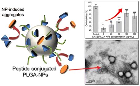
Xiong N, Dong X-Y, Zheng J, et al. (2015) Design of LVFFARK and LVFFARK-functionalized nanoparticles for inhibiting amyloid β-protein fibrillation and cytotoxicity. ACS Appl Mater Interfaces 7: 5650-5662. doi: 10.1021/acsami.5b00915
    [68] Taylor M, Moore S, Mourtas S, et al. (2011) Effect of curcumin-associated and lipid ligand-functionalized nanoliposomes on aggregation of the Alzheimer's Abeta peptide. Nanomedicine 7: 541-550. doi: 10.1016/j.nano.2011.06.015
    [69] Armstrong RA (2014) A critical analysis of the 'amyloid cascade hypothesis'. Folia Neuropathol 52: 211-225.
    [70] Castello MA, Soriano S (2014) On the origin of Alzheimer's disease. Trials and tribulations of the amyloid hypothesis. Ageing Res Rev 13: 10-12.
    [71] Chen Y, Liu L (2012) Modern methods for delivery of drugs across the blood-brain barrier. Adv Drug Deliv Rev 64: 640-665. doi: 10.1016/j.addr.2011.11.010
    [72] Pardridge WM (2003) Blood-brain barrier drug targeting: the future of brain drug development. Mol Interv 3: 90-105, 151. doi: 10.1124/mi.3.2.90
    [73] 
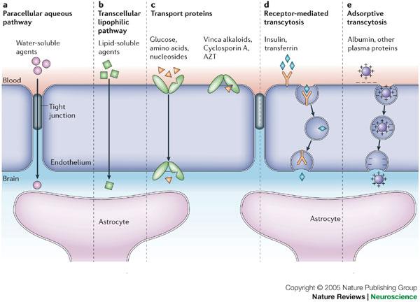
Abbott NJ, Patabendige AA, Dolman DE, et al. (2010) Structure and function of the blood-brain barrier. Neurobiol Dis 37: 13-25. doi: 10.1016/j.nbd.2009.07.030
    [74] Abbott NJ, Ronnback L, Hansson E (2006) Astrocyte-endothelial interactions at the blood-brain barrier. Nat Rev Neurosci 7: 41-53. doi: 10.1038/nrn1824
    [75] Mehta DC, Short JL, Nicolazzo JA (2013) Memantine transport across the mouse blood-brain barrier is mediated by a cationic influx H+ antiporter. Mol Pharm 10: 4491-4498. doi: 10.1021/mp400316e
    [76] Kim MH, Maeng HJ, Yu KH, et al. (2010) Evidence of carrier-mediated transport in the penetration of donepezil into the rat brain. J Pharm Sci 99: 1548-1566. doi: 10.1002/jps.21895
    [77] Cirrito JR, Deane R, Fagan AM, et al. (2005) P-glycoprotein deficiency at the blood-brain barrier increases amyloid-beta deposition in an Alzheimer disease mouse model. J Clin Invest 115: 3285-3290. doi: 10.1172/JCI25247
    [78] Deane R, Wu Z, Zlokovic BV (2004) RAGE (yin) versus LRP (yang) balance regulates alzheimer amyloid beta-peptide clearance through transport across the blood-brain barrier. Stroke 35: 2628-2631. doi: 10.1161/01.STR.0000143452.85382.d1
    [79] Guillemin GJ, Brew BJ (2004) Microglia, macrophages, perivascular macrophages, and pericytes: a review of function and identification. J Leukoc Biol 75: 388-397.
    [80] Bowman GL, Kaye JA, Moore M, et al. (2007) Blood-brain barrier impairment in Alzheimer disease: stability and functional significance. Neurology 68: 1809-1814. doi: 10.1212/01.wnl.0000262031.18018.1a
    [81] Claudio L (1996) Ultrastructural features of the blood-brain barrier in biopsy tissue from Alzheimer's disease patients. Acta Neuropathol 91: 6-14.
    [82] Kalaria RN (1992) The blood-brain barrier and cerebral microcirculation in Alzheimer disease. Cerebrovasc Brain Metab Rev 4: 226-260.
    [83] Marco S, Skaper SD (2006) Amyloid beta-peptide1-42 alters tight junction protein distribution and expression in brain microvessel endothelial cells. Neurosci Lett 401: 219-224. doi: 10.1016/j.neulet.2006.03.047
    [84] Zlokovic BV (2005) Neurovascular mechanisms of Alzheimer's neurodegeneration. Trends Neurosci 28: 202-208. doi: 10.1016/j.tins.2005.02.001
    [85] Wisniewski HM, Vorbrodt AW, Wegiel J (1997) Amyloid angiopathy and blood-brain barrier changes in Alzheimer's disease. Ann N Y Acad Sci 826: 161-172. doi: 10.1111/j.1749-6632.1997.tb48468.x
    [86] Kalaria RN, Sromek SM, Grahovac I, et al. (1992) Transferrin receptors of rat and human brain and cerebral microvessels and their status in Alzheimer's disease. Brain Res 585: 87-93. doi: 10.1016/0006-8993(92)91193-I
    [87] Jones AR, Shusta EV (2007) Blood-brain barrier transport of therapeutics via receptor-mediation. Pharm Res 24: 1759-1771. doi: 10.1007/s11095-007-9379-0
    [88] Gatter KC, Brown G, Trowbridge IS, et al. (1983) Transferrin receptors in human tissues: their distribution and possible clinical relevance. J Clin Pathol 36: 539-545. doi: 10.1136/jcp.36.5.539
    [89] Lee HJ, Engelhardt B, Lesley J, et al. (2000) Targeting rat anti-mouse transferrin receptor monoclonal antibodies through blood-brain barrier in mouse. J Pharmacol Exp Ther 292: 1048-1052.
    [90] 
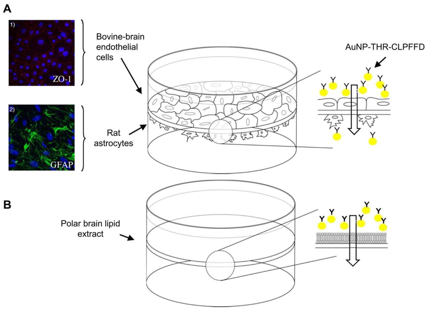
Prades R, Guerrero S, Araya E, et al. (2012) Delivery of gold nanoparticles to the brain by conjugation with a peptide that recognizes the transferrin receptor. Biomaterials 33: 7194-7205. doi: 10.1016/j.biomaterials.2012.06.063
    [91] Zhao WQ, De Felice FG, Fernandez S, et al. (2008) Amyloid beta oligomers induce impairment of neuronal insulin receptors. Faseb J 22: 246-260.
    [92] Xie L, Helmerhorst E, Taddei K, et al. (2002) Alzheimer's beta-amyloid peptides compete for insulin binding to the insulin receptor. J Neurosci 22: Rc221.
    [93] Markoutsa E, Papadia K, Giannou AD, et al. (2014) Mono and dually decorated nanoliposomes for brain targeting, in vitro and in vivo studies. Pharm Res 31: 1275-1289. doi: 10.1007/s11095-013-1249-3
    [94] Re F, Cambianica I, Sesana S, et al. (2010) Functionalization with ApoE-derived peptides enhances the interaction with brain capillary endothelial cells of nanoliposomes binding amyloid-beta peptide. J Biotechnol 156: 341-346.
    [95] Gaillard PJ, Visser CC, de Boer AG (2005) Targeted delivery across the blood-brain barrier. Expert Opin Drug Deliv 2: 299-309. doi: 10.1517/17425247.2.2.299
    [96] Lentz TL (1990) Rabies virus binding to an acetylcholine receptor alpha-subunit peptide. J Mol Recognit 3: 82-88. doi: 10.1002/jmr.300030205
    [97] Liu Y, Huang R, Han L, et al. (2009) Brain-targeting gene delivery and cellular internalization mechanisms for modified rabies virus glycoprotein RVG29 nanoparticles. Biomaterials 30: 4195-4202. doi: 10.1016/j.biomaterials.2009.02.051
    [98] Couch JA, Yu YJ, Zhang Y, et al. (2013) Addressing safety liabilities of TfR bispecific antibodies that cross the blood-brain barrier. Sci Transl Med 5: 183ra157, 181-112.
    [99] Bien-Ly N, Yu YJ, Bumbaca D, et al. (2014) Transferrin receptor (TfR) trafficking determines brain uptake of TfR antibody affinity variants. J Exp Med 211: 233-244. doi: 10.1084/jem.20131660
    [100] Yu YJ, Atwal JK, Zhang Y, et al. (2014) Therapeutic bispecific antibodies cross the blood-brain barrier in nonhuman primates. Sci Transl Med 6: 261ra154.
    [101] Niewoehner J, Bohrmann B, Collin L, et al. (2014) Increased brain penetration and potency of a therapeutic antibody using a monovalent molecular shuttle. Neuron 81: 49-60. doi: 10.1016/j.neuron.2013.10.061
    [102] 
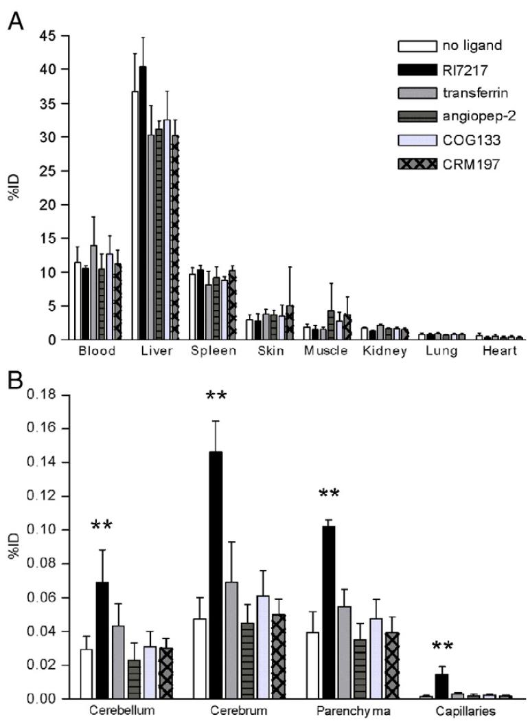
van Rooy I, Mastrobattista E, Storm G, et al. (2011) Comparison of five different targeting ligands to enhance accumulation of liposomes into the brain. J Control Release 150: 30-36. doi: 10.1016/j.jconrel.2010.11.014
    [103] Zhang C, Wan X, Zheng X, et al. (2014) Dual-functional nanoparticles targeting amyloid plaques in the brains of Alzheimer's disease mice. Biomaterials 35: 456-465. doi: 10.1016/j.biomaterials.2013.09.063
    [104] Zhang C, Zheng X, Wan X, et al. (2014) The potential use of H102 peptide-loaded dual-functional nanoparticles in the treatment of Alzheimer's disease. J Control Release 192: 317-324. doi: 10.1016/j.jconrel.2014.07.050
    [105] Salvati E, Re F, Sesana S, et al. (2013) Liposomes functionalized to overcome the blood-brain barrier and to target amyloid-beta peptide: the chemical design affects the permeability across an in vitro model. Int J Nanomedicine 8: 1749-1758.
    [106] Markoutsa E, Papadia K, Clemente C, et al. (2012) Anti-Abeta-MAb and dually decorated nanoliposomes: effect of Abeta1-42 peptides on interaction with hCMEC/D3 cells. Eur J Pharm Biopharm 81: 49-56. doi: 10.1016/j.ejpb.2012.02.006
    [107] Herve F, Ghinea N, Scherrmann JM (2008) CNS delivery via adsorptive transcytosis. AAPS J 10: 455-472. doi: 10.1208/s12248-008-9055-2
    [108] Pardridge WM, Buciak JL, Kang YS, et al. (1993) Protamine-mediated transport of albumin into brain and other organs of the rat. Binding and endocytosis of protamine-albumin complex by microvascular endothelium. J Clin Invest 92: 2224-2229.
    [109] Bickel U (1995) Antibody delivery through the blood-brain barrier. Adv Drug Deliv Rev 15: 53-72. doi: 10.1016/0169-409X(95)00005-R
    [110] Bechara C, Sagan S (2013) Cell-penetrating peptides: 20 years later, where do we stand? FEBS Lett 587: 1693-1702. doi: 10.1016/j.febslet.2013.04.031
    [111] Jaruszewski KM, Ramakrishnan S, Poduslo JF, et al. (2012) Chitosan enhances the stability and targeting of immuno-nanovehicles to cerebro-vascular deposits of Alzheimer's disease amyloid protein. Nanomedicine 8: 250-260. doi: 10.1016/j.nano.2011.06.008
    [112] Parhamifar L, Sime W, Yudina Y, et al. (2010) Ligand-induced tyrosine phosphorylation of cysteinyl leukotriene receptor 1 triggers internalization and signaling in intestinal epithelial cells. PLoS One 5: e14439. doi: 10.1371/journal.pone.0014439
    [113] Malhotra M, Tomaro-Duchesneau C, Prakash S (2013) Synthesis of TAT peptide-tagged PEGylated chitosan nanoparticles for siRNA delivery targeting neurodegenerative diseases. Biomaterials 34: 1270-1280. doi: 10.1016/j.biomaterials.2012.10.013
    [114] Sarvaiya J, Agrawal YK (2015) Chitosan as a suitable nanocarrier material for anti-Alzheimer drug delivery. Int J Biol Macromol 72: 454-465. doi: 10.1016/j.ijbiomac.2014.08.052
    [115] Gao Y, Wang ZY, Zhang J, et al. (2014) RVG-peptide-linked trimethylated chitosan for delivery of siRNA to the brain. Biomacromolecules 15: 1010-1018. doi: 10.1021/bm401906p
    [116] Sharma G, Modgil A, Layek B, et al. (2013) Cell penetrating peptide tethered bi-ligand liposomes for delivery to brain in vivo: Biodistribution and transfection. J Control Release 167: 1-10. doi: 10.1016/j.jconrel.2013.01.016
  • This article has been cited by:

    1. Jai Pal Sharma, Sapna Thakur, Parul Sharma, Anamika Thakur, Genetic structure and gene flow among populations of willow (Salix species), 2024, 32, 24059854, 100283, 10.1016/j.egg.2024.100283
    2. Anand Mishra, R.K. Lal, Pankhuri Gutpa, Sunita Singh Dhawan, Analysis of genetic divergence and heterogeneity in Vetiver (Chrysopogon zizanioides (L.) Roberty) using SCoT and ISSR molecular markers, 2024, 24059854, 100312, 10.1016/j.egg.2024.100312
  • Reader Comments
  • © 2015 the Author(s), licensee AIMS Press. This is an open access article distributed under the terms of the Creative Commons Attribution License (http://creativecommons.org/licenses/by/4.0)
通讯作者: 陈斌, bchen63@163.com
  • 1. 

    沈阳化工大学材料科学与工程学院 沈阳 110142

  1. 本站搜索
  2. 百度学术搜索
  3. 万方数据库搜索
  4. CNKI搜索

Metrics

Article views(10165) PDF downloads(1974) Cited by(16)

/

DownLoad:  Full-Size Img  PowerPoint
Return
Return

Catalog