Mini review Special Issues

Protein aggregation in neurodegenerative disease: the nucleolar connection

  • Protein- and sometimes RNA-containing aggregates are a hallmark of many age-related neurodegenerative diseases. Aggregate depositions can be cytoplasmic, nuclear and even extracellular. This article focuses on nuclear aggregation and the potential role of a specific compartment—the nucleolus, in the process. The nucleolus is a formation site of nucleolar aggresomes—protein and RNA aggregates formed in vitro by hampered proteasome function. Whether the nucleolar aggresomes are connected to nuclear aggregation involved in certain neurodegenerative diseases is an intriguing question for future studies. In addition, recent evidence connecting aggregation and aggregate sorting in the cytoplasm to membrane-enveloped organelles, namely ER and mitochondria, raises the question whether nuclear aggregation and aggregate positioning is controlled by different mechanisms or by the only membrane available—the nuclear membrane.

    Citation: Leena Latonen. Protein aggregation in neurodegenerative disease: the nucleolar connection[J]. AIMS Molecular Science, 2015, 2(3): 324-331. doi: 10.3934/molsci.2015.3.324

    Related Papers:

    [1] Isabel Saez, David Vilchez . Protein clearance mechanisms and their demise in age-related neurodegenerative diseases. AIMS Molecular Science, 2015, 1(1): 1-21. doi: 10.3934/molsci.2015.1.1
    [2] Rosanna Parlato, Holger Bierhoff . Role of nucleolar dysfunction in neurodegenerative disorders: a game of genes?. AIMS Molecular Science, 2015, 2(3): 211-224. doi: 10.3934/molsci.2015.3.211
    [3] Atsushi Tsubota, Hidenori Ichijo, Kengo Homma . Mislocalization, aggregation formation and defect in proteolysis in ALS. AIMS Molecular Science, 2016, 3(2): 246-268. doi: 10.3934/molsci.2016.2.246
    [4] Giulia Ambrosi, Pamela Milani . Endoplasmic reticulum, oxidative stress and their complex crosstalk in neurodegeneration: proteostasis, signaling pathways and molecular chaperones. AIMS Molecular Science, 2017, 4(4): 424-444. doi: 10.3934/molsci.2017.4.424
    [5] Ashok Chakraborty, Anil Diwan . Molecular mechanisms of neurodegenerative disease (NDD). AIMS Molecular Science, 2023, 10(3): 171-185. doi: 10.3934/molsci.2023012
    [6] Joanne L. Hopper, Natasha Begum, Laura Smith, Thomas A. Hughes . The role of PSMD9 in human disease: future clinical and therapeutic implications. AIMS Molecular Science, 2015, 2(4): 476-484. doi: 10.3934/molsci.2015.4.476
    [7] Mohamed A. Eldeeb . Aging: when the ubiquitin–proteasome machinery collapses. AIMS Molecular Science, 2017, 4(2): 219-223. doi: 10.3934/molsci.2017.2.219
    [8] Yuri L. Lyubchenko . Amyloid misfolding, aggregation, and the early onset of protein deposition diseases: insights from AFM experiments and computational analyses. AIMS Molecular Science, 2015, 2(3): 190-210. doi: 10.3934/molsci.2015.3.190
    [9] Marco Feligioni, Serena Marcelli, Erin Knock, Urooba Nadeem, Ottavio Arancio, Paul E. Fraser . SUMO modulation of protein aggregation and degradation. AIMS Molecular Science, 2015, 2(4): 382-410. doi: 10.3934/molsci.2015.4.382
    [10] Morgan Robinson, Brenda Yasie Lee, Zoya Leonenko . Drugs and drug delivery systems targeting amyloid-β in Alzheimer's disease. AIMS Molecular Science, 2015, 2(3): 332-358. doi: 10.3934/molsci.2015.3.332
  • Protein- and sometimes RNA-containing aggregates are a hallmark of many age-related neurodegenerative diseases. Aggregate depositions can be cytoplasmic, nuclear and even extracellular. This article focuses on nuclear aggregation and the potential role of a specific compartment—the nucleolus, in the process. The nucleolus is a formation site of nucleolar aggresomes—protein and RNA aggregates formed in vitro by hampered proteasome function. Whether the nucleolar aggresomes are connected to nuclear aggregation involved in certain neurodegenerative diseases is an intriguing question for future studies. In addition, recent evidence connecting aggregation and aggregate sorting in the cytoplasm to membrane-enveloped organelles, namely ER and mitochondria, raises the question whether nuclear aggregation and aggregate positioning is controlled by different mechanisms or by the only membrane available—the nuclear membrane.


    1. Introduction

    Physiological health and life span depend on maintenance of protein homeostasis, proteostasis [1]. Adaptive protein quality control system detects non-functional and potentially harmful misfolded proteins and promotes their refolding by chaperones and/or their degradation by proteolysis [2,3]. Protein degradation of misfolded proteins takes place by two main mechanisms: ubiquitin-proteome system (UPS) and autophagy [4,5], with UPS degrading misfolded proteins and small aggregates, as autophagy is considered to take care of larger aggregates and cytoplasmic aggresomes.

    Certain proteins have an intrinsic tendency to aggregate, which often involves naturally unstructured domains [6]. In addition, the tendency of a certain protein to aggregate may be increased through disorder-promoting changes in the amino acid sequence. Thus, protein aggregation is in most cases increased due to stress or mutation. Aggregation can be cytoprotective by preventing harmful functions of misfolded proteins. Presence of aggregated proteins, however, has detrimental effects to cells by e.g. decreasing cell proliferation [7]. The aggregates also inhibit the function of the UPS [8]. This may be due to both physical clogging of the proteasomes by aggregated substrates that cannot be degraded [9] and indirect inhibition of the proteasomes [10,11]. This further magnifies the problem, and the aggregates increase. Often, when the capacity of the proteostasis system is exceeded, aggregation is harmful and the aggregates can become cytotoxic [2,5].

    Ability of cells and tissues to maintain proteostasis declines with age. Furthermore, proteostasis failures contribute to a large group of protein misfolding, as aggregation diseases that are often age-related [1,12]. Although still debatable, disease aggregates are increasingly recognized as the cause of cytotoxicity in several neurodegenerative disorders including Alzheimer’s, Parkinson’s, and Huntington’s diseases [12,13].

    2. Composition of aggregates

    Most often the neurodegenerative disease aggregates harbor a primary aggregating substance, such as amyloid beta in Alzheimer’s disease or α-synuclein in Parkinson’s disease. Several mutation mechanisms are known to promote aggregation formation and thus contribute to disease formation. These include e.g. polyglutamine (polyQ) repeats (involved in e.g. Huntington’s disease (HD), spinocerebellar ataxias (SCAs), and spinal bulbar muscular atrophy (SBMA)), other amino acid repeats (e.g. polyalanine extensions in PABPN1 in oculopharyngeal muscular dystrophy (OPMD)), and even point mutations (e.g. SOD1 mutations in Amyotrophic lateral sclerosis (ALS)) [14,15].

    Even when the aggregation is occurring due to mutation in one gene, the aggregates are not solely composed of the mutated protein. Rather, the aggregate composition is complex and most likely varies both due to mutating or stressful condition and the extent of aggregate/inclusion formation. The toxic inclusions formed by aggregating proteins often, if not always, also capture heat shock proteins and other proteins. Ubiquitin is frequently found in aggregates and impairment of UPS is likely connected to several of these diseases [13,14,15].

    In certain inclusion diseases, the primary aggregating agent is not protein, but RNA [6,14]. The mechanism is similar to polyQ diseases in that the repeat expansion in the gene involved leads to a repeat-expanded product, often in a non-coding part of the RNA. These diseases include Fragile X tremor/atasia syndrome (FXTAS), Friedreich’s ataxia, and Myotonic dystrophies (DM1, DM2). RNA inclusions, so called ribonuclear foci, have been implicated also in SCAs 8, 10, and 31. Recently, it has also been recognized that species of RNA are included in many aggregates induced by expanded proteins [6]. Thus, a theme of combined protein-RNA aggregate is emerging [6,14,16].

    Proteotoxic stress agents, such as proteasome inhibitors (PIs), can be used in cell culture assays to mimic the cellular aggregates in inclusion diseases [14]. In S. cerevisiae, it has been reported that only the persistence of protein aggregation was increased by proteasome inhibition, not the formation or sorting of the aggregates [17]. In mammalian cells, both cytoplasmic and nuclear PI-induced aggresomes harbor ubiquitin, proteasome components and heat shock proteins [14]. In addition, at least the nucleolar PI-induced aggregates contain nuclear proteasome client proteins and RNA [14]. However, there may be differences in the formation of nuclear inclusions and their export between in vivo and in vitro cells. For example, differences may exist due to proliferation state and 2D- vs. 3D-environment of the cells.

    3. Nuclear protein quality control

    Disease-associated inclusions are often cytoplasmic, as in Alzheimer’s or Parkinson’s disease. However, more than 15 human degenerative diseases are known to involve nuclear inclusions, including polyQ repeat diseases, polyA repeat diseases, RNA-mediated diseases, subtypes of frontotemperal dementia, neuronal intranuclear inclusion disease, multiple system atrophy, and neuroferritinopathy [15]. Nuclear inclusions are detected in diseases such as Huntington’s disease and certain SCAs [14].

    Cytoplasmic aggregates and their clearance by autophagy have been intensively studied recently [4], but not much is known about the clearance of nuclear aggregates. Autophagy is not present in the nucleus, and the primary machinery for nuclear protein degradation is UPS. Chaperones, ubiquitin-protein ligases, and proteasomes are present in the nucleus, through which nuclear protein quality control functions [2,5]. Several nuclear compartments have been studied in relation to protein quality control and degradation, especially PML-bodies [5,15]. PML-bodies, named after promyelocytic leukemia protein, have several implicated roles in DNA repair, transcriptional regulation, senescence and apoptosis [18]. They also contain proteasomal particles, ubiquitin, multiple chaperones, and misfolded or metastable proteins, and are implicated in sequestering protein aggregates from the nucleoplasm [5]. When protein quality control system is exceeded, proteins accumulate to stress granules in the cytoplasm, but the corresponding events in the nucleus are not as well resolved [5]. Recently, a possible role for the nucleolus in nuclear protein accumulation events has been indicated.

    In S. cerevisiae, a juxtanuclear quality control compartment (JUNQ) is associated with the nucleus and considered to localize within the cytoplasm, more specifically, within an indentation of nuclear membrane [19]. Recently, however, this was described to be in fact an intranuclear quality control compartment (INQ) to serve for deposition of both nuclear and cytosolic misfolded proteins [17]. In fact, there is data in yeast to suggest that some misfolded proteins are trafficked to nucleus for degradation [2]. A similar nuclear pathway for degradation of misfolded cytosolic proteins seems to exist also in mammalian cells [20]. Thus, intercompartmental organization of aggregated protein deposits seems to occur and this may be relevant for inclusion disease pathology. For example, Huntingtin is primarily localized in the cytoplasm, however, a polyQ-expanded form it localizes to nuclear inclusions [2].

    Interestingly, high levels of polyQ expression and large aggresomes are also connected with abnormal nuclear morphology, such as lobed and fragmented nuclei [21]. Expression of Q72-Huntingtin in mammalian cells induced formation of aggregates, as well as long fibrous structures associated with nuclear envelope [21,22], and induces focal distortion of the nuclear envelope [23]. Toxic effects of expanded proteins may also be mediated by increased membrane fragility and distort the regulation of ionic pathways in the nuclear membrane, as indicated for ataxin-1 [24].

    4. Nucleoli in protein homeostasis

    Nucleoli (Figure 1) are the ribosome factories of cells, formed around the transcription and maturation of ribosomal RNA. Recently, it has become clear that the nucleolus takes part in regulation of multiple cellular functions, which include reacting to several forms of cellular stress [25]. In normal growth conditions the nucleoli are thought to lack proteasomes, and the nucleolus has not been directly linked to protein degradation. However, several chaperones and UPS components localize to nucleolus [5,26].

    Figure 1. The appearance of nucleolar aggresomes in cultured cells. Human fibroblasts were either growing in normal culture conditions (A) or treated with proteasome inhibitor (MG132, 10uM, for 12 hours) (B). Bright-field images reveal changes in nucleolar structure by formation of an aggregate, a nucleolar aggresome, within the nucleolus by proteasome inhibition (compare A and B). Outlines of different structures in A and B are presented in A’ and B’, respectively.

    Several pieces of evidence indicate that the nucleolus takes part in aggregate formation and/or regulation in the nucleus [14]. In S. cerevisiae, nuclear INQ aggregates provoked by overexpression of aggregation prone proteins were found to always localize adjacent to nucleoli [17]. In mammalian cells, many repeat disease-associated proteins accumulate to nucleoli when overexpressed in vitro, and several disease-related inclusion proteins translocate to nucleolar aggresomes formed upon proteasome inhibition (Figure 1) [27]. The nucleolar aggresomes do not colocalize with nucleolar markers. Instead, they are surrounded by the normal components of the nucleoli [27], and are thus sometimes referred as being formed in nucleolar cavities [28,29,30]. In addition, nucleolar accumulation of ubiquitin, RNA, and proteasome components occurs in certain in vitro models for muscular dystrophies [14]. Proteasome inhibitor-provoked aggresomes also form in ex vivo prostate tissue [27], indicating that nucleolar aggresomes are not merely an in vitro-artifact, but can occur in a tissue environment.

    It is so far unclear why aggregation takes place in the nucleolus. Interestingly, in heat shock experiments, Hsp70, a chaperone often detected in aggregates, drives heat-unfolded nuclear proteins to the nucleolus during stress. This may allow refolding at permissive conditions in the nucleolus, and thus random aggregation of thermolabile proteins within the nucleoplasm might be prevented [31]. Another option is that the nucleolus somehow governs early aggregate formation in the nucleus.

    in vivo, some nuclear disease inclusions (e.g. in ALS [32] and SBMA [33]) have been noted as often localizing adjacent to the nucleoli. The conventional 2D histology most often used is not suited to draw conclusions on 3D connections of cellular compartments, as with 2D sections any two components may reside in entirely different areas of the sections depending on their relative orientation to the section cut. It is possible that the cut orientation, single slices usually visualized, and lack of nucleolar markers have not allowed detection of possible nucleolar connections for most of the aggregates. Thus, it is currently unresolved whether in tissue material the nuclear aggregates form in connection with nucleoli.

    5. The functional consequences of nucleolar protein aggregation

    A variety of changes in nucleolar morphology and functions have been detected in neurodegenerative diseases, including reduced nucleolar size, reduced rRNA transcription, increased oxidation of rRNA, and silencing of rDNA [33,35]. Whether some of these changes are related to protein aggregates is often indirect or lacking. Treatments with proteasome inhibitors inducing nucleolar aggresomes can alter nucleolar morphology, but what is their effect on nucleolar activity? Measurements of nuclear activity have been performed in cultured cells treated by proteasome inhibition, resulting in indication that interference with proteasome degradation induces the accumulation of 90S preribosomes, alters the dynamic properties of a number of processing factors, slows the release of mature rRNA from the nucleolus, and leads to the depletion of 18S and 28S rRNAs [36]. Modulating levels or activities of certain proteins aggregating to nucleoli upon proteasome inhibition have an effect to rRNA biogenesis [29]. However, for example siRNA downregulation experiments target cannot distinguish between effects of aggregated and non-aggregated populations of the proteins in question. Thus, the functional effects of nucleolar aggregates and the relationship between the aggregates and altered ribosome biogenesis remain important subjects of investigation.

    6. Getting rid of aggregates

    In replicating cells, to decrease the damaging and growth-slowing effects of aggregates in a population of cells, aggregates are asymmetrically inherited. This means that in cell division the aggregates are destined to only one daughter cell, which leaves the other daughter cell healthier. Recent studies in yeast have shed light on asymmetrical inheritance of protein aggregates [5,37,38]. In yeast, it has recently been studied how aggregates are directed to stay in the mother cell and not to end up in the daughter cell. An actin-based movement pushing the aggregates away from the bud in yeast was reported [39], however, this was recently challenged by a surprising link of aggregates with mitochondria directed to stay in the mother cell [40]. Connected to the recent finding in a mammalian stem cell model that aged and potentially damaged mitochondria are directed to the daughter cell not inheriting stemness [41]. This paints a picture where asymmetric inheritance of aggregates is, if not directed by, at least connected with that of used mitochondria [42]. Zhou et al. [40] also showed that aggregate formation in S. cerevisiae requires de novo protein synthesis and the aggregates form tethered to ER. Some of the aggregates are already tethered to mitochondria in this early phase, and most are anchored to mitochondria later after initial formation [40].

    In mammals, problems arising from aggregate depositions manifest most severely in postmitotic cells that lack the ability to remove aggregates during cell division. This, and their supposed relatively high metabolic activity, is why mostly neurons are the ones giving symptoms. Post-replicative cells have, according to current knowledge, no way to get rid of the aggregates as soon as they have reached a size or other qualities beyond the capacity of both UPS and autophagocytosis. Moreover, whether aggregates formed initially in the nucleus are somehow transported to cytoplasm - or the other way around - in attempts to degrade them or protect certain compartments from the deleterious effects is still very much unresolved. Whether nuclear aggregates also tether to membranes is uninvestigated. As the nuclei lack membrane-enveloped organelles, the main suspect would be the nuclear membrane.

    7. Concluding remarks

    It is evident that the role and mechanisms of the nucleoli as aggregation sites are far from resolved. Whether the nucleoli take part in the protein and/or RNA quality control in a manner that is relevant for neurodegenerative diseases remains to be thoroughly investigated. Especially the recent evidence of mitochondria taking part in aggregate sorting during asymmetric cell division and connections with cytoplasmic aggregates with ER and nuclear membrane pose questions to how these events are controlled in the nucleus. The membrane component is lacking from the nucleoli, indicating a separate mechanism to anchor nucleolar aggregates than the cytoplasmic ones, unless connections of nuclear membrane and nucleoli prove systematic and lie behind the aggresome tethering in the nucleoli.

    Conflict of Interest

    Author declares no conflicts of interest in this paper.

    [1] Labbadia J, Morimoto RI (2015) The Biology of Proteostasis in Aging and Disease. Annu Rev Biochem 84: 435-464.
    [2] Gallagher PS, Oeser ML, Abraham AC, et al. (2014) Cellular maintenance of nuclear protein homeostasis. Cell Mol Life Sci 71: 1865-1879. doi: 10.1007/s00018-013-1530-y
    [3] Hart FU, Bracher A, Hayer-Hartl M (2011) Molecular chaperones in protein folding and proteostasis. Nature 475: 324-332. doi: 10.1038/nature10317
    [4] Lamark T, Johansen T (2012) Aggrephagy: selective disposal of protein aggregates by macroautophagy. Int J Cell Biol 2012: 736905.
    [5] Shibata Y, Morimoto RI (2014) How the nucleus copes with proteotoxic stress. CurrBiol 24: R463-474.
    [6] Gray DA, Woulfe J (2013) Structural disorder and the loss of RNA homeostasis in aging and neurodegenerative disease. Front Genet 4: 149.
    [7] Arslan MA, Chikina M, Csermely P, et al. (2012) Misfolded proteins inhibit proliferation and promote stress-induced death in SV40-transformed mammalian cells. FASEB J 26: 766-777. doi: 10.1096/fj.11-186197
    [8] Bence NF, Sampat RM, Kopito RR (2001) Impairment of the ubiquitin-proteasome system by protein aggregation. Science 292: 1552-1555. doi: 10.1126/science.292.5521.1552
    [9] Holmberg CI, Staniszewski KE, Mensah KN, et al. (2004) Inefficient degradation of truncated polyglutamine proteins by the proteasome. EMBO J 23: 4307-4318. doi: 10.1038/sj.emboj.7600426
    [10] Bennett EJ, Bence NF, Jayakumar R, et al. (2005) Global impairment of the ubiquitin-proteasome system by nuclear or cytoplasmic protein aggregates precedes inclusion body formation. Mol Cell 17: 351-365. doi: 10.1016/j.molcel.2004.12.021
    [11] Hipp MS, Patel CN, Bersuker K, et al. (2012) Indirect inhibition of 26S proteasome activity in a cellular model of Huntington’s disease. J Cell Biol 196: 573-857. doi: 10.1083/jcb.201110093
    [12] Hipp MS, Park SH, Hartl FU (2014) Proteostasis impairment in protein-misfolding and -aggregation diseases. Trends Cell Biol 24: 506-514. doi: 10.1016/j.tcb.2014.05.003
    [13] Takalo M, Salminen A, Soininen H, et al. (2013) Protein aggregation and degradation mechanisms in neurodegenerative diseases. Am J Neurodegener Dis 2: 1-14.
    [14] 
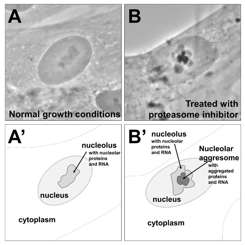
Latonen L (2011) Nucleolar aggresomes as counterparts of cytoplasmic aggresomes in proteotoxic stress. Bioessays 33: 386-395. doi: 10.1002/bies.201100008
    [15] Woulfe J (2008) Nuclear bodies in neurodegenerative disease. Biochim BiophysActa 1783: 2195-2206.
    [16] Ramaswami M, Taylor JP, Parker R (2013) RNA-Protein Granules in Degenerative Disorders. Cell 154: 727-736. doi: 10.1016/j.cell.2013.07.038
    [17] Miller SB, Ho CT, Winkler J, et al. (2015) Compartment-specific aggregases direct distinct nuclear and cytoplasmic aggregate deposition. EMBO J 34: 778-797. doi: 10.15252/embj.201489524
    [18] Dundr M (2012) Nuclear bodies: multifunctional companions of the genome. Curr Opin Cell Biol 24: 415-422. doi: 10.1016/j.ceb.2012.03.010
    [19] Kaganovich D, Kopito R, Frydman J (2008) Misfolded proteins partition between two distinct quality control compartments. Nature 454: 1088-1095. doi: 10.1038/nature07195
    [20] Park SH, Kukushkin Y, Gupta R, et al. (2013) PolyQ proteins interfere with nuclear degradation of cytosolic proteins by sequestering the Sis1p chaperone. Cell 154: 134-145. doi: 10.1016/j.cell.2013.06.003
    [21] Lu M, Williamson N, Boschetti C, et al. (2015) Expression-level dependent perturbation of cell proteostasis and nuclear morphology by aggregation-prone polyglutamine proteins. Biotechnol Bioeng. doi: 10.1002/bit.25606.
    [22] Caron NS, Hung CL, Atwal RS, et al. (2014) Live cell imaging and biophotonic methods reveal two types of mutant huntingin inclusions. Hum Mol Genet 23: 2324-2338. doi: 10.1093/hmg/ddt625
    [23] Chapple JP, Bros-Facer V, Butler R, et al. (2008) Focal distortion of the nuclear envelope by huntingtin aggregates revealed by lamin immunostaining. Neurosci Lett 447: 172-174. doi: 10.1016/j.neulet.2008.09.075
    [24] Mapelli L, Canale C, Pesci D, et al. (2012) Toxic effects of expanded ataxin-1 involve mechanical instability of the nuclear membrane. Biochim Biophys Acta 1822: 906-917. doi: 10.1016/j.bbadis.2012.01.016
    [25] Lindström MS, Latonen L (2013) The nucleolus as a stress respose organelle. In: Proteins of the Nucleolus. Regulation, Translocation and Biomedical Functions. Springer, 251-273.
    [26] Andersen JS, Lam YW, Leung AK, et al. (2005) Nucleolar proteome dynamics. Nature 433: 77-83. doi: 10.1038/nature03207
    [27] Latonen L, Moore HM, Bai B, et al. (2011) Proteasome inhibitors induce nucleolar aggregation of proteasome target proteins and polyadenylated RNA by altering ubiquitin availability. Oncogene 30: 790-805. doi: 10.1038/onc.2010.469
    [28] Krüger T, Scheer U (2010) p53 localizes to intranucleolar regions distinct from the ribosome production compartments. J Cell Sci 123: 1203-1208. doi: 10.1242/jcs.062398
    [29] Vilotti S, Biagioli M, Foti R, et al. (2012) The PML nuclear bodies-associated protein TTRAP regulates ribosome biogenesis in nucleolar cavities upon proteasome inhibition. Cell Death Differ 19: 488-500. doi: 10.1038/cdd.2011.118
    [30] Ehm P, Nalaskowski MM, Wundenberg T, et al. (2015) The tumor suppressor SHIP1 colocalizes in nucleolar cavities with p53 and components of PML nuclear bodies. Nucleus 6: 154-164. doi: 10.1080/19491034.2015.1022701
    [31] Nollen EA, Salomons FA, Brunsting JF, et al. (2001) Dynamic changes in the localization of thermally unfolded nuclear proteins associated with chaperone-dependent protection. Proc Natl AcadSci USA 98: 12038-12043. doi: 10.1073/pnas.201112398
    [32] Forsberg K, Andersen PM, Marklund SL, et al. (2011) Glial nuclear aggregates of superoxide dismutase-1 are regularly present in patients with amyotrophic lateral sclerosis. Acta Neuropathol 121: 623-634. doi: 10.1007/s00401-011-0805-3
    [33] Li M, Nakagomi Y, Kobayashi Y, et al. (1998) Nonneural nuclear inclusions of androgen receptor protein in spinal and bulbar muscular atrophy. Am J Pathol 153: 695-701.
    [34] Hetman M, Pietrzak M (2012) Emerging roles of the neuronal nucleolus. Trends Neurosci 35: 305-314. doi: 10.1016/j.tins.2012.01.002
    [35] Parlato R, Kreiner G (2013) Nucleolar activity in neurodegenerative diseases: a missing piece of the puzzle? J Mol Med (Berl) 91: 541-547. doi: 10.1007/s00109-012-0981-1
    [36] Stavreva DA, Kawasaki M, Dundr M, et al. (2006) Potential roles for ubiquitin and the proteasome during ribosome biogenesis. Mol Cell Biol 26: 5131-5145 doi: 10.1128/MCB.02227-05
    [37] Nyström T, Liu B (2014) The mystery of aging and rejuvenation—a budding topic. Curr Opin Microbiol 18: 61-67. doi: 10.1016/j.mib.2014.02.003
    [38] Song J, Yang Q, Yang J, et al. (2014) Essential genetic interactors of SIR2 required for spatial sequestration and asymmetrical inheritance of protein aggregates. PLoS Genet 10: e1004539. doi: 10.1371/journal.pgen.1004539
    [39] Liu B, Larsson L, Caballero A, et al. (2010) The polarisome is required for segregation and retrograde transport of protein aggregates. Cell 140: 257-267. doi: 10.1016/j.cell.2009.12.031
    [40] Zhou C, Slaughter BD, Unruh JR, et al. (2014) Organelle-based aggregation and retention of damaged proteins in asymmetrically dividing cells. Cell 159: 530-542. doi: 10.1016/j.cell.2014.09.026
    [41] Katajisto P, Döhla J, Chaffer CL, et al. (2015) Stem cells. Asymmetric apportioning of aged mitochondria between daughter cells is required for stemness. Science 348: 340-343.
    [42] Mogk A, Bukau B (2014) Mitochondria tether protein trash to rejuvenate cellular environments. Cell 159: 471-472. doi: 10.1016/j.cell.2014.10.007
  • This article has been cited by:

    1. Jifeng Ren, Yongshu Li, Shuhuan Hu, Yi Liu, Sai Wah Tsao, Denvid Lau, Guannan Luo, Chi Man Tsang, Raymond H. W. Lam, Nondestructive quantification of single-cell nuclear and cytoplasmic mechanical properties based on large whole-cell deformation, 2020, 20, 1473-0197, 4175, 10.1039/D0LC00725K
  • Reader Comments
  • © 2015 the Author(s), licensee AIMS Press. This is an open access article distributed under the terms of the Creative Commons Attribution License (http://creativecommons.org/licenses/by/4.0)
通讯作者: 陈斌, bchen63@163.com
  • 1. 

    沈阳化工大学材料科学与工程学院 沈阳 110142

  1. 本站搜索
  2. 百度学术搜索
  3. 万方数据库搜索
  4. CNKI搜索

Metrics

Article views(7112) PDF downloads(1404) Cited by(1)

Article outline

Other Articles By Authors

/

DownLoad:  Full-Size Img  PowerPoint
Return
Return

Catalog