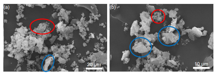
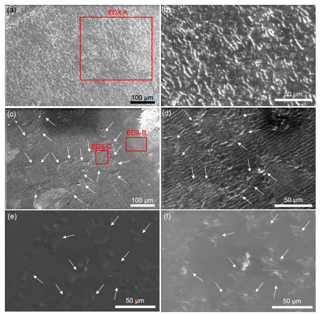
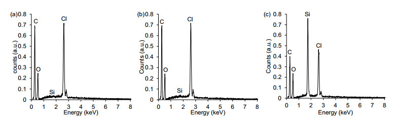
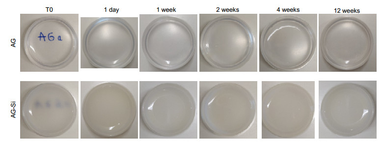
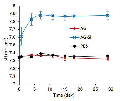
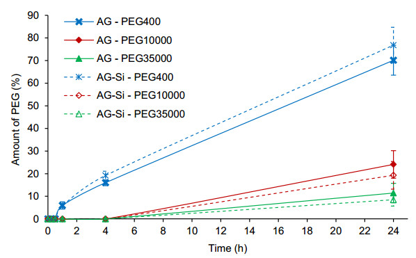
Over the last decades, different materials have been investigated to overcome some flaws of bone substitutes. Even though various materials have been proposed for this conception, the in vivo assessments have still highlighted a lack of bioactivity and integration. In this context, this work focuses on the development of hybrid gel with surface properties specifically designed to promote bone regeneration by a sustained local delivery of active agents. We propose a new approach using modified-silica with high specific surface area and superior hydrophilicity dispersed in agarose hydrogel. In this optic, silica particles were dispersed in agarose solutions before the gelation of the composite upon cooling. The dispersion of the silica particles in the agarose gel was determined via scanning electronic microscopy. The degradation of the silica/agarose gels was also studied over a period of 12 weeks. Finally, the influence of the addition of silica on the permeability of the agarose gel was assessed via a diffusion test. The results showed that modified-silica particles exhibit a wide size distribution (500 nm and 10 µm) and can form clusters with higher size after their dispersion in agarose (up to 100 µm). The hybrid gel was stable over 12 weeks in aqueous solution. Moreover, no difference in permeability was noted between the hybrid gel and agarose hydrogel, allowing molecules up to 3 nm in diameter to diffuse freely within 1 mm thick agarose gels in less than 24 h. The present results indicate that hybrid agarose gel could represent an attractive matrix to disperse silica for scaffold applications.
Citation: Rémi G. Tilkin, Ana P. F. Monteiro, Julien G. Mahy, Jérome Hurlet, Nicolas Régibeau, Christian Grandfils, Stéphanie D. Lambert. Hybrid agarose gel for bone substitutes[J]. AIMS Materials Science, 2022, 9(3): 430-445. doi: 10.3934/matersci.2022025
Over the last decades, different materials have been investigated to overcome some flaws of bone substitutes. Even though various materials have been proposed for this conception, the in vivo assessments have still highlighted a lack of bioactivity and integration. In this context, this work focuses on the development of hybrid gel with surface properties specifically designed to promote bone regeneration by a sustained local delivery of active agents. We propose a new approach using modified-silica with high specific surface area and superior hydrophilicity dispersed in agarose hydrogel. In this optic, silica particles were dispersed in agarose solutions before the gelation of the composite upon cooling. The dispersion of the silica particles in the agarose gel was determined via scanning electronic microscopy. The degradation of the silica/agarose gels was also studied over a period of 12 weeks. Finally, the influence of the addition of silica on the permeability of the agarose gel was assessed via a diffusion test. The results showed that modified-silica particles exhibit a wide size distribution (500 nm and 10 µm) and can form clusters with higher size after their dispersion in agarose (up to 100 µm). The hybrid gel was stable over 12 weeks in aqueous solution. Moreover, no difference in permeability was noted between the hybrid gel and agarose hydrogel, allowing molecules up to 3 nm in diameter to diffuse freely within 1 mm thick agarose gels in less than 24 h. The present results indicate that hybrid agarose gel could represent an attractive matrix to disperse silica for scaffold applications.

| [1] |
Brydone AS, Meek D, MacLaine S (2010) Bone grafting, orthopaedic biomaterials, and the clinical need for bone engineering. P I Mech Eng H 224: 1329–1343. https://doi.org/10.1243/09544119JEIM770 doi: 10.1243/09544119JEIM770

|
| [2] |
Campana V, Milano G, Pagano E, et al. (2014) Bone substitutes in orthopaedic surgery: from basic science to clinical practice. J Mater Sci-Mater M 25: 2445–2461. https://doi.org/10.1007/s10856-014-5240-2 doi: 10.1007/s10856-014-5240-2
|
| [3] | US Census Bureau (2012) Statistical abstracts of the United States: 2012-Section 1. Population. Available from: www.census.gov/library/publications/2011/compendia/statab/131ed/population.html. |
| [4] |
Dimitriou R, Jones E, McGonagle D, et al. (2011) Bone regeneration: current concepts and future directions. BMC Med 9: 66. https://doi.org/10.1186/1741-7015-9-66 doi: 10.1186/1741-7015-9-66
|
| [5] |
Wang M, Yang N (2017) A review of bioregulatory and coupled mechanobioregulatory mathematical models for secondary fracture healing. Med Eng Phys 48: 90–102. https://doi.org/10.1016/j.medengphy.2017.06.031 doi: 10.1016/j.medengphy.2017.06.031
|
| [6] |
Baumhauer J, Pinzur MS, Donahue R, et al. (2014) Site selection and pain outcome after autologous bone graft harvest. Foot Ankle Int 35: 104–107. https://doi.org/10.1177/1071100713511434 doi: 10.1177/1071100713511434
|
| [7] |
Bolland BJRF, Wilson MJ, Howell JR, et al. (2017) An analysis of reported cases of fracture of the universal exeter femoral stem prosthesis. J Arthroplasty 32: 1318–1322. https://doi.org/10.1016/j.arth.2016.09.032 doi: 10.1016/j.arth.2016.09.032
|
| [8] |
Othmani M, Aissa A, Bac CG, et al. (2013) Surface modification of calcium hydroxyapatite by grafting of etidronic acid. Appl Surf Sci 274: 151–157. https://doi.org/10.1016/j.apsusc.2013.03.002 doi: 10.1016/j.apsusc.2013.03.002
|
| [9] |
Szubert M, Adamska K, Szybowicz M, et al. (2014) The increase of apatite layer formation by the poly(3-hydroxybutyrate) surface modification of hydroxyapatite and β-tricalcium phosphate. Mater Sci Eng C-Mater 34: 236–244. https://doi.org/10.1016/j.msec.2013.09.023 doi: 10.1016/j.msec.2013.09.023
|
| [10] | Tonon G, Morpurgo M (2006) Sol-gel derived silica polymers for the sustained release of proteins. |
| [11] |
Chen YC, Liu CP, Yang CK, et al. (2013) Preparation and release properties of sol-gel encapsulated proteins. JASMI 3: 11–16. https://doi.org/10.4236/jasmi.2013.33A002 doi: 10.4236/jasmi.2013.33A002
|
| [12] |
Zdarta J, Sałek K, Kołodziejczak-radzimska A, et al. (2015) Immobilization of Amano Lipase A onto Stöber silica surface: Process characterization and kinetic studies. Open Chem 13: 138–148. https://doi.org/10.1515/chem-2015-0017 doi: 10.1515/chem-2015-0017
|
| [13] |
Yang W, Hellner B, Baneyx F (2016) Self-immobilization of Car9 fusion proteins within high surface area silica sol-gels and dynamic control of protein release. Bioconjug Chem 27: 2450–2459. https://doi.org/10.1021/acs.bioconjchem.6b00406 doi: 10.1021/acs.bioconjchem.6b00406
|
| [14] |
Chen Y-C, Smith T, Hicks RH, et al. (2017) Thermal stability, storage and release of proteins with tailored fit in silica. Sci Rep 7: 46568. https://doi.org/10.1038/srep46568 doi: 10.1038/srep46568
|
| [15] |
Vlasenkova MI, Dolinina ES, Parfenyuk EV (2019) Preparation of mesoporous silica microparticles by sol-gel/emulsion route for protein release. Pharm Dev Technol 24: 243–252. https://doi.org/10.1080/10837450.2018.1457051 doi: 10.1080/10837450.2018.1457051
|
| [16] | Andreani T, Souza ALRD, Silva AM, et al. (2012) Sol-gel carrier system: A novel controlled drug delivery, In: Souto EB, Patenting Nanomedicines: Legal Aspects, Intellectual Property and Grant Opportunities, Berlin, Heidelberg: Springer Berlin Heidelberg, 151–166. https://doi.org/10.1007/978-3-642-29265-1_5 |
| [17] |
Pirard SL, Mahy JG, Pirard JP, et al. (2015) Development by the sol-gel process of highly dispersed Ni-Cu/SiO2 xerogel catalysts for selective 1, 2-dichloroethane hydrodechlorination into ethylene. Micropor Mesopor Mat 209: 197–207. https://doi.org/10.1016/j.micromeso.2014.08.015 doi: 10.1016/j.micromeso.2014.08.015
|
| [18] |
Wang X, Ahmed N Ben, Alvarez GS, et al. (2015) Sol-gel encapsulation of biomolecules and cells for medicinal applications. Curr Top Med Chem 15: 223–244. https://doi.org/10.2174/1568026614666141229112734 doi: 10.2174/1568026614666141229112734
|
| [19] |
Reiner T, Kababya S, Gotman I (2008) Protein incorporation within Ti scaffold for bone ingrowth using sol-gel SiO2 as a slow release carrier. J Mater Sci-Mater M 19: 583–589. https://doi.org/10.1007/s10856-007-3194-3 doi: 10.1007/s10856-007-3194-3
|
| [20] |
Ukmar T, Planinsek O (2010) Ordered mesoporous silicates as matrices for controlled release of drugs. Acta Pharmceut 60: 373–385. https://doi.org/10.2478/v1007-010-0037-4 doi: 10.2478/v1007-010-0037-4
|
| [21] |
Zhou Y, Quan G, Wu Q, et al. (2018) Mesoporous silica nanoparticles for drug and gene delivery. Acta Pharm Sin B 8: 165–177. https://doi.org/10.1016/j.apsb.2018.01.007 doi: 10.1016/j.apsb.2018.01.007
|
| [22] |
Kang S, Hong SI, Choe CR, et al. (2001) Preparation and characterization of epoxy composites filled with functionalized nanosilica particles obtained via sol-gel process. Polymer 42: 879–887. https://doi.org/10.1016/S0032-3861(00)00392-X doi: 10.1016/S0032-3861(00)00392-X
|
| [23] |
Bravo J, Zhai L, Wu Z, et al. (2007) Transparent superhydrophobic films based on silica nanoparticles. Langmuir 23: 7293–7298. https://doi.org/10.1021/la070159q doi: 10.1021/la070159q
|
| [24] |
Elias L, Fenouillot F, Majesté JC, et al. (2008) Immiscible polymer blends stabilized with nano-silica particles: Rheology and effective interfacial tension. Polymer 49: 4378–4385. https://doi.org/10.1016/j.polymer.2008.07.018 doi: 10.1016/j.polymer.2008.07.018
|
| [25] |
Ghanbari A, Attar MM (2015) A study on the anticorrosion performance of epoxy nanocomposite coatings containing epoxy-silane treated nano-silica on mild steel substrate. J Ind Eng Chem 23: 145–153. https://doi.org/10.1016/j.jiec.2014.08.008 doi: 10.1016/j.jiec.2014.08.008
|
| [26] |
Jiang R, Kunz HR, Fenton JM (2006) Composite silica/Nafion ® membranes prepared by tetraethylorthosilicate sol-gel reaction and solution casting for direct methanol fuel cells. J Membrane Sci 272: 116–124. https://doi.org/10.1016/j.memsci.2005.07.026 doi: 10.1016/j.memsci.2005.07.026
|
| [27] |
Faustini M, Louis B, Albouy PA, et al. (2010) Preparation of sol-gel films by dip-coating in extreme conditions. J Phys Chem C 114: 7637–7645. https://doi.org/10.1021/jp9114755 doi: 10.1021/jp9114755
|
| [28] |
Rocha CR, Chávez-Flores D, Zuverza-Mena N, et al. (2020) Surface organo-modification of hydroxyapatites to improve PLA/HA compatibility. J Appl Polym 137: 1–9. https://doi.org/10.1002/app.49293 doi: 10.1002/app.49293
|
| [29] |
Mano JF, Silva GA, Azevedo HS, et al. (2007) Natural origin biodegradable systems in tissue engineering and regenerative medicine: present status and some moving trends. J R Soc Interface 4: 999–1030. https://doi.org/10.1098/rsif.2007.0220 doi: 10.1098/rsif.2007.0220
|
| [30] |
Buckley CT, Thorpe SD, Brien FJO, et al. (2009) The effect of concentration, thermal history and cell seeding density on the initial mechanical properties of agarose hydrogels. J Mech Behav Biomed 2: 512–521. https://doi.org/10.1016/j.jmbbm.2008.12.007 doi: 10.1016/j.jmbbm.2008.12.007
|
| [31] |
Miguel SP, Ribeiro MP, Brancal H, et al. (2014) Thermoresponsive chitosan-agarose hydrogel for skin regeneration. Carbohyd Polym 111: 366–373. https://doi.org/10.1016/j.carbpol.2014.04.093 doi: 10.1016/j.carbpol.2014.04.093
|
| [32] |
Griess GA, Moreno ET, Herrmann R, et al. (1990) The sieving of rod-shaped viruses during agarose gel electrophoresis. I. comparison with the sieving of spheres. Biopolymers 29: 1277–1287. https://doi.org/10.1002/bip.360290816 doi: 10.1002/bip.360290816
|
| [33] |
Pluen A, Netti PA, Jain RK, et al. (1999) Diffusion of macromolecules in agarose gels: comparison of linear and globular configurations. Biophys J 77: 542–552. https://doi.org/10.1016/S0006-3495(99)76911-0 doi: 10.1016/S0006-3495(99)76911-0
|
| [34] |
Paris JL, Lafuente-Gómez N, Cabañas MV, et al. (2019) Fabrication of a nanoparticle-containing 3D porous bone scaffold with proangiogenic and antibacterial properties. Acta Biomater 86: 441–449. https://doi.org/10.1016/j.actbio.2019.01.013 doi: 10.1016/j.actbio.2019.01.013
|
| [35] |
Kazimierczak P, Benko A, Palka K, et al. (2020) Novel synthesis method combining a foaming agent with freeze-drying to obtain hybrid highly macroporous bone scaffolds. J Mater Sci Technol 43: 52–63. https://doi.org/10.1016/j.jmst.2020.01.006 doi: 10.1016/j.jmst.2020.01.006
|
| [36] |
Tilkin RG, Colle X, Argento Finol A, et al. (2020) Protein encapsulation in functionalized sol-gel silica: Effect of the encapsulation method on the release kinetics and the activity. Micropor Mesopor Mater 308: 110502. https://doi.org/10.1016/j.micromeso.2020.110502 doi: 10.1016/j.micromeso.2020.110502
|
| [37] |
Tilkin RG, Mahy JG, Régibeau N, et al. (2021) Protein encapsulation in functionalized sol-gel silica: influence of organosilanes and main silica precursors. J Mater Sci 56: 14234–14256. https://doi.org/10.1007/s10853-021-06182-9 doi: 10.1007/s10853-021-06182-9
|
| [38] |
Rozaini MNH, Semail N-F, Saad B, et al. (2019) Molecularly imprinted silica gel incorporated with agarose polymer matrix as mixed matrix membrane for separation and preconcentration of sulfonamide antibiotics in water samples. Talanta 199: 522–531. https://doi.org/10.1016/j.talanta.2019.02.096 doi: 10.1016/j.talanta.2019.02.096
|
| [39] |
Pertoft H, Hallen A (1976) Preparation of silica-agarose beads for gel chromatography. J Chromatogr A 128: 125–131. https://doi.org/10.1016/S0021-9673(00)84038-8 doi: 10.1016/S0021-9673(00)84038-8
|
| [40] | Lecloux A (1971) Exploitation des isothermes d'adsorption et de désorption d'azote pour l'étude de la texture des solides poreux. Mémoires Société des Sci Liege 4: 169–209. |
| [41] | Lecloux A (1981) Texture of catalysts, In: Anderson JR, Boudart M, Catalysis: Science and Technology, Berlin, 171. https://doi.org/10.1007/978-3-642-93171-0_4 |
| [42] |
Pirard R, Heinrichs B, Cantfort OVAN, et al. (1998) Mercury porosimetry applied to low density xerogels; relation between structure and mechanical properties. JSST 13: 335–339. https://doi.org/10.1023/A:1008676211157 doi: 10.1023/A:1008676211157
|
| [43] |
Lambert S, Alié C, Pirard JP, et al. (2004) Study of textural properties and nucleation phenomenon in Pd/SiO2, Ag/SiO2 and Cu/SiO2 cogelled xerogel catalysts. J Non Cryst Solids 342: 70–81. https://doi.org/10.1016/j.jnoncrysol.2004.06.005 doi: 10.1016/j.jnoncrysol.2004.06.005
|
| [44] |
Park J, Regalbuto JR (1995) A simple, accurate determination of oxide PZC and the strong buffering effect of oxide surfaces at incipient wetness. J Colloid Interface Sci 175: 239–252. https://doi.org/10.1006/jcis.1995.1452 doi: 10.1006/jcis.1995.1452
|
| [45] |
Lambert S, Job N, Souza LD, et al. (2009) Synthesis of very highly dispersed platinum catalysts supported on carbon xerogels by the strong electrostatic adsorption method. J Catal 261: 23–33. https://doi.org/10.1016/j.jcat.2008.10.014 doi: 10.1016/j.jcat.2008.10.014
|
| [46] |
Van Der Voort P, Esquivel D, et al. (2013) Periodic mesoporous organosilicas: from simple to complex bridges; a comprehensive overview of functions, morphologies and applications. Chem Soc Rev 42: 3913–3955. https://doi.org/10.1039/C2CS35222B doi: 10.1039/C2CS35222B
|
| [47] |
Manayil JC, Lee AF, Wilson K (2019) Functionalized periodic mesoporous organosilicas: tunable hydrophobic solid acids for biomass conversion. Molecules 24: 239. https://doi.org/10.3390/molecules24020239 doi: 10.3390/molecules24020239
|
| [48] | te Nijenhuis K (1997) Agarose, Thermoreversible Networks: Viscoelastic Properties and Structure of Gels, Heidelberg, Springer Berlin Heidelberg, 194–202. https://doi.org/10.1007/BFb0008710 |
| [49] | Lonza Appendix B: Agarose Physical Chemistry. Available from: http://www.lonzabio.jp/catalog/pdf/pd/PD029.pdf. |
| [50] |
Régibeau N, Tilkin RG, Compère P, et al. (2020) Preparation of PDLLA based nanocomposites with modified silica by in situ polymerization: Study of molecular, morphological, and mechanical properties. Mater Today Commun 25: 101610. https://doi.org/10.1016/j.mtcomm.2020.101610 doi: 10.1016/j.mtcomm.2020.101610
|
| [51] |
Weng L, Liang S, Zhang L, et al. (2005) Transport of glucose and poly(ethylene glycol)s in agarose gels studied by the refractive index method. Macromolecules 38: 5236–5242. https://doi.org/10.1021/ma047337w doi: 10.1021/ma047337w
|
| [52] |
Devanand K, Selser JC (1991) Asymptotic behavior and long-range interactions in aqueous solutions of poly(ethylene oxide). Macromolecules 24: 5943–5947. https://doi.org/10.1021/ma00022a008 doi: 10.1021/ma00022a008
|
| [53] |
Fee CJ, Van Alstine JM (2004) Prediction of the viscosity radius and the size exclusion chromatography behavior of PEGylated proteins. Bioconjug Chem 15: 1304–1313. https://doi.org/10.1021/bc049843w doi: 10.1021/bc049843w
|
| [54] |
Ogston AG, Preston BN, Wells JD (1973) On the transport of compact particles through solutions of chain-polymers. Proc R Soc A 333: 297–316. https://doi.org/10.1098/rspa.1973.0064 doi: 10.1098/rspa.1973.0064
|
| [55] |
Karlsson D, Zacchi G, Axelsson A (2002) Electronic speckle pattern interferometry: A tool for determining diffusion and partition coefficients for proteins in gels. Biotechnol Prog 18: 1423–1430. https://doi.org/10.1021/bp0255659 doi: 10.1021/bp0255659
|
| [56] |
Monteiro APF, Idczak G, Tilkin RG, et al. (2021) Evaluation of hydroxyapatite texture using CTAB template and effects on protein adsorption. Surf Interfaces 27: 101565. https://doi.org/10.1016/j.surfin.2021.101565 doi: 10.1016/j.surfin.2021.101565
|
Figures(6)

Rémi G. Tilkin, Ana P. F. Monteiro, Julien G. Mahy, Jérome Hurlet, Nicolas Régibeau, Christian Grandfils, Stéphanie D. Lambert. Hybrid agarose gel for bone substitutes[J]. AIMS Materials Science, 2022, 9(3): 430-445. doi: 10.3934/matersci.2022025


 DownLoad: