Loading [Contrib]/a11y/accessibility-menu.js
Review Special Issues

MYC function and regulation in flies: how Drosophila has enlightened MYC cancer biology

  • Progress in our understanding of the complex signaling events driving human cancer would have been unimaginably slow without discoveries from Drosophila genetic studies. Significantly, many of the signaling pathways now synonymous with cancer biology were first identified as a result of elegant screens for genes fundamental to metazoan development. Indeed the name given to many core cancer-signaling cascades tells of their history as developmental patterning regulators in flies—e.g. Wingless (Wnt), Notch and Hippo. Moreover, astonishing insight has been gained into these complex signaling networks, and many other classic oncogenic signaling networks (e.g. EGFR/RAS/RAF/ERK, InR/PI3K/AKT/TOR), using sophisticated fly genetics. Of course if we are to understand how these signaling pathways drive cancer, we must determine the downstream program(s) of gene expression activated to promote the cell and tissue over growth fundamental to cancer. Here we discuss one commonality between each of these pathways: they are all implicated as upstream activators of the highly conserved MYC oncogene and transcription factor. MYC can drive all aspects of cell growth and cell cycle progression during animal development. MYC is estimated to be dysregulated in over 50% of all cancers, underscoring the importance of elucidating the signals activating MYC. We also discuss the FUBP1/FIR/FUSE system, which acts as a ‘cruise control’ on the MYC promoter to control RNA Polymerase II pausing and, therefore, MYC transcription in response to the developmental signaling environment. Importantly, the striking conservation between humans and flies within these major axes of MYC regulation has made Drosophila an extremely valuable model organism for cancer research. We therefore discuss how Drosophila studies have helped determine the validity of signaling pathways regulating MYC in vivo using sophisticated genetics, and continue to provide novel insight into cancer biology.

    Citation: Jue Er Amanda Lee, Linda May Parsons, Leonie M. Quinn. MYC function and regulation in flies: how Drosophila has enlightened MYC cancer biology[J]. AIMS Genetics, 2014, 1(1): 81-98. doi: 10.3934/genet.2014.1.81

    Related Papers:

    [1] Tin Tin Su . Non-autonomous consequences of cell death and other perks of being metazoan. AIMS Genetics, 2015, 2(1): 54-69. doi: 10.3934/genet.2015.1.54
    [2] Michael J. Murray . Drosophila models of metastasis. AIMS Genetics, 2015, 2(1): 25-53. doi: 10.3934/genet.2015.1.25
    [3] Xianjue Ma . Context-dependent interplay between Hippo and JNK pathway in Drosophila. AIMS Genetics, 2014, 1(1): 20-33. doi: 10.3934/genet.2014.1.20
    [4] Dawei Liu, Zeeshan Shaukat, Rashid Hussain, Mahwish Khan, Stephen L. Gregory . Drosophila as a model for chromosomal instability. AIMS Genetics, 2015, 2(1): 1-12. doi: 10.3934/genet.2015.1.1
    [5] John E. La Marca, Wayne Gregory Somers . The Drosophila gonads: models for stem cell proliferation, self-renewal, and differentiation. AIMS Genetics, 2014, 1(1): 55-80. doi: 10.3934/genet.2014.1.55
    [6] Helena E. Richardson . Drosophila models of cancer. AIMS Genetics, 2015, 2(1): 97-103. doi: 10.3934/genet.2015.1.97
    [7] Francesca Froldi, Milán Szuperák, Louise Y. Cheng . Neural stem cell derived tumourigenesis. AIMS Genetics, 2015, 2(1): 13-24. doi: 10.3934/genet.2015.1.13
    [8] Carlos Estella, Antonio Baonza . Cell proliferation control by Notch signalling during imaginal discs development in Drosophila. AIMS Genetics, 2015, 2(1): 70-96. doi: 10.3934/genet.2015.1.70
    [9] Tú Nguyen-Dumont, Jenna Stewart, Ingrid Winship, Melissa C. Southey . Rare genetic variants: making the connection with breast cancer susceptibility. AIMS Genetics, 2015, 2(4): 281-292. doi: 10.3934/genet.2015.4.281
    [10] Valeriia Mikhailova, Valeriia Gulaia, Vladlena Tiasto, Stanislav Rybtsov, Margarita Yatsunskaya, Alexander Kagansky . Towards an advanced cell-based in vitro glioma model system. AIMS Genetics, 2018, 5(2): 91-112. doi: 10.3934/genet.2018.2.91
  • Progress in our understanding of the complex signaling events driving human cancer would have been unimaginably slow without discoveries from Drosophila genetic studies. Significantly, many of the signaling pathways now synonymous with cancer biology were first identified as a result of elegant screens for genes fundamental to metazoan development. Indeed the name given to many core cancer-signaling cascades tells of their history as developmental patterning regulators in flies—e.g. Wingless (Wnt), Notch and Hippo. Moreover, astonishing insight has been gained into these complex signaling networks, and many other classic oncogenic signaling networks (e.g. EGFR/RAS/RAF/ERK, InR/PI3K/AKT/TOR), using sophisticated fly genetics. Of course if we are to understand how these signaling pathways drive cancer, we must determine the downstream program(s) of gene expression activated to promote the cell and tissue over growth fundamental to cancer. Here we discuss one commonality between each of these pathways: they are all implicated as upstream activators of the highly conserved MYC oncogene and transcription factor. MYC can drive all aspects of cell growth and cell cycle progression during animal development. MYC is estimated to be dysregulated in over 50% of all cancers, underscoring the importance of elucidating the signals activating MYC. We also discuss the FUBP1/FIR/FUSE system, which acts as a ‘cruise control’ on the MYC promoter to control RNA Polymerase II pausing and, therefore, MYC transcription in response to the developmental signaling environment. Importantly, the striking conservation between humans and flies within these major axes of MYC regulation has made Drosophila an extremely valuable model organism for cancer research. We therefore discuss how Drosophila studies have helped determine the validity of signaling pathways regulating MYC in vivo using sophisticated genetics, and continue to provide novel insight into cancer biology.


    1. The history of MYC

    Since the identification of the oncogenic potential of c-MYC (MYC) in the early 1980s [1] the mammalian MYC family, which also includes MYCN, and MYCL, has been the focus of extensive investigation. Early studies demonstrated MYC drives cell cycle progression [2,3,4], which is central to MYC’s capacity as an oncoprotein [5]. Despite considerable efforts using mammalian models, Drosophila genetic studies were first to demonstrate that the sole MYC ortholog, dMYC [6] drives acquisition of biomass, or cell growth, via its ability to upregulate ribosome biogenesis during animal development [7,8,9]. dMYCmutant flies are small, with reduced cell growth [7] and decreased rDNA transcription [9]. Conversely, increased dMYC is sufficient to drive increased rDNA transcription, cell growth and proliferation [7,9]. Function al conservation between fly and human MYC has been demonstrated by classic functional rescue experiments i. e. dMYC can transform primary mammalian cells and rescue proliferation defects in MYC null fibroblasts [10], whilst MYC rescues lethal dMYC mutations in Drosophila [11]. Thus, the targets and pathways impacted by MYC are conserved between flies and humans, particularly regarding the ability to drive ribosome biogenesis, cell growth and tumour progression.

    Dissecting essential roles for MYC using loss of function approaches in mammals has been complicated by functional redundancy between the 3 MYC genes. For example, in adult bone marrow both MYC and MYCN are endogenously expressed and MYC appears to act redundantly with MYCN to achieve hematopoietic stem cell maintenance [12]. Moreover, although much has been discovered regarding the molecular and pathological mechanisms that drive cells to become malignant, dissecting the physiological/molecular mechanisms linking upstream signaling to MYC-driven cancers requires further investigation using in vivo genetic models. In flies the presence of a single MYC gene and available genetic tools have provided clear connections between developmental pathways and dMYC regulation. In this review, we will focus on research undertaken in Drosophila demonstrating how MYC is regulated during development and provide parallels with work carried out in vertebrates.

    2. MYC function

    MYC is a basic helix-loop-helix zipper (bHLHZ) transcriptional factor that heterodimerizes with a second bHLHZ protein MAX [13] todirectly bind DNA sequences, called E-boxes, within the promoter regions of target genes [13,14,15,16] (reviewed in [17]). However, in vivo MAX loss-of-function studies in Drosophila have demonstrated that MYC can also elicit many of its functions independently of MAX [18]. For example, transcriptional activation of 5S rRNA and tRNA via RNA Polymerase III can be achieved in a complete loss-of-function MAX mutant background [18].

    MYC interacts with a range of chromatin modifying enzymes and transcription factors to control expression of an extensive network of downstream transcriptional targets [9,19,20,21,22,23,24,25,26,27,28]. Early studies in Drosophila [9,23,24] and human cells [29], revealed MYC bound 10-15% of the genome (reviewed in [30,31]). More recently, next generation sequencing (NGS), including ChIP-seq, RNA-seq and GRO-seq nuclear run-on assays, have provided deeper insight into MYC-targets [27,28,32,33]. Broadly, these studies suggest that MYC acts not so much to establish a fixed signature of gene expression, but rather enhances programs of transcriptional activity, which are based on cell type and/or developmental context [27,28]. For instance, at target promoters in activated lymphocytes [27]increased MYC was detected on all active promoters, i. e those enriched for RNA Polymerase II (Pol II) and with active chromatin marks. Thus amplification of MYC dependent gene expression was only associated with active genes already marked as MYC targets, not additional genes. Furthermore, these studies confirmed the strong correlation between E-boxes within target promoters, and their vicinity to the transcription start site (TSS), for optimal MYC loading and amplification of active genes. Additionally, analysis from both studies indicated enhanced transcriptional elongation upon increased MYC occupancy [27,28] (reviewed in [34]). Consistent with this MYC bound sites had increased enrichment of the transcription elongation factor p-TEFb and elongating RNA Pol II [27,28]. Together, these studies suggest elevated levels of MYC in tumour cells intensifie s MYCs normal function by amplifying established MYC-targets, rather than initiating new sites of gene activation.

    These observations are consistent with earlier studies, which demonstrated that promoters targeted by MYC are associated with an active chromatin profile, including methylation marks at H3K4 (me1, me2, me3), acetylation of H3K27 (ac), and DNA methylation across the CpG island, a hallmark of MYC binding sites [29,35]. The demonstration that MYC binding is not detected on inactive heterochromatin [27,28,36], suggested MYC was unlikely to establish gene activation in the context of overexpression. Rather, MYC requires prior opening of chromatin for binding site recognition, consistent with the DNA ampl ification model. That overexpression of MYC in Drosophila is sufficient to drive a program of ribosome biogenesis and cell growth [7,8,9] would be expected under the amplification model, since the tissues used in these studies were all primed for cell growth (e.g. proliferating wing imaginal discs and endoreplicating salivary glands). Thus increased MYC amplifies pre-existing cell growth transcriptional networks, much as it does in a cancer setting [37,38].

    3. MYC in Cancer

    As stated above, MYC proteins drive tumourigenesis primarily by activating transcription of target genes required for growth and proliferation [31,39,40,41,42,43]. MYCN is predominantly expressed in the brain and amplification has been associated with neuroblastoma, glioblastoma, medulloblastoma and astrocytoma [44]. MYCL, as the name suggests, has been mostly associated with lung cancer [45]. Although MYC is frequently overexpressed in cancer via gene translocation e.g. in Burkitt’s B-cell lymphoma [46,47], MYC amplification is broadly observed in many tumours including, but not limited to other leukaemias, breast, prostate, cervical, bone, brain, colon, lung and liver cancers [43,45]. Thus, all members of the MYC gene family are linked to the aetiology of a wide range of human cancers.

    MYC driven cell growth is fundamental to cancer progression

    MYC drives cell growth by increasing activity from all three RNA polymerases (I, II and III) to escalate ribosome production [9,37,38,48,49,50,51,52,53,54]. Ribosomes form around the actively transcribing rDNA repeat genes within the nucleoli, and ribosome biogenesis is orchestrated by MYC [38]. In fact, the capacity to drive ribosome production appears to be central to MYC's powerful cell growth effects, as ribosome biogenesis is rate-limiting for progression of MYC-driven lymphoma [39,42]. The first evidence that MYC drives RNA Pol I activity and, therefore, rDNA transcription was the observation that MYC increases expression of the homodimeric upstream activation binding factor (UBF), a crucial component of the RNA Pol I pre-initiation complex [51,55]. Subsequently, studies in both Drosophila [9,23] and mammals [37], revealed MYC stimulates Pol I transcriptional activity indirectly via its ability to regulate a large number of RNA polymerase II-transcribed genes.

    In mammals MYC directly interacts with the Pol I recruitment complex SL-1 to assist in initiation of Pol I driven rDNA transcription to form the 47S pre-rRNA, which is processed into mature 18S, 5.8S and 28S rRNAs. In mammals [54] and flies [18,53] MYC activates RNA Pol III for 5S rRNA production for assembly of the 60S ribosomal subunit and tRNA transcription directly through an interaction with TFIIB [54]. MYC also upregulates ribosomal protein production at the level of Pol II-dependent transcription [9,37]. Thus MYC activity increases abundance of all components required to assemble the 40S and 60S ribosomal subunits. [38,56]. In addition to increasing ribosomal components, MYC also increases upstream growth signaling proteins, factors involved in ribosome assembly and processing (e.g. nucleolin and fibrillarin), components of the RNA Pol I machinery and translation factors [9,37]. Indeed evidence suggests it is MYC’s role in harnessing and driving ribosome biogenesis that is central to MYC’s malignant function. In particular, drug development studies have demonstrated that targeting the ribosome biogenesis program might provide a platform for therapeutic intervention of a broad range of MYC-driven cancers. Specifically, small molecule inhibition of RNA Polymerase I, the enzyme responsible for rDNA transcription, selectively kills MYC-driven lymphoma in vivo [38,42].

    Further to MYC’s oncogenic potential, MYC couples growth with cell cycle progression by promoting G1 progression. MYC drives S phase by directly upregulating the S phase cyclins, Cyclin D and Cyclin E [24]. The upregulation of G1 cyclins leads to phosphorylation and inactivation of the key inhibitor of S phase progression, Rbf, a member of the Drosophila Retinoblastoma family. Inhibition of Rbf results in release of the E2F1 transcription factor from the inhibitory complex with Rbf, permitting upregulation of E2F1 dependent S phase genes [57,58]. Like MYC, early in G1 phase, dMYC activates genes required for DNA replication and progression through S phase [7,9,59].

    The ability of MYC to amplify active gene expression further stresses the importance of maintaining tight control of MYC activity. Consequently, several mechanisms act to ensure MYC is tightly regulated at the level of mRNA transcription [60,61], mRNA stability [62,63,64,65], and protein turnover [66,67]. In addition, to allow organ and tissue growth in response to nutrients and other external cues, the levels of MYC need to be responsive to grow th and developmental signals [68]. Despite the large number of transcriptional targets and the associated oncogenic potential of MYCdysregulation, how MYC transcription is regulated in response to developmental growth signals is largely unknown. As most signaling pathways and core components of the transcriptional machinery required for development are well characterized and conserved between Drosophila and humans [68,69,70], fly genetic models remain an excellent means of rapidly identifying and dissecting pathways impacting MYC activity.

    4. MYC regulation

    FUBP1-FUSE-FIR activate MYC transcription

    Multiple upstream signaling pathways funnel into MYC transcription by modulating the complement of transcription factors and enhancers on the MYC promoter [60,71,72,73]. Previous reviews have focused on canonical transcription factors (e.g. E2F, B-catenin/TCF, Smad3, STAT3) in MYC transcription [60], here we will focus on two non-canonical MYC regulators (FUBP1 and FIR), which we believe will be required for “fine tuning” activated MYC transcription in response to multiple inputs from the extracellular signaling environment.

    The FUSE binding protein (FBP/FUBP1) and the FBP Interacting Repressor (FIR) bind the Far Upstream Sequence Element (FUSE) region of singled stranded DNA in the “activated” MYC promoter (Figure 1) [71,72,74,75]. We predict that the FUBP1/FIR/FUSE system acts as a “cruise-control” by controlling RNA Polymerase II pausing and, therefore, MYC transcription in response to the developmental signaling environment [73]. Specifically, mammalian tissue culture studies suggests that RNA Pol II re lease and MYC transcription in response to serum stimulation requires interactions between the XPB helicase component of the Transcription Factor IIH (TFIIH) complex and FUBP1 and FIR complexes [76,77]. In response to binding of chromatin remodeling complexes and canonical MYC transcription factors, TFIIH/XPB stimulates MYC promoter melting, which is required for physical interaction between FUBP1 and FIR at the single stranded FUSE [76,77]. The “pulse” of MYC transcription stimulated by serum in culture correlates with decreased enrichment for RNA Pol II across the MYC Transcription Start Site (TSS) and sequential recruitment of FUBP1 and FIR to the FUSE element [77]. Enrichment for FUBP1 is detected prior to a peak in MYC mRNA expression, consistent with FUBP1 promoting release of paused RNA Pol II to activate MYC transcription. Conversely, FIR is detected at FUSE subsequent to the peak in MYC mRNA and is proposed to inactivate RNA Pol II thereby returning MYC transcription to basal levels (Figure 1) [77]. Consistent with a repressive function of FIR, loss-of-function FIR mutations are associated with increased MYC mRNA levels in colorectal cancer [78].

    Figure 1. The FUSE binding protein (FBP/FUBP) and the FBP Interacting Repressor (FIR) bind the Far Upstream Sequence Element (FUSE) region of singled stranded DNA in the “activated” MYC promoter. Prior to the peak in MYC expression, FUBP is recruited promoting release of poised RNA Pol II to activate MYC transcription. Subsequently, FIR is recruited to the MYC promoter following to the peak in MYC and is proposed to inactivate Pol II to return MYC transcription to basal levels. Thus the FUSE/FUBP/FIR axis has been speculated to be a “cruise-control” for controlling stimulated MYC transcription.

    Drosophila Hfp is functionally homologous to mammalian FIR, being implicated in the repression of MYC transcription in Drosophila. Specifically, Hfp behaves as a tumour suppressor, as loss of Hfp results in overproliferation and tissue overgrowth [79]. RNA interference to deplete Hfp results in increased dMYC expression and cell growth [80]. Drosophila studies have also demonstrated enrichment for Hfp on the dMYC promoter and that Hfp is essential for repression of dMYC expression in vivo. Moreover, Hfp-dependent repression requires interaction between Hfp and the Drosophila homolog of the XPB DNA helicase component of TFIIH, Haywire [80], demonstrating the mechanism proposed for transcriptional repression of MYC mediated by FIR-XPB-RNA Pol II is conserved in flies. Given FIR and Hfp both act as transcriptional repressors of MYC [77,80], we predict that the homolog of FUBP1, Psi, will also activate dMYC transcription in vivo. Furthermore, we predict that FUBP1-stimulated and FIR-mediated repression of MYC transcription will be responsive to developmental pathways patterning MYC (Wg/Wnt, Notch and BMP/TGF-B/Dpp) and growth regulating pathways (EGFR/RAS/MAPK and PI3K/AKT/TOR), discussed in the context of MYC below.

    5. Signaling inputs to MYC

    5.1. The WNT and Notch developmental pathways

    In mammals, MYC is a target of Wnt/Wg signaling in both normal intestine and in colorectal carcinomas [81,82,83]. In vivo, Wnt signalling is required for MYC expression and proliferation of intestinal crypt stem cells in mice [84,85]. MYC can prevent cell cycle arrest normally induced by blocking the Wnt pathway in vitro [86] and MYC is required for Wnt-dependent tumourigenesis [87]. Therefore, in the context of the intestinal crypt, MYC normally acts downstream of Wnt to drive proliferation and chronic Wnt activity is required for tumourigenesis. In mammals, the Wnt-responsive transcription factor LEF/TCF, TCF4 binds and directly regulates MYC [81,86,87,88], however, to date evidence for direct TCF binding to the dMYC promoter has not been reported for Drosophila.

    Intriguingly, accumulated evidence suggests Wnt proteins behave as tumour suppressors in certain contexts, being required to inhibit cell transformation by growth inhibition and differentiation [89,90,91,92,93]. In Drosophila the primordial Wnt family member Wingless/Wg differentially regulates proliferation in the wing imaginal disc. In the wing pouch, Wg inhibits cell division and drives differentiation across the dorsal-ventral boundary by down regulating MYC [7], whilst in the neighbouring hinge tissue at the same developmental time point Wg promotes proliferation [94], however whether this is mediated by MYC requires further investigation. Similar differential effects of Wnt have been reported in the intestinal crypt, where Paneth cells require Wnt signals to achieve terminal differentiation. Thus Wnt signals in the crypt can separately drive stem cell proliferation and paneth cell differentiation [95,96]. Together the fly and mammalian studies demonstrate that Wg/Wnt can behave as either a tumour suppressor or oncogene within the same tissue/organ, and cell fate will almost certainly depend on the local signaling environment. The fact that both gene programs are deregulated simultaneously in invasive colorectal cancer [97] reinforces the importance of understanding the diverse biological effects of the Wnt pathway on MYC regulation in vivo, using developmental models.

    Like Wnt, Notch signaling has been implicated in many developmental processes such as cell fate specification, stem cell maintenance and initiation of differentiation [98]. Similar to Wnt/Wg the effects of Notch signaling on cell proliferation are context dependent [99,100]. Crosstalk between Wg and Notch signaling is key to cell cycle arrest and differentiation across the dorsal-ventral boundary of the wing imaginal disc, where activation of Notch is required for the expression of Wg [101]. Notch also acts downstream of Wg to repress MYC [102,103], however, the molecular mechanism(s) for altered MYC protein abundance are yet to be elucidated. As mentioned above, Hfp is the Drosophila FIR homolog and an essential repressor of dMYC transcription [79,80]. Although upstream factors regulating mammalian FIR have not been reported, in Drosophila Wg signaling regulates Hfp in the wing imaginal disc. Specifically, activation of the Wg pathway results in upregulation of Hfp, while cells with reduced levels of Wg signaling show reduced Hfp protein [79]. Thus, Hfp may provide a connection between Wg signaling and the regulation of dMYC transcription in Drosophila. Moreover, identification of FIR mutant isoforms unable to repress MYC in colorectal cancer [78], suggest dysregulation of Wnt signaling might further promote colorectal cancer by disruption of FIR-mediated MYC repression.

    5.2. TGFB/BMP pathway

    The TGFβ/BMP superfamily of growth factor signals can behave as tumour-suppressors or oncogenes depending on the tissue microenvironment [104]. Initial studies suggested TGFβ represses MYC expression [105] via a downstream transcriptional regulatory complex including Smad3, Smad4, the repressor E2F4/5, and p107 [106]. In Drosophila lymph glands, the BMP ortholog Dpp also exhibits MYC-dependent tumour suppressor behaviour [107]. Downregulation of Dpp/BMP signaling drives over proliferation and prevents differentiation of precursor cells into mature blood cells, and MYC knockdown abrogated overproliferation caused by Dpp/BMP inhibition, suggesting MYC is the major mediator of Dpp/BMP pathway driven progenitor proliferation in the lymph gland.

    Conversely, TGFβ has been implicated in driving an epithelial-mesenchymal transition (EMT) and melanoma metastasis [108]. Specifically, TGFβ treatment indirectly increases phosphorylation of the known oncogene AKT1, which in turn results in MYC phosphorylation, increased protein stability and MYC accumulation (Figure 2, discussed further below) [108]. In the Drosophila wing disc Dpp/BMP also drives MYC-dependent cell proliferation [109,110,111]. Specifically, Dpp represses transcription at the brinker locus (brk) via its downstream transcriptional effector p-Mad. Brk is a transcriptional repressor, which normally limits MYC expression in the wing [111]. The net effect is that Dpp/BMP signaling upregulates MYC at the level of transcription to drive cell growth in the wing. Therefore, using different molecular mechanisms to target MYC, the TGFβ/BMP pathway can be oncogenic in some contexts and a tumour suppressor in others (Figure 2).

    Figure 2. Signaling inputs to MYC. WNT/Wg signaling drives relocation of β-catenin to the nucleus, which can activate MYC expression via canonical transcription factors TCF/ LEF. Alternatively, Wnt/Wg signaling can repress MYC, via the ssDNA binding MYC-repressor FIR. TGFβ/BMP can repress MYC via the Smad transcription factor complex, which alternatively can activate MYC by repressing the transcriptional repressor Brk. Inactivation of Hippo signaling drives MYC expression by phosphorylating Yki to activate its interaction with the transcriptional coactivator Sd. The RAS and PI3K pathways modulate MYC protein stability. ERK stabilises MYC via phosphorylation of Ser62. Decreased activity of the PI3K pathway leads to increased GSK3β-driven phosphorylation of Thr58, which targets MYC for turnover. All pathways are conserved between mammals and Drosophila. * Indicts pathway components only elucidated in mammals, # Indicts pathway components only determined in Drosophila.

    5.3. The Hippo pathway

    The Hippo tumour suppressor pathway is a central controller of organ size [112,113], which not surprisingly has been implicated in MYC regulation in flies [114] and mammals [115]. Hippo inactivation allows downstream pathway component Yorkie (Yki; YAP in vertebrates) to enter the nucleus and, in conjunction with transcription factors such as Scalloped (Sd; TEAD/TEF in vertebrates), activates the expression of genes that promote cellular growth and proliferation and prevent apoptosis [116]. In Drosophila, MYC transcription is upregulated in vivo in response to inactivation of the Hpo pathway or ectopic expression of Yki [114]. Conversely, reducing Yki activity reduces dMYC transcription. ChIP analysis revealed enrichment for both Sd and Yki proximal to the transcription start site of the dMYC gene, suggesting that dMYC is a direct target of Yki/Sd transcriptional complex (Figure 2) [114]. Furthermore, Yki protein levels were elevated in cells with reduced dMYC function, whilst overexpression of dMYC resulted in lower Yki levels, suggesting a regulatory feedback mechanism might exist between MYC and Yki to maintain organ size. Consistent with this a second study demonstrated that Yki-driven overproliferation and competitive activity requires MYC in wing imaginal disc clones [117]. In mammals, MYC is transcriptionally activated in livers from mice expressing YAP, raising the possibility that part of YAP/Yki’s functions are regulated by MYC [116,118]. However, it remains to be determined whether a regulatory feedback relationship between YAP and MYC also exists in mammals.

    More recently, studies in mammalian tissue culture revealed that over activation of YAP globally suppresses miRNAs, some of which are normally associated with translational repression of MYC [115]. Hippo signaling is, therefore, likely to exploit both transcriptional and miRNA-mediated silencing mechanisms to modulate MYC abundance, and therefore, organ growth. Thus determining whether the transcriptional mechanisms uncovered using Drosophila translate into mammalian systems and, moreover, whether Hippo-dependent MYC regulation is disrupted in cancer is critical to gaining a full understanding of MYC dysregulation in tumourigenesis.

    5.4. RAS/RAF/MAPK, PI3K/AKT/TOR and WNT/GSK3β regulate MYC protein stability

    Signaling downstream of the EGF Receptor (EGFR) can proceed through the RAS/RAF/MAPK and/or PI3K/AKT/TOR pathway, and activation of either pathway is strongly implicated in many cancers. Interactions between these mitogenic signaling pathways are required for ribosome biogenesis, growth and proliferation in flies and mammals [119,120,121,122,123,124,125,126,127]. Gain of PI3K/AKT/TOR signaling drives a range of cancers, with many pathway components implicated as oncogenes or tumour suppressors [128]. The PI3K pathway can signal in part through the AKT/TOR pathway to increase protein translation and cell growth [121,122,125] and nutrient-dependent TOR signalling controls ribosomal RNA (rRNA) transcription, via the conserved RNA Pol I initiation factor, TIF-IA [123].

    In Drosophila, MYC not only controls expression of ribosome synthesis genes, but also mediates signalling via the InR/PI3K/AKT/TOR signalling pathway to further drive ribosome biogenesis [122,129,130]. Imaginal disc cells lacking TSC activity (i. e with increased TOR signaling) overproliferate in a MYC-dependent manner [131]. Moreover, in mammalian MYC-driven lymphoma mouse models (Eμ-Myc) the AKT/TOR pathway is required for maximal MYC-dependent ribosome biogenesis [132]. Specifically, inhibition of the AKT/TOR pathway leads to lymphoma cell death via apoptosis, which suggests therapeutic strategies targeting AKT/TOR will be viable for treatment of MYC-driven malignancies.

    The importance of rapid MYC turnover to normal growth control is highlighted by its short half-life (around 30 min) [133,134] and is evidenced by the detection of stabilising mutations in transforming retroviruses and Burkitt’s lymphoma [135]. Early in vitro mammalian cell culture studies also demonstrated co-operative “oncogenesis” between EGFR/RAS and MYC [136], which may occur, at least in part, via the ability of the RAS pathway to stabilise MYC and further increase MYC protein levels [62,63,64,137,138]. Drosophila studies suggest that the RAS pathway will also likely regulate MYC turnover in vivo, as RAS activation in wing imaginal discs promotes cell growth by increasing MYC protein levels, and RAS is required to maintain endogenous levels of MYC [120].

    In mammalian cell culture systems, RAS-driven MAP kinase activity mediates phosphorylation of MYC (Ser62), which stabilizes MYC and is also required for subsequent phosphorylation of MYC (Thr58) by GSK3β and ubiquitination-mediated MYC degradation [135,137,139,140,141]. In the context of MYC-driven S phase progression, this fits with the observation that early in G1 phase RAS-mediated activation of the PI3K/AKT pathway phosphorylates and inhibits GSK3β, which will stabilise MYC. Later in G1 phase, AKT activity will decline to activate GSK3β, which in turn will phosphorylate MYC on Thr58, to drive MYC turnover [142]. This sets up the potential for cross talk between the PI3K/AKT pathway and the WNT pathway in the regulation of MYC. Indeed, the AXIN1 tumour suppressor, a negative regulator of the WNT pathway, provides a scaffold (that includes GSK3β, Pin1, and PP2A-B56α), which drives MYC turnover [146]. Specifically, knockdown of AXIN decreases the abundance of MYC in the AXIN/GSK3β/Pin1/PP2A-B56α degradation complex, which is associated with decreased MYC-Thr58 and enhanced MYC-Ser62 phosphorylation, thus stabilising MYC [146]. Conversely, AXIN over expression drives MYC turnover. Thus, canonical WNT signaling has the potential to increase MYC at the level of transcription [81,86,87,88] and protein stability [146].

    In mammals, phosphorylation of MYC at Thr58 by GSK3β, the recognition site for the F-box component of the SCF-ubiquitin ligase (Fbw7/hCDC4), drives MYC turnover via the proteasome pathway [67,143]. Aputative conserved domain similar to Thr58 of MYC as also been identified in flies [144] and the Drosophila Fbw7/hCDC4 homolog Archipelago (Ago) drives dMYC degradation [145]. In addition, the InR/TOR pathway induces dMYC accumulation in Drosophila S2 cells, and this correlates with decreased GSK3β suggesting that nutrient pathways also converge at GSK3β to control MYC stability in flies [130].

    6. MYC in cell competition: implications from fly studies to cancer

    Animal development not only requires coordinate regulation of cell growth, proliferation and cell survival, but also depends on competitive cell-cell interactions [147]. Pioneering studies in Drosophila revealed cell competition is dictated by dMYC abundance i. e. cells with relatively higher levels of dMYC out-compete their neighbours, which are eliminated via apoptosis [148,149]. Interestingly, cell competition was not observed for other growth regulators including PI3K/Dp110 or Cyclin D [148].

    The question remains how these intriguing discoveries made a decade ago in Drosophila relate to mammalian biology and, in particular, whether MYC directs cell competition in the context of cancer [150,151,152]? There is some evidence that MYC-dependent cell competition does occur in mammalian tissues, as conditional deletion of MYC from intestinal cells leads to regeneration from the wild-type tissue and elimination of the MYC depleted cells [87]. Furthermore, in line with MYC-driven cancer being dependent on ribosome biogenesis [42], Drosophila studies have shown that competition was eliminated by reduced translational capacity (via heterozygosity for ribosomal protein RpL19) [149].

    Together the Drosophila studies predict elevated MYC expression drives tumour progression, which is a feature of most cancers, by enabling tumour cells to out-compete their “weaker” neighbours with lower MYC abundance. Determining how tumour cells compete with the surrounding normal tissues will therefore be of critical importance to our understanding of how cancer progresses to more lethal stages. Elegant Drosophila co-culture studies suggest cells with higher levels of dMYC achieve competition via secreted soluble factors [153]. Thus, identification of the secreted factors driving MYC-dependent competition has fantastic therapeutic potential, particularly the possibility of using drugs to inhibit the activity of these secreted factors to prevent the loss of healthy normal cells and, therefore, reduce the progression of MYC-driven tumours.

    7. Conclusion

    MYC is upregulated in most human cancers, with mis-regulation leading to cancer initiation and malignancy. This review has highlighted the commonality between the function and regulation of MYC between Drosophila and mammals. The picture that is emerging is a complex array of inputs from major developmental signaling pathways (e.g. Wnt, Notch, TGF/BMP, Hippo) and established oncogenic networks (e.g. EGFR/RAS/RAF/ERK, InR/PI3K/AKT/TOR), which act at multiple levels (e.g. transcription, translation and protein stability) to modulate MYC abundance, the key determinant of cell and tissue growth. The knowledge we have gained from Drosophila is, therefore, not only useful for understanding fly growth and development, but also translates to the study of MYC in mammals. Indeed, it was in flies that MYC’s capacity to drive ribosome biogenesis was first reported and, recently, pre-clinical mammalian drug discovery trails have demonstrated efficacy for small molecules inhibiting MYC driven ribosome biogenesis [42], [154]. Thus Drosophila studies have defined the physiological function of MYC and indubitably hastened the progress of the MYC field to a point where therapies targeting MYC-driven cancers are plausible.

    Conflict of Interest

    Authors declare no conflictsof interest in this paper.

    [1] Vennstrom B, Sheiness D, Zabielski J, et al. (1982) Isolation and characterization of c-myc, a cellular homolog of the oncogene (v-myc) of avian myelocytomatosis virus strain 29. J Virol 42: 773-779.
    [2] Bouchard C, Staller P, Eilers M (1998) Control of cell proliferation by Myc. Trends Cell Biol 8: 202-206. doi: 10.1016/S0962-8924(98)01251-3
    [3] Schmidt EV (1999) The role of c-myc in cellular growth control. Oncogene 18: 2988-2996. doi: 10.1038/sj.onc.1202751
    [4] Trumpp A, Refaeli Y, Oskarsson T, et al. (2001) c-Myc regulates mammalian body size by controlling cell number but not cell size. Nature 414: 768-773. doi: 10.1038/414768a
    [5] Dang CV (2010) Enigmatic MYC Conducts an Unfolding Systems Biology Symphony. Genes Cancer 1: 526-531. doi: 10.1177/1947601910378742
    [6] Gallant P, Shiio Y, Cheng PF, et al. (1996) Myc and Max homologs in Drosophila. Science 274: 1523-1527. doi: 10.1126/science.274.5292.1523
    [7] Johnston LA, Prober DA, Edgar BA, et al. (1999) Drosophila myc regulates cellular growth during development. Cell 98: 779-790. doi: 10.1016/S0092-8674(00)81512-3
    [8] Pierce SB, Yost C, Britton JS, et al. (2004) dMyc is required for larval growth and endoreplication in Drosophila. Development 131: 2317-2327. doi: 10.1242/dev.01108
    [9] Grewal SS, Li L, Orian A, et al. (2005) Myc-dependent regulation of ribosomal RNA synthesis during Drosophila development. Nat Cell Biol 7: 295-302. doi: 10.1038/ncb1223
    [10] Schreiber-Agus N, Stein D, Chen K, et al. (1997) Drosophila Myc is oncogenic in mammalian cells and plays a role in the diminutive phenotype. Proc Natl Acad Sci U S A 94: 1235-1240. doi: 10.1073/pnas.94.4.1235
    [11] Benassayag C, Montero L, Colombie N, et al. (2005) Human c-Myc isoforms differentially regulate cell growth and apoptosis in Drosophila melanogaster. Mol Cell Biol 25: 9897-9909. doi: 10.1128/MCB.25.22.9897-9909.2005
    [12] Laurenti E, Varnum-Finney B, Wilson A, et al. (2008) Hematopoietic stem cell function and survival depend on c-Myc and N-Myc activity. Cell Stem Cell 3: 611-624. doi: 10.1016/j.stem.2008.09.005
    [13] Blackwood EM, Eisenman RN (1991) Max: a helix-loop-helix zipper protein that forms a sequence-specific DNA-binding complex with Myc. Science 251: 1211-1217. doi: 10.1126/science.2006410
    [14] Amati B, Dalton S, Brooks MW, et al. (1992) Transcriptional activation by the human c-Myc oncoprotein in yeast requires interaction with Max. Nature 359: 423-426. doi: 10.1038/359423a0
    [15] Amati B, Land H (1994) Myc-Max-Mad: a transcription factor network controlling cell cycle progression, differentiation and death. Curr Opin Genet Dev 4: 102-108. doi: 10.1016/0959-437X(94)90098-1
    [16] Grandori C, Mac J, Siebelt F, et al. (1996) Myc-Max heterodimers activate a DEAD box gene and interact with multiple E box-related sites in vivo. EMBO J, 15: 4344-4357.
    [17] Eilers M, Eisenman RN (2008) Myc's broad reach. Genes Dev 22: 2755-2766. doi: 10.1101/gad.1712408
    [18] Steiger D, Furrer M, Schwinkendorf D (2008) Max-independent functions of Myc in Drosophila melanogaster. Nat Genet 40: 1084-1091. doi: 10.1038/ng.178
    [19] Bellosta P, Hulf T, Balla Diop S, et al. (2008) Myc interacts genetically with Tip48/Reptin and Tip49/Pontin to control growth and proliferation during Drosophila development. Proc Natl Acad Sci U S A 102: 11799-11804.
    [20] Wood MA, McMahon SB, Cole MD (2000) An ATPase/helicase complex is an essential cofactor for oncogenic transformation by c-Myc. Mol Cell 5: 321-330. doi: 10.1016/S1097-2765(00)80427-X
    [21] Rahl PB, Lin CY, Seila AC, et al. (2010) c-Myc Regulates Transcriptional Pause Release. Cell 141: 432-445. doi: 10.1016/j.cell.2010.03.030
    [22] Wasylishen AR, Penn LZ (2010) Myc: The Beauty and the Beast. Genes Cancer 1: 532-541. doi: 10.1177/1947601910378024
    [23] Orian A, Grewal SS, Knoepfler PS, et al. (2005) Genomic binding and transcriptional regulation by the Drosophila Myc and Mnt transcription factors. Cold Spring Harb Sym 70: 299-307. doi: 10.1101/sqb.2005.70.019
    [24] Orian A, van Steensel B, Delrow J, et al. (2003) Genomic binding by the Drosophila Myc, Max, Mad/Mnt transcription factor network. Genes Dev 17: 1101-1114. doi: 10.1101/gad.1066903
    [25] Grandori C, Cowley SM, James LP, et al. (2000) The Myc/Max/Mad network and the transcriptional control of cell behavior. Annu Rev Cell Dev Bi 16: 653-699. doi: 10.1146/annurev.cellbio.16.1.653
    [26] Coller HA, Grandori C, Tamayo P, et al. (2000) Expression analysis with oligonucleotide microarrays reveals that MYC regulates genes involved in growth, cell cycle, signaling, and adhesion. Proc Natl Acad Sci U S A 97: 3260-3265. doi: 10.1073/pnas.97.7.3260
    [27] Nie Z, Hu G, Wei G, et al. (2012) c-Myc Is a Universal Amplifier of Expressed Genes in Lymphocytes and Embryonic Stem Cells. Cell 151: 68-79. doi: 10.1016/j.cell.2012.08.033
    [28] Lin CY, Lovén J, Rahl PB, et al. (2012) Transcriptional Amplification in Tumor Cells with Elevated c-Myc. Cell 151: 56-67. doi: 10.1016/j.cell.2012.08.026
    [29] Fernandez PC, Frank SR, Wang L, et al. (2003) Genomic targets of the human c-Myc protein, Genes Dev 17: 1115-1129.
    [30] Levens DL (2003) Reconstructing MYC. Genes Dev 17: 1071-1077. doi: 10.1101/gad.1095203
    [31] Dang CV, O'Donnell KA, Zeller KI, et al. (2006) The c-Myc target gene network. Semin Cancer Biol 16: 253-264. doi: 10.1016/j.semcancer.2006.07.014
    [32] Ji H, Wu G, Zhan X, et al. (2011) Cell-type independent MYC target genes reveal a primordial signature involved in biomass accumulation. PLoS ONE 6: e26057. doi: 10.1371/journal.pone.0026057
    [33] Sabò A, Amati B (2014) Genome recognition by MYC. Cold Spring Harb Perspect Med 4.
    [34] Littlewood TD, Kreuzaler P, Evan GI (2012) All things to all people. Cell 151: 11-13. doi: 10.1016/j.cell.2012.09.006
    [35] Zeller KI, Zhao X, Lee CWH, et al. (2006) Global mapping of c-Myc binding sites and target gene networks in human B cells. Proc Natl Acad Sci U S A 103: 17834-17839. doi: 10.1073/pnas.0604129103
    [36] Guccione E, Martinato F, Finocchiaro G, et al. (2006) Myc-binding-site recognition in the human genome is determined by chromatin context. Nat Cell Biol 8: 764-770. doi: 10.1038/ncb1434
    [37] Poortinga G, Wall M, Sanij E, et al. (2011) c-MYC coordinately regulates ribosomal gene chromatin remodeling and Pol I availability during granulocyte differentiation. Nucleic Acids Res 39: 3267-3281. doi: 10.1093/nar/gkq1205
    [38] Poortinga G, Quinn LM, Hannan RD (2014) Targeting RNA polymerase I to treat MYC-driven cancer. Oncogene Epub ahead of print.
    [39] Barna M, Pusic A, Zollo O, et al. (2008) Suppression of Myc oncogenic activity by ribosomal protein haploinsufficiency. Nature 456: 971-975. doi: 10.1038/nature07449
    [40] Ruggero D (2009) The role of Myc-induced protein synthesis in cancer. Cancer Res 69: 8839-8843. doi: 10.1158/0008-5472.CAN-09-1970
    [41] Ruggero D (2012) Revisiting the nucleolus: from marker to dynamic integrator of cancer signaling. Sci Signal 5: pe38.
    [42] Bywater MJ, Poortinga G, Sanij E, et al. (2012) Inhibition of RNA Polymerase I as a Therapeutic Strategy to Promote Cancer-Specific Activation of p53. Cancer Cell 22: 51-65. doi: 10.1016/j.ccr.2012.05.019
    [43] Dang CV (2012) MYC on the path to cancer. Cell 149: 22-35. doi: 10.1016/j.cell.2012.03.003
    [44] Schwab M (2004) MYCN in neuronal tumours. Cancer Lett 204: 179-187. doi: 10.1016/S0304-3835(03)00454-3
    [45] Wahlström T, Arsenian Henriksson M (2014) Impact of MYC in regulation of tumor cell metabolism. Biochim Biophys Acta S1874-9399: 00192-00198.
    [46] Dalla-Favera R, Bregni M, Erikson J, et al. (1982) Human c-myc onc gene is located on the region of chromosome 8 that is translocated in Burkitt lymphoma cells. Proc Natl Acad Sci U S A 79: 7824-7827. doi: 10.1073/pnas.79.24.7824
    [47] Taub R, Kirsch I, Morton C, et al. (1982) Translocation of the c-myc gene into the immunoglobulin heavy chain locus in human Burkitt lymphoma and murine plasmacytoma cells. Proc Natl Acad Sci U S A 79l: 7837-7841.
    [48] Arabi A, Wu S, Ridderstråle K, et al. (2005) c-Myc associates with ribosomal DNA and activates RNA polymerase I transcription. Nat Cell Biol 7: 303-310. doi: 10.1038/ncb1225
    [49] Grandori C, Gomez-Roman N, Felton-Edkins ZA, et al. (2005) c-Myc binds to human ribosomal DNA and stimulates transcription of rRNA genes by RNA polymerase I. Nat Cell Biol 7: 311-318. doi: 10.1038/ncb1224
    [50] Schlosser I, Hölzel M, Mürnseer M, et al. (2003) A role for c-Myc in the regulation of ribosomal RNA processing. Nucleic Acids Res 31: 6148-6156. doi: 10.1093/nar/gkg794
    [51] Poortinga G, Hannan KM, Snelling H, et al. (2004) MAD1 and c-MYC regulate UBF and rDNA transcription during granulocyte differentiation. EMBO J 23: 3325-3335. doi: 10.1038/sj.emboj.7600335
    [52] van Riggelen J, Yetil A, Felsher DW (2010) MYC as a regulator of ribosome biogenesis and protein synthesis. Nat Rev 10: 301-309. doi: 10.1038/nrc2819
    [53] Oskarsson T, Trumpp A (2005) The Myc trilogy: lord of RNA polymerases. Nat cell Biol 7: 215-217. doi: 10.1038/ncb0305-215
    [54] Gomez-Roman N, Grandori C, Eisenman RN, et al. (2003) Direct activation of RNA polymerase III transcription by c-Myc. Nature 421: 290-294. doi: 10.1038/nature01327
    [55] Sanij E, Poortinga G, Sharkey K, et al. (2008) UBF levels determine the number of active ribosomal RNA genes in mammals. J Cell Biol 183: 1259-1274. doi: 10.1083/jcb.200805146
    [56] Hein N, Hannan KM, George AJ, et al. (2013) The nucleolus: an emerging target for cancer therapy. Trends Mol Med 19: 643-654. doi: 10.1016/j.molmed.2013.07.005
    [57] Giacinti C, Giordano A (2006) RB and cell cycle progression. Oncogene 25: 5220-5227. doi: 10.1038/sj.onc.1209615
    [58] 
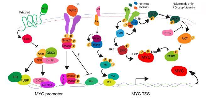
Duman-Scheel M, Johnston LA, Du W (2004) Repression of dMyc expression by Wingless promotes Rbf-induced G1 arrest in the presumptive Drosophila wing margin. Proc Natl Acad Sci U S A 101: 3857-3862. doi: 10.1073/pnas.0400526101
    [59] Maines JZ (2004) Drosophila dMyc is required for ovary cell growth and endoreplication. Development 131: 775-786. doi: 10.1242/dev.00932
    [60] Wierstra I, Alves J (2008) Advances in Cancer Research, 99. Elsevier. 113-333.
    [61] Levens D (2010) You Don't Muck with MYC. Genes Cancer 1: 547-554. doi: 10.1177/1947601910377492
    [62] Sears R, Leone G, DeGregori J, et al. (1999) Ras enhances Myc protein stability. Mol Cell 3: 169-179. doi: 10.1016/S1097-2765(00)80308-1
    [63] Salghetti SE, Kim SY, Tansey WP (1999) Destruction of Myc by ubiquitin-mediated proteolysis: cancer-associated and transforming mutations stabilize Myc. EMBO J 18: 717-726. doi: 10.1093/emboj/18.3.717
    [64] Sears RC (2004) The life cycle of C-myc: from synthesis to degradation. Cell Cycle 3: 1133-1137.
    [65] Daneshvar K, Nath S, Khan A, et al. (2013) MicroRNA miR-308 regulates dMyc through a negative feedback loop in Drosophila. Biol Open 2: 1-9. doi: 10.1242/bio.20122725
    [66] Amati B, Sanchez-Arevalo Lobo VJ (2007) MYC degradation: deubiquitinating enzymes enter the dance. Nat Cell Biol 9: 729-731. doi: 10.1038/ncb0707-729
    [67] Yada M, Hatakeyama S, Kamura T, et al. (2004) Phosphorylation-dependent degradation of c-Myc is mediated by the F-box protein Fbw7. EMBO J 23: 2116-2125. doi: 10.1038/sj.emboj.7600217
    [68] Siddall NA, Lin JI, Hime GR, et al. (2009) Myc--what we have learned from flies. Curr Drug Targets 10: 590-601. doi: 10.2174/138945009788680400
    [69] Cranna N, Quinn L (2009) Impact of steroid hormone signals on Drosophila cell cycle during development. Cell Div 4: 3. doi: 10.1186/1747-1028-4-3
    [70] Baker NE (2007) Patterning signals and proliferation in Drosophila imaginal discs. Curr Opin Genet Dev 17: 287-293. doi: 10.1016/j.gde.2007.05.005
    [71] Chung HJ, Levens D (2005) c-myc expression: keep the noise down!. Mol Cells 20: 157-166.
    [72] Liu J, Levens D (2006) Making myc. Curr Top Microbiol 302: 1-32.
    [73] Levens D, Gupta A (2010) Molecular biology. Reliable noise. Science 327: 1088-1089.
    [74] Avigan MI, Strober B, Levens D (1990) A far upstream element stimulates c-myc expression in undifferentiated leukemia cells. J Biol Chem 265: 18538-18545.
    [75] Michelotti GA, Michelotti EF, Pullner A, et al. (1996) Multiple single-stranded cis elements are associated with activated chromatin of the human c-myc gene in vivo. Mol Cell Biol 16: 2656-2669.
    [76] Liu J, Akoulitchev S, Weber A, et al. (2001) Defective interplay of activators and repressors with TFIH in xeroderma pigmentosum. Cell 104: 353-363. doi: 10.1016/S0092-8674(01)00223-9
    [77] 
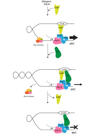
Liu J, Kouzine F, Chung HJ, et al. (2006) The FUSE/FBP/FIR/TFIIH system is a molecular machine programming a pulse of c-myc expression. EMBO J 25: 2119-2130. doi: 10.1038/sj.emboj.7601101
    [78] Matsushita K, Tomonaga T, Shimada H, et al. (2006) An essential role of alternative splicing of c-myc suppressor FUSE-binding protein-interacting repressor in carcinogenesis. Cancer Res 66: 1409-1417. doi: 10.1158/0008-5472.CAN-04-4459
    [79] Quinn LM, Dickins RA, Coombe M, et al. (2004) Drosophila Hfp negatively regulates dmyc and stg to inhibit cell proliferation. Development 131:1411-1423. doi: 10.1242/dev.01019
    [80] Mitchell NC, Johanson TM, Cranna NJ, et al. (2010) Hfp inhibits Drosophila myc transcription and cell growth in a TFIIH/Hay-dependent manner. Development 137: 2875-2884. doi: 10.1242/dev.049585
    [81] He TC, Sparks AB, Rago C, et al. (1998) Identification of c-MYC as a target of the APC pathway. Science 281: 1509-1512.
    [82] Korinek V, Barker N, Morin PJ, et al. (1997) Constitutive transcriptional activation by a beta-catenin-Tcf complex in APC-/- colon carcinoma. Science 275: 1784-1787. doi: 10.1126/science.275.5307.1784
    [83] Morin PJ, Sparks AB, Korinek V, et al. (1997) Activation of beta-catenin-Tcf signaling in colon cancer by mutations in beta-catenin or APC. Science 275: 1787-1790. doi: 10.1126/science.275.5307.1787
    [84] Korinek V, Barker N, Moerer P, et al. (1998) Depletion of epithelial stem-cell compartments in the small intestine of mice lacking Tcf-4. Nat Genet 19: 379-383. doi: 10.1038/1270
    [85] Pinto D, Gregorieff A, Begthel H, et al. (2003) Canonical Wnt signals are essential for homeostasis of the intestinal epithelium. Genes Dev 17: 1709-1713. doi: 10.1101/gad.267103
    [86] van de Wetering M, Sancho E, Verweij C, et al. (2002) The beta-catenin/TCF-4 complex imposes a crypt progenitor phenotype on colorectal cancer cells. Cell 111: 241-250. doi: 10.1016/S0092-8674(02)01014-0
    [87] Muncan V, Sansom OJ, Tertoolen L, et al. (2006) Rapid loss of intestinal crypts upon conditional deletion of the Wnt/Tcf-4 target gene c-Myc. Mol Cell Biol 26: 8418-8426. doi: 10.1128/MCB.00821-06
    [88] Sansom OJ, Meniel VS, Muncan V, et al. (2007) Myc deletion rescues Apc deficiency in the small intestine. Nature 446: 676-679. doi: 10.1038/nature05674
    [89] Jonsson M, Dejmek J, Bendahl PO, et al. (2002) Loss of Wnt-5a protein is associated with early relapse in invasive ductal breast carcinomas. Cancer Res 62: 409-416.
    [90] Winn RA, Marek L, Han SY, et al. (2005) Restoration of Wnt-7a expression reverses non-small cell lung cancer cellular transformation through frizzled-9-mediated growth inhibition and promotion of cell differentiation. J Biol Chem 280: 19625-19634. doi: 10.1074/jbc.M409392200
    [91] Kremenevskaja N, von Wasielewski R, Rao AS, et al. (2005) Wnt-5a has tumor suppressor activity in thyroid carcinoma. Oncogene 24: 2144-2154. doi: 10.1038/sj.onc.1208370
    [92] Ross SE, Erickson RL, Gerin I, et al. (2002) Microarray analyses during adipogenesis: understanding the effects of Wnt signaling on adipogenesis and the roles of liver X receptor alpha in adipocyte metabolism. Mol Cell Biol 22: 5989-5999. doi: 10.1128/MCB.22.16.5989-5999.2002
    [93] Hirabayashi Y, Itoh Y, Tabata H, et al. (2004) The Wnt/beta-catenin pathway directs neuronal differentiation of cortical neural precursor cells. Development 131: 2791-2801. doi: 10.1242/dev.01165
    [94] Johnston LA, Sanders AL (2003) Wingless promotes cell survival but constrains growth during Drosophila wing development. Nat Cell Biol 5: 827-833. doi: 10.1038/ncb1041
    [95] van Es JH, Jay P, Gregorieff A, et al. (2005) Wnt signalling induces maturation of Paneth cells in intestinal crypts. Nat Cell Biol 7: 381-386. doi: 10.1038/ncb1240
    [96] Fodde R, Brabletz T (2007) Wnt/beta-catenin signaling in cancer stemness and malignant behavior. Curr Opin Cell Biol 19: 150-158. doi: 10.1016/j.ceb.2007.02.007
    [97] Atlasi Y, Looijenga L, Fodde R (2014) Cancer stem cells, pluripotency, and cellular heterogeneity: a WNTer perspective. Curr Top Dev Biol 107: 373-404. doi: 10.1016/B978-0-12-416022-4.00013-5
    [98] Sancho E, Batlle E, Clevers H (2004) Signaling pathways in intestinal development and cancer. Annu Rev Cell Dev Bi 20: 695-723. doi: 10.1146/annurev.cellbio.20.010403.092805
    [99] Kelleher FC, Fennelly D, Rafferty M (2006) Common critical pathways in embryogenesis and cancer, Acta Oncol 45: 375-388.
    [100] Hopfer O, Zwahlen D, Fey MF, et al. (2005) The Notch pathway in ovarian carcinomas and adenomas. Brit J Cancer 93: 709-718. doi: 10.1038/sj.bjc.6602719
    [101] de Celis JF, Garcia-Bellido A, Bray SJ (1996) Activation and function of Notch at the dorsal-ventral boundary of the wing imaginal disc. Development 122: 359-369.
    [102] Johnston LA, Edgar BA (1998) Wingless and Notch regulate cell-cycle arrest in the developing Drosophila wing. Nature 394: 82-84. doi: 10.1038/27925
    [103] Herranz H, Pérez L, Martín FA, et al. (2008) A Wingless and Notch double-repression mechanism regulates G1-S transition in the Drosophila wing. EMBO J 27: 1633-1645. doi: 10.1038/emboj.2008.84
    [104] Massague J (2008) TGFbeta in Cancer. Cell 134: 215-230. doi: 10.1016/j.cell.2008.07.001
    [105] Coffey RJ, Bascom CC, Sipes NJ, et al. (1988) Selective inhibition of growth-related gene expression in murine keratinocytes by transforming growth factor beta. Mol Cell Biol 8: 3088-3093.
    [106] Chen CR, Kang Y, Siegel PM, et al. (2002) E2F4/5 and p107 as Smad cofactors linking the TGFbeta receptor to c-myc repression. Cell 110: 19-32. doi: 10.1016/S0092-8674(02)00801-2
    [107] Pennetier D, Oyallon J, Morin-Poulard I, et al. (2012) Size control of the Drosophila hematopoietic niche by bone morphogenetic protein signaling reveals parallels with mammals. PNAS 109: 3389-3394. doi: 10.1073/pnas.1109407109
    [108] Qu X, Shen L, Zheng Y, et al. (2014) A signal transduction pathway from TGF-β1 to SKP2 via Akt1 and c-Myc and its correlation with progression in human melanoma. J Invest Dermatol 134: 159-167. doi: 10.1038/jid.2013.281
    [109] Burke R, Basler K (1996) Hedgehog-dependent patterning in the Drosophila eye can occur in the absence of Dpp signaling. Dev Biol 179: 360-368. doi: 10.1006/dbio.1996.0267
    [110] Martin-Castellanos C, Edgar BA (2002) A characterization of the effects of Dpp signaling on cell growth and proliferation in the Drosophila wing. Development 129: 1003-1013.
    [111] Doumpas N, Ruiz-Romero M, Blanco E, et al. (2013) Brk regulates wing disc growth in part via repression of Myc expression. EMBO Rep 14: 261-268. doi: 10.1038/embor.2013.1
    [112] Lawrence PA, Casal J (2013) The mechanisms of planar cell polarity, growth and the Hippo pathway: some known unknowns. Dev Biol 377: 1-8. doi: 10.1016/j.ydbio.2013.01.030
    [113] Pan D (2010) The hippo signaling pathway in development and cancer. Dev cell 19: 491-505. doi: 10.1016/j.devcel.2010.09.011
    [114] Neto-Silva RM, de Beco S, Johnston LA (2010) Evidence for a Growth-Stabilizing Regulatory Feedback Mechanism between Myc and Yorkie, the Drosophila Homolog of Yap. Dev Cell 19: 507-520. doi: 10.1016/j.devcel.2010.09.009
    [115] Mori M, Triboulet R, Mohseni M, et al. (2014) Hippo signaling regulates microprocessor and links cell-density-dependent miRNA biogenesis to cancer. Cell 156: 893-906. doi: 10.1016/j.cell.2013.12.043
    [116] Pan D (2007) Hippo signaling in organ size control. Genes Dev 21: 886-897. doi: 10.1101/gad.1536007
    [117] Ziosi M, Baena-López LA, Grifoni D, et al. (2010) dMyc functions downstream of Yorkie to promote the supercompetitive behavior of hippo pathway mutant cells. PLoS Genet 6: e1001140. doi: 10.1371/journal.pgen.1001140
    [118] Xiao W, Wang J, Ou C, et al. (2013) Mutual interaction between YAP and c-Myc is critical for carcinogenesis in liver cancer. Biochem Bioph Res Co 439: 167-172. doi: 10.1016/j.bbrc.2013.08.071
    [119] Orme MH, Alrubaie S, Bradley GL, et al. (2006) Input from Ras is required for maximal PI(3)K signalling in Drosophila. Nat Cell Biol 8: 1298-1302. doi: 10.1038/ncb1493
    [120] Prober DA, Edgar BA (2002) Interactions between Ras1, dMyc, and dPI3K signaling in the developing Drosophila wing. Genes Dev 16:2286-2299. doi: 10.1101/gad.991102
    [121] Leevers SJ, Weinkove D, MacDougall LK, et al. (1996) The Drosophila phosphoinositide 3-kinase Dp110 promotes cell growth. EMBO J 15: 6584-6594.
    [122] Hall DJ, Grewal SS, de la Cruz AF, et al. (2007) Rheb-TOR signaling promotes protein synthesis, but not glucose or amino acid import, in Drosophila. BMC Biol 5: 1. doi: 10.1186/1741-7007-5-1
    [123] Grewal SS, Evans JR, Edgar BA (2007) Drosophila TIF-IA is required for ribosome synthesis and cell growth and is regulated by the TOR pathway. J Cell Biol 179: 1105-1113. doi: 10.1083/jcb.200709044
    [124] Grewal SS (2009) Insulin/TOR signaling in growth and homeostasis: a view from the fly world. Int J Biochem Cell Biol 41: 1006-1010. doi: 10.1016/j.biocel.2008.10.010
    [125] Li L, Edgar BA, Grewal SS (2010) Nutritional control of gene expression in Drosophila larvae via TOR, Myc and a novel cis-regulatory element. BMC Cell Biol 11: 7.
    [126] Edgar BA (2006) How flies get their size: genetics meets physiology. Nat Rev 7: 907-916.
    [127] Britton JS, Lockwood WK, Li L, et al. (2002) Drosophila's insulin/PI3-kinase pathway coordinates cellular metabolism with nutritional conditions. Dev Cell 2: 239-249. doi: 10.1016/S1534-5807(02)00117-X
    [128] Jiang BH, Liu LZ (2009) PI3K/PTEN signaling in angiogenesis and tumorigenesis. Adv Cancer Res 102: 19-65. doi: 10.1016/S0065-230X(09)02002-8
    [129] Teleman AA, Hietakangas V, Sayadian AC, et al. (2008) Nutritional control of protein biosynthetic capacity by insulin via Myc in Drosophila. Cell Metab 7: 21-32. doi: 10.1016/j.cmet.2007.11.010
    [130] Parisi F, Riccardo S, Daniel M, et al. (2011) Drosophila insulin and target of rapamycin (TOR) pathways regulate GSK3 beta activity to control Myc stability and determine Myc expression in vivo. BMC Biol 9: 65. doi: 10.1186/1741-7007-9-65
    [131] Amcheslavsky A, Ito N, Jiang J, et al. (2011) Tuberous sclerosis complex and Myc coordinate the growth and division of Drosophila intestinal stem cells. J Cell Biol 193: 695-710. doi: 10.1083/jcb.201103018
    [132] Chan JC, Hannan KM, Riddell K, et al. (2011) AKT promotes rRNA synthesis and cooperates with c-MYC to stimulate ribosome biogenesis in cancer. Sci Signal 4: ra56.
    [133] Hann SR, Eisenman RN (1984) Proteins encoded by the human c-myc oncogene: differential expression in neoplastic cells. Mol Cellular Biol 4: 2486-2497.
    [134] Gregory MA, Hann SR (2000) c-Myc proteolysis by the ubiquitin-proteasome pathway: stabilization of c-Myc in Burkitt's lymphoma cells. Mol Cellular Biol 20: 2423-2435. doi: 10.1128/MCB.20.7.2423-2435.2000
    [135] Farrell AS, Sears RC (2014) MYC degradation. Cold Spring Harb Perspect Med 4: 3.
    [136] Wang C, Lisanti MP, Liao DJ (2011) Reviewing once more the c-myc and Ras collaboration: converging at the cyclin D1-CDK4 complex and challenging basic concepts of cancer biology. Cell Cycle 10: 57-67. doi: 10.4161/cc.10.1.14449
    [137] Sears R, Nuckolls F, Haura E, et al. (2000) Multiple Ras-dependent phosphorylation pathways regulate Myc protein stability, Genes Dev 14: 2501-2514.
    [138] Papas TS, Lautenberger JA (1985) Sequence curiosity in v-myc oncogene. Nature 318: p. 237.
    [139] Gregory MA, Qi Y, Hann SR (2003) Phosphorylation by Glycogen Synthase Kinase-3 Controls c-Myc Proteolysis and Subnuclear Localization. J Biol Chem 278: 51606-51612. doi: 10.1074/jbc.M310722200
    [140] Yeh E, Cunningham M, Arnold H, et al. (2004) A signalling pathway controlling c-Myc degradation that impacts oncogenic transformation of human cells. Nat Cell Biol 6: 308-318. doi: 10.1038/ncb1110
    [141] Davis MA, Larimore EA, Fissel BM, et al. (2013) The SCF-Fbw7 ubiquitin ligase degrades MED13 and MED13L and regulates CDK8 module association with Mediator. Genes Dev 27: 151-156. doi: 10.1101/gad.207720.112
    [142] Cross DA, Alessi DR, Cohen P, et al. (1995) Inhibition of glycogen synthase kinase-3 by insulin mediated by protein kinase B. Nature 378: 785-789. doi: 10.1038/378785a0
    [143] Welcker M, Orian A, Jin J, et al. (2004) The Fbw7 tumor suppressor regulates glycogen synthase kinase 3 phosphorylation-dependent c-Myc protein degradation. Proc Natl Acad Sci U S A 101: 9085-9090. doi: 10.1073/pnas.0402770101
    [144] Galletti M, Riccardo S, Parisi F, et al. (2009) Identification of domains responsible for ubiquitin-dependent degradation of dMyc by glycogen synthase kinase 3beta and casein kinase 1 kinases. Mol Cell Biol 29: 3424-3434. doi: 10.1128/MCB.01535-08
    [145] Moberg KH, Mukherjee A, Veraksa A, et al. (2004) The Drosophila F box protein archipelago regulates dMyc protein levels in vivo. Curr Biol 14: 965-974. doi: 10.1016/j.cub.2004.04.040
    [146] Arnold HK, Zhang X, Daniel CJ, et al. (2009) The Axin1 scaffold protein promotes formation of a degradation complex for c-Myc. EMBO J 28: 500-512. doi: 10.1038/emboj.2008.279
    [147] Johnston LA (2009) Competitive Interactions Between Cells: Death, Growth, and Geography. Science 324: 1679-1682. doi: 10.1126/science.1163862
    [148] de la Cova C, Abril M, Bellosta P, et al (2004) Drosophila myc regulates organ size by inducing cell competition. Cell 117: 107-116. doi: 10.1016/S0092-8674(04)00214-4
    [149] Moreno E, Basler K (2004) dMyc transforms cells into super-competitors. Cell 117: 117-129. doi: 10.1016/S0092-8674(04)00262-4
    [150] Baker NE, Li W. (2008) Cell competition and its possible relation to cancer. Cancer Res 68: 5505-5507. doi: 10.1158/0008-5472.CAN-07-6348
    [151] Moreno E (2008) Is cell competition relevant to cancer?. Nat Rev 8: 141-147. doi: 10.1038/nrc2252
    [152] Johnston LA (2014) Socializing with MYC: cell competition in development and as a model for premalignant cancer. Cold Spring Harb Perspect Med 4: a014274.
    [153] Senoo-Matsuda N, Johnston LA (2007) Soluble factors mediate competitive and cooperative interactions between cells expressing different levels of Drosophila Myc. Proc Natl Acad Sci U S A 104: 18543-18548. doi: 10.1073/pnas.0709021104
    [154] Bywater MJ, Pearson RB, McArthur GA, et al. (2013) Dysregulation of the basal RNA polymerase transcription apparatus in cancer. Nat Rev 13: 299-314. doi: 10.1038/nrc3496
  • This article has been cited by:

    1. Chunhua Cao, Jianhua Chen, Chengqi Lyu, Jia Yu, Wei Zhao, Yi Wang, Derong Zou, Marinus F.W. te Pas, Bioinformatics Analysis of the Effects of Tobacco Smoke on Gene Expression, 2015, 10, 1932-6203, e0143377, 10.1371/journal.pone.0143377
    2. Helena E. Richardson, Drosophila models of cancer, 2015, 02, 2377-1143, 097, 10.3934/genet.2015.1.97
    3. Luisa Mori, Katharine Jenike, Yang-Hui Jimmy Yeh, Benoît Lacombe, Chuan Li, Adam J. Getzler, Sonia Mediouni, Michael E. Cameron, Matthew E. Pipkin, Ya-Chi Ho, Bertha Cecilia Ramirez, Susana T. Valente, Guido Silvestri, The XPB Subunit of the TFIIH Complex Plays a Critical Role in HIV-1 Transcription, and XPB Inhibition by Spironolactone Prevents HIV-1 Reactivation from Latency, 2021, 95, 0022-538X, 10.1128/JVI.01247-20
    4. Olga Zaytseva, Leonie M. Quinn, DNA Conformation Regulates Gene Expression: The MYC Promoter and Beyond, 2018, 40, 0265-9247, 10.1002/bies.201700235
    5. Coral G. Warr, Katherine H. Shaw, Arani Azim, Matthew D. W. Piper, Linda M. Parsons, Using Mouse and Drosophila Models to Investigate the Mechanistic Links between Diet, Obesity, Type II Diabetes, and Cancer, 2018, 19, 1422-0067, 4110, 10.3390/ijms19124110
    6. Leonie M. Quinn, FUBP/KH domain proteins in transcription: Back to the future, 2017, 8, 2154-1264, 185, 10.1080/21541264.2017.1293595
    7. Olga Zaytseva, Leonie Quinn, Controlling the Master: Chromatin Dynamics at the MYC Promoter Integrate Developmental Signaling, 2017, 8, 2073-4425, 118, 10.3390/genes8040118
    8. Naomi C. Mitchell, Elissaveta B. Tchoubrieva, Arjun Chahal, Simone Woods, Amanda Lee, Jane I. Lin, Linda Parsons, Katarzyna Jastrzebski, Gretchen Poortinga, Katherine M. Hannan, Richard B. Pearson, Ross D. Hannan, Leonie M. Quinn, S6 Kinase is essential for MYC-dependent rDNA transcription in Drosophila, 2015, 27, 08986568, 2045, 10.1016/j.cellsig.2015.07.018
  • Reader Comments
  • © 2014 the Author(s), licensee AIMS Press. This is an open access article distributed under the terms of the Creative Commons Attribution License (http://creativecommons.org/licenses/by/4.0)
通讯作者: 陈斌, bchen63@163.com
  • 1. 

    沈阳化工大学材料科学与工程学院 沈阳 110142

  1. 本站搜索
  2. 百度学术搜索
  3. 万方数据库搜索
  4. CNKI搜索

Metrics

Article views(7360) PDF downloads(1225) Cited by(8)

Article outline

/

DownLoad:  Full-Size Img  PowerPoint
Return
Return

Catalog