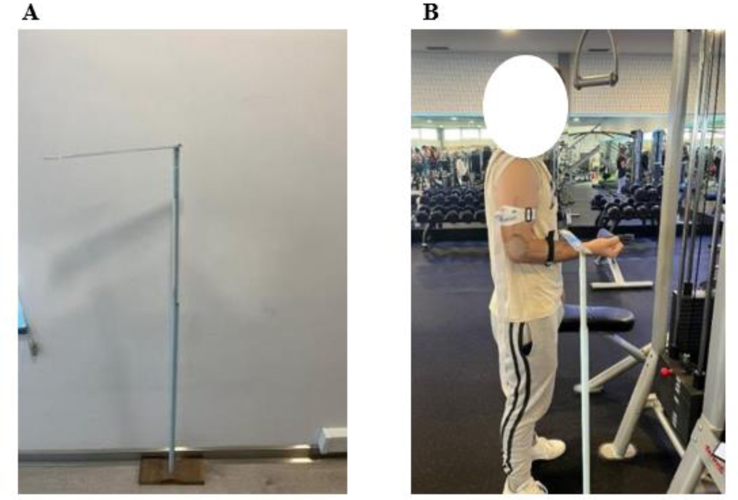
Range of motion in exercises is one of the foundations for greater activation of a muscle group. The objective of this investigation was to compare the structural and functional capacity of the triceps brachii between three groups with different angles (90°, 110°, and 130°) in a unilateral elbow extension exercise. The sample consisted of 25 subjects with a mean age of 24.12 ± 3.83 years, mean height of 1.78 ± 0.10 m and mean body weight of 78.01 ± 15.70 kg. The following variables were collected pre- and post-intervention: triceps brachii circumference, one repetition maximum, and electromyography during dynamic exercise. Over eight weeks, subjects performed this exercise, performing 3 sets of 12 repetitions for each arm, with days of rest in between. The results showed that the 110° angle provided greater muscle activation compared to the other angles. There was no difference between the triceps brachii circumference and the root mean square (RMS) between the groups. It was concluded that, although the 110° angle showed a tendency for greater muscle activation, the RMS and arm perimeter data did not show significant differences between all the angles evaluated (90°, 110°, 130°).
Citation: Luís M. Ferreira, Luís Ferreira, Joana Ribeiro, Luís Branquinho, Rafael Peixoto, Luciano Bernardes Leite, Pedro Forte. The influence of range of motion on the functional and structural capacity of the triceps brachii—an experimental study with electromyography[J]. AIMS Biophysics, 2024, 11(4): 445-454. doi: 10.3934/biophy.2024024
Range of motion in exercises is one of the foundations for greater activation of a muscle group. The objective of this investigation was to compare the structural and functional capacity of the triceps brachii between three groups with different angles (90°, 110°, and 130°) in a unilateral elbow extension exercise. The sample consisted of 25 subjects with a mean age of 24.12 ± 3.83 years, mean height of 1.78 ± 0.10 m and mean body weight of 78.01 ± 15.70 kg. The following variables were collected pre- and post-intervention: triceps brachii circumference, one repetition maximum, and electromyography during dynamic exercise. Over eight weeks, subjects performed this exercise, performing 3 sets of 12 repetitions for each arm, with days of rest in between. The results showed that the 110° angle provided greater muscle activation compared to the other angles. There was no difference between the triceps brachii circumference and the root mean square (RMS) between the groups. It was concluded that, although the 110° angle showed a tendency for greater muscle activation, the RMS and arm perimeter data did not show significant differences between all the angles evaluated (90°, 110°, 130°).

| [1] | Fleck SJ, Kraemer WJ Fundamentos Do Treinamento de Força Muscular (2017). |
| [2] |
Lakka TA, Laaksonen DE, Lakka HM, et al. (2003) Sedentary lifestyle, poor cardiorespiratory fitness, and the metabolic syndrome. Med Sci Sport Exer 35: 1279-1286. https://doi.org/10.1249/01.mss.0000079076.74931.9a 
|
| [3] |
Gordon KD, Pardo RD, Johnson JA, et al. (2004) Electromyographic activity and strength during maximum isometric pronation and supination efforts in healthy adults. J Orthop Res 22: 208-213. https://doi.org/10.1016/S0736-0266(03)00115-3
|
| [4] | Moritani T, Devries HA (1979) Neural factors versus hypertrophy in the time course of muscle strength gain. Am J Phys Med Rehab 58: 115-130. |
| [5] |
Häkkinen K, Alén M, Komi PV (1985) Changes in isometric force- and relaxation-time, electromyographic and muscle fibre characteristics of human skeletal muscle during strength training and detraining. Acta Physiol Scand 125: 573-585. https://doi.org/10.1111/j.1748-1716.1985.tb07759.x
|
| [6] |
Barsanti RR, Fonseca BPA, Silvatti AP, et al. (2021) Descriptive electromyography signals analysis of equine longissimus dorsi, rectus abdominis and gluteus medius muscles during maneuvers used to activate the core. Arq Bras Med Vet Zoo 73: 843-852. https://doi.org/10.1590/1678-4162-11309
|
| [7] |
Cardoso JR, Prado AI, Iriya HK, et al. (2008) Atividade eletromiográfica dos músculos do joelho em indivíduos com reconstrução do ligamento cruzado anterior sob diferentes estímulos sensório-motores: relato de casos. Fisioter Pesqui 15: 78-85. https://doi.org/10.1590/S1809-29502008000100013
|
| [8] | Cogley RM, Archambault TA, Fibeger JF, et al. (2005) Comparison of muscle activation using various hand positions during the push-up exercise. J Strength Cond Res 19: 628-633. https://doi.org/10.1519/15094.1 |
| [9] | Huang J, Tian F, Zhang Z, et al. (2020) Reliability and concurrent validity of angle measurements in lower limb: EOS 3D goniometer versus 2D manual goniometer. J Orthop Transl 24: 96-102. https://doi.org/10.1016/j.jot.2020.05.002 |
| [10] |
Larsen MN, Krustrup P, Araújo Póvoas SC, et al. (2021) Accuracy and reliability of the InBody 270 multi-frequency body composition analyser in 10-12-year-old children. PLoS One 16: e0247362. https://doi.org/10.1371/journal.pone.0247362
|
| [11] |
Ntineri A, Theodosiadi A, Menti A, et al. (2023) A novel professional automated auscultatory blood pressure monitor with visual display of Korotkoff sounds: InBody BPBIO480KV validation according to the Association for the Advancement of Medical Instrumentation/European Society of Hypertension/International Organization for Standardization Universal Standard. J Hypertens 41: 356-361. https://doi.org/10.1097/HJH.0000000000003341
|
| [12] | Buscà B, Font A (2011) A low-cost contact system to assess load displacement velocity in a resistance training machine. J Sports Sci Med 10: 472. |
| [13] |
Tereso D, Paulo R, Petrica J, et al. (2021) Assessment of body composition, lower limbs power, and anaerobic power of senior soccer players in Portugal: differences according to the competitive level. Int J Env Res Pub He 18: 8069. https://doi.org/10.3390/ijerph18158069
|
| [14] |
Molina-Molina A, Ruiz-Malagón EJ, Carrillo-Pérez F, et al. (2020) Validation of mDurance, a wearable surface electromyography system for muscle activity assessment. Front Physiol 11: 606287. https://doi.org/10.3389/fphys.2020.606287
|
| [15] |
Schoenfeld BJ, Grgic J, Van Every DW, et al. (2021) Loading recommendations for muscle strength, hypertrophy, and local endurance: a re-examination of the repetition continuum. Sports 9: 32. https://doi.org/10.3390/sports9020032
|
| [16] |
Androulakis Korakakis P, Wolf M, Coleman M, et al. (2024) Optimizing resistance training technique to maximize muscle hypertrophy: a narrative review. J Funct Morphol Kinesiol 9: 9. https://doi.org/10.3390/jfmk9010009
|
| [17] |
Westcott WL, Winett RA, Annesi JJ, et al. (2009) Prescribing physical activity: applying the ACSM protocols for exercise type, intensity, and duration across 3 training frequencies. Physcian Sportsmed 37: 51-58. https://doi.org/10.3810/psm.2009.02.1882
|
| [18] |
Haskell WL, Lee IM, Pate RR, et al. (2007) Physical activity and public health: updated recommendation for adults from the American College of Sports Medicine and the American Heart Association. Circulation 116: 1081-1093. https://doi.org/10.1161/CIRCULATIONAHA.107.185649
|
| [19] |
Schoenfeld BJ, Peterson MD, Ogborn D, et al. (2015) Effects of low-vs. high-load resistance training on muscle strength and hypertrophy in well-trained men. J Strength Cond Res 29: 2954-2963. https://doi.org/10.1519/JSC.0000000000000958
|
| [20] |
Ogasawara R, Loenneke JP, Thiebaud RS, et al. (2013) Low-load bench press training to fatigue results in muscle hypertrophy similar to high-load bench press training. Int J Clin Med 4: 114-121. https://doi.org/10.4236/ijcm.2013.42022
|
| [21] |
Schoenfeld BJ, Wilson JM, Lowery RP, et al. (2016) Muscular adaptations in low-versus high-load resistance training: a meta-analysis. Eur J Sport Sci 16: 1-10. https://doi.org/10.1080/17461391.2014.989922
|
| [22] |
Alegre LM, Aguado X, Rojas-Martín D, et al. (2015) Load-controlled moderate and high-intensity resistance training programs provoke similar strength gains in young women. Muscle Nerve 51: 92-101. https://doi.org/10.1002/mus.24271
|
| [23] |
Kukić F, Mrdaković V, Stanković A, et al. (2022) Effects of knee extension joint angle on quadriceps femoris muscle activation and exerted torque in maximal voluntary isometric contraction. Biology 11: 1490. https://doi.org/10.3390/biology11101490
|
| [24] |
Akima H, Maeda H, Koike T, et al. (2021) Effect of elbow joint angles on electromyographic activity versus force relationships of synergistic muscles of the triceps brachii. PLoS One 16: e0252644. https://doi.org/10.1371/journal.pone.0252644
|
| [25] |
Hager R, Poulard T, Nordez A, et al. (2020) Influence of joint angle on muscle fascicle dynamics and rate of torque development during isometric explosive contractions. J Appl Physiol 129: 569-579. https://doi.org/10.1152/japplphysiol.00143.2019
|
| [26] |
Sieck GC (1989) Recruitment and frequency coding of diaphragm motor units during ventilatory and non-ventilatory behaviors. Respiratory Control: A Modeling Perspective . Boston: Springer 441-450. https://doi.org/10.1007/978-1-4613-0529-3_48
|
| [27] | Na'aim N, Chen CK, Ooi FK, et al. (2022) Combined effects of bee pollen supplementation and resistance training on aerobic capacity, muscular performance, antioxidant status, and bone metabolism markers in young men: a randomised controlled trial. Malays J Nutr 28: 239-251. https://doi.org/10.31246/mjn-2021-0072 |
| [28] |
Firdaus W, Kuan G, Krasilshchikov O (2018) The effects of using complex training method on muscular strength among male weightlifters. Jurnal Sains Sukan dan Pendidikan Jasmani 7: 1-12. https://doi.org/10.37134/jsspj/vol7.1.1.2018
|
| [29] |
Molsted S, Andersen JL, Eidemak I, et al. (2013) Increased rate of force development and neuromuscular activity after high-load resistance training in patients undergoing dialysis. Nephrology 18: 770-776. https://doi.org/10.1111/nep.12145
|
Figures(1) / Tables(2)

Luís M. Ferreira, Luís Ferreira, Joana Ribeiro, Luís Branquinho, Rafael Peixoto, Luciano Bernardes Leite, Pedro Forte. The influence of range of motion on the functional and structural capacity of the triceps brachii—an experimental study with electromyography[J]. AIMS Biophysics, 2024, 11(4): 445-454. doi: 10.3934/biophy.2024024


 DownLoad: