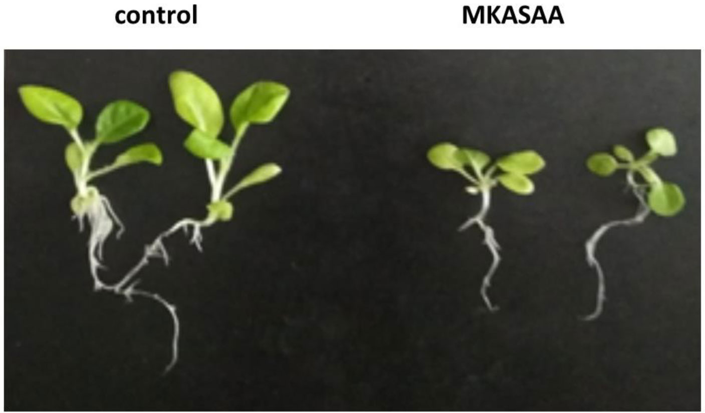
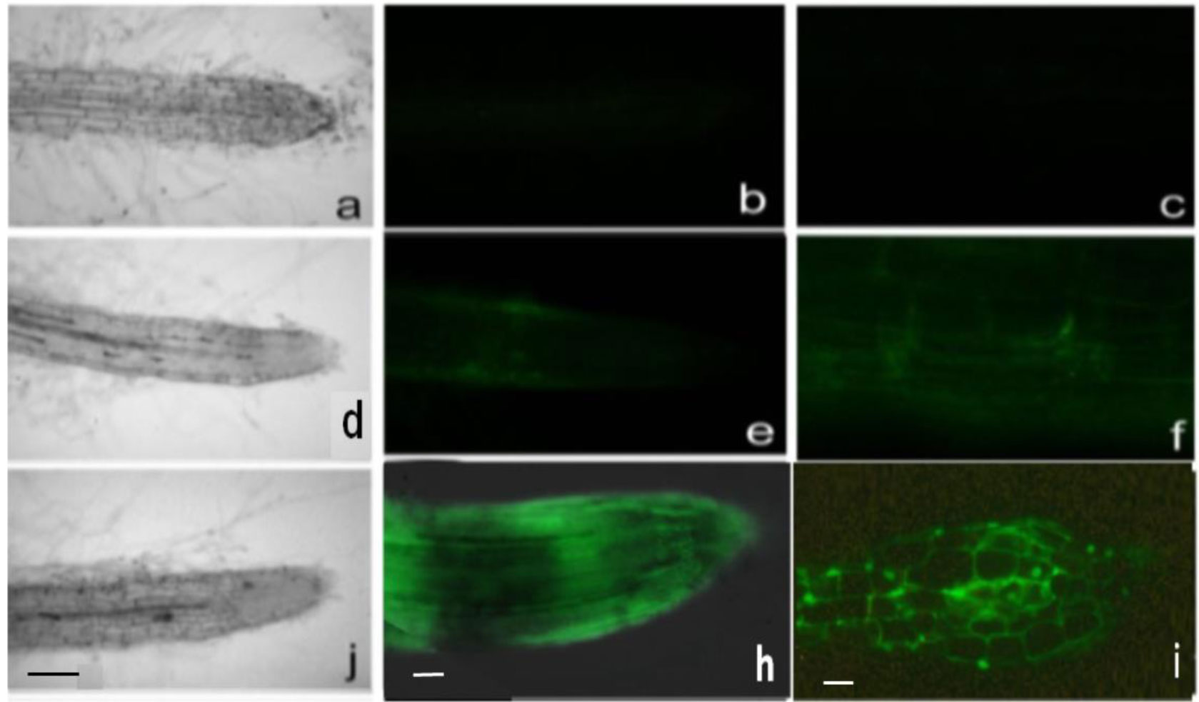
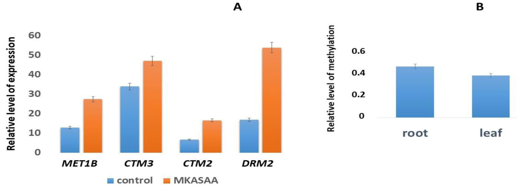
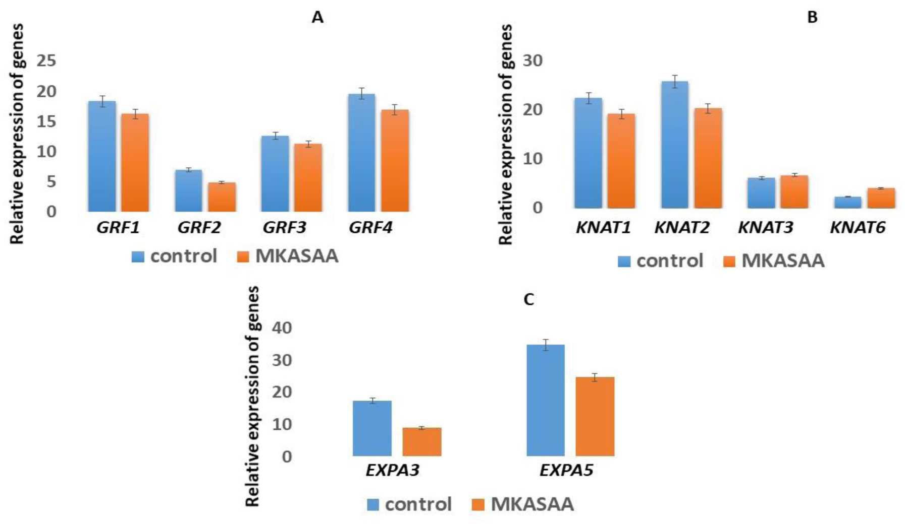
1.
Introduction
Plants are exposed to the environment. In response to environmental influences, plants have evolved by changing gene expression without changing the original DNA sequence, creating a “memory process” that is defined as epigenetic. This epigenetic memory can be achieved through covalent modifications of nucleotides and chromatin, including DNA cytosine methylation, modification of basic histone proteins, RNA-mediated gene silencing, and chromatin remodeling [1]. Chromatin is a dynamic DNA-protein structure that can exist as transcriptionally permissive euchromatin or repressive heterochromatin. The structure of chromatin in eukaryotic organisms can change during growth and development, as well as in response to environmental influences. Chromatin remodeling controls important molecular processes such as gene transcription, replication, repair, and recombination [2].
There are two main players that regulate chromatin dynamics: (1) chromatin remodelers, which alter DNA-histone interactions with energy derived from ATP hydrolysis, and (2) nucleosome-modifying enzymes, which modulate DNA and histone residues by specifically adding or removal of covalent modifications [3]. DNA methylation is usually associated with long-term gene silencing, while histone modifications promote both activation and repression of gene transcription and can be removed after several cell cycles [4],[5].
Post-translational modification of histones plays a key role in the regulation of chromatin dynamics. Methylation of Lys residues in the H3 histone tail is a key regulator of chromatin state and gene expression, which is provided by a large family of enzymes containing the evolutionarily conserved SET domain. Transcriptionally active chromatins typically contain trimethylated histone H3K4 and highly acetylated histone H3 and H4. In contrast, transcriptionally silent chromatins are enriched in lysine 9 and/or 27 histone H3 methylation [6],[7].
DNA methylation is the covalent modification of nucleotides in DNA. Methylation of eukaryotic DNA is carried out by DNA methyltransferases (DMT) enzymes, which transfer a methyl group from S-adenosyl methionine (SAM) to the cytosine carbon at the fifth position. These enzymes are classified into maintenance and de novo DMT, depending on whether the recognition site is already methylated or not. There are two types of maintaining DMT in plants: DNA methyltransferase (MET) and chromomethyltransferase (CMT) [8]. Plant genomes have three types of methylation sites: CG, CNG, and CNN [9], [10].
Methylation of previously unmethylated DNA, de novo, is carried out by a family of DNA methyltransferases called DRM (Domains rearranged methyltransferases). DRMs are known to primarily methylate unsymmetrical CHH sites, (where H is any nucleotide except G), but are capable of de novo methylation of cytosines in any sequence context in a process called RNA-directed DNA methylation (RdDM). RdDM is a biological process in which non-coding RNA molecules direct the addition of DNA methylation to specific DNA sequences. The RdDM pathway is unique to plants. DNA methylation added by RdDM is usually associated with transcriptional repression of genetic sequences where de novo methylation occurs. [11].
Since DNA methylation and repressive histone modifications together define heterochromatin, most DNA methylation pathways in plants recognize and interact with repressive histone marks and vice versa, forming positive feedback loops that help maintain the state of repressive chromatin [12]. Ultimately, the constant increase in silencing modifications of chromatin at heterochromatic loci creates a repressive chromatin state in which DNA and histones (nucleosomes) become densely packed together. This helps to suppress gene expression by blocking access to DNA, preventing transcription initiation by RNA polymerase II, transcription factors and other proteins [13].
In addition to cellular regulation of chromatin dynamics, including modifications of DNA, histone tails, and nucleosome-binding proteins, its structure is affected by external stress factors such as temperature, drought, high salt concentration, and infection. RdDM helps plants respond to a number of abiotic stresses, such as heat stress, drought, phosphate starvation, salt stress, and others [14]. Many transposable element TEs are upregulated under conditions of abiotic stress [15],[16], and thus one of the functions of RdDM in response to stress is to help counteract this activation.
Peptides play a key role in many physiological processes by coordinating developmental and environmental signals between different cells. Small peptides are important signaling molecules that coordinate comprehensive cell-to-cell communication in many aspects of plant development. Hormonal peptides play a crucial role in plant growth and development, including defense mechanisms in response to pest damage, control of cell division and expansion, and pollen incompatibility [17].
The aim of this work was to study the regulation of DNA methyltransferase gene expression in tobacco by the MKASAA peptide, de nova DNA methylation, and the effect of de nova DNA methylation on the development of tobacco seedlings.
2.
Materials and methods
2.1. Plant material
Seeds of tobacco (Nicotiana tabacum L.) cultivar Samsun (collection variety of the Agricultural Academy of the Russian Federation) were placed in flasks containing hormone-free Murashige–Skoog (MS) medium supplemented with or without 10–7 M MKASAA. Experiments were carried out in four replicates. After 28 days, the seedling fresh weight, seedling height, and root length were evaluated [18]. The last parameter was measured under an Olympus BX51 microscope (Japan) furnished with the Cell program. The calculation of the main statistical parameters was carried out according to standard methods, and Statistica 6.0 and STATAN 2.0 programs for statistical data processing were used.
2.2. Synthetic preparations
Oligoribonucleotides and the MKASAA peptide were synthesized by Sintol LLC (Russia). The purity of the synthetic preparations was checked by chromatography on a BioLogic DuoFlow chromatograph on a C-18 column in a concentration gradient of acetonitrile (0–60%) containing 1% trifluoroacetic acid.
2.3. Preparation of FITC-labeled peptides
A solution of FITC (1 µg in 10 µl of 0.5 M sodium bicarbonate) was added to a solution of MKASAA in 0.01 M Tris-HCl buffer (pH 7.0) at a ratio of 1.2: 1 [18]. The reaction mixture was incubated at room temperature for 30 min with constant shaking. The obtained fluorescently labeled peptides were analyzed by chromatography on a BioLogic DuoFlow chromatograph on a C-18 column in a concentration gradient of acetonitrile (0–100%) containing 1% trifluoroacetic acid.
2.4. Fluorescence microscopy
To determine the localization of peptides in root cells, the FITC-labeled dipeptides and Gly were used at a concentration of 10–5 M; control FITC and FITC without peptides were used as controls [19]. The incubation time was 20 h. Root tips (4–5 mm) were excised from tobacco roots, and fixed in 4% paraformaldehyde (Sigma-Aldrich, USA) for 1.5 h at room temperature. After three washes with phosphate-buffered saline (PBS), the root tips were mounted on glass slides and embedded in Moviol. Fluorescence was analyzed at a wavelength of 490 nm using Olympus BX51 microscope (Japan) with 10X and 20X objective lens. Photographs were obtained using Color View digital camera (Germany) [20].
2.5. Light microscopy
Roots stained with FITC-labeled peptides were fixed in 4% paraformaldehyde and embedded in LRW, as described previously [20]. Then, semi-thin root sections (1,000 µm) were obtained using the LKB-III microtome (LKB, Sweden). Fluorescence was analyzed at a wavelength of 490 nm using Olympus BX51 microscope (Japan) with 10X and 20X objective lens. Photographs were captured using Color View digital camera (Germany). Photographs of root and determination of their length along the main root were taken with a digital camera Rising View (China).
2.6. Fluorescence
Fluorescence spectra were recorded using a PerkinElmer LS 55 spectrofluorometer (USA). Titration of FITC-labeled MKASAA with deoxyribooligonucleotides (oligos) was carried out as described in [21]. This allows us to use the Stern–Volmer constant to evaluate the binding of peptides to various oligos [22].
2.7. Light scattering
The dynamic light scattering of samples at a concentration of 10–5 M was determined on a Zetaseizer Nano-zs “Malvern” instrument (UK).
2.8. DNA extraction
Total DNA was isolated from the roots and leaves of tobacco seedlings according to the standard DNA from tobacco regenerants was isolated using a standard method, utilizing a HigherPurity Plant DNA Purification Kit (Canvax, Spain). The purity of the preparations were determined by electrophoresis in 1% agarose. The mass of the obtained DNA was determined relative to DNA markers.
2.9. Global DNA methylation status
Global DNA methylation status was determined using the MethylFlash Methylated DNA Quantification kit (Epigentek). Total DNA was sequentially processed by a complex of enzymes to mononucleotides. The mixture was further analyzed on a chromatography on a BioLogic DuoFlow chromatograph on a C-18 column in a concentration gradient of acetonitrile (0–100%) containing 1% trifluoroacetic acid. 5-methylcytosine was used as an internal standard.
2.10. Real-time PCR
Total RNA was isolated from tobacco, from which root hairs were carefully removed, using RNA-Extran RNA isolation reagent kit (Syntol, Russia), according to the manufacturer's instructions. The concentration of isolated RNA preparations was determined on NanoPhotometer IMPLEN. Then, cDNA was obtained using a set of reverse transcription reagents (Syntol, Russia), according to the manufacturer's instructions.
Information on the primary structure of Nicotiana tabacum genes was obtained from the National Center for Biotechnology Information (NCBI). Primers for these genes were designed using the NCBI Primer-BLAST web tool, and synthesized at Syntol (Table 1,2). Real-time PCR was performed on CFX96 Touch Real-Time PCR Detection System (Bio-Rad) using a set of RT-PCR reagents and SYBR Green (Syntol). The relative expression level of genes was calculated using a calibration curve constructed with PCR products derived from the GAPDH gene. The calculation of the main statistical parameters was carried out according to STATAN program on a significance level p < 0.05.
2.11. Statistical processing of results
All experiments were repeated three or five times. The results of multiple experiments are presented as the mean ± SE. Statistical analysis was carried out using analysis of variance (ANOVA) followed by least significant difference (LSD) test. A p-value of < 0.05 was considered for a statistically significant result.
The equipment of the Center for Collective Use of the Federal State Budgetary Scientific Institution VNIISB RAS was used in this study.
3.
Results
3.1. Morphometric parameters
MKASAA peptide at a concentration of 10−7 M inhibits the growth and development of Nicotiana tabacum seedlings (Figure 1).
This is confirmed by morphometric parameters such as seedling weight, main root length and seedling height (Table 3). Thus, under the action of histone, the fresh weight of seedlings is reduced by 4 times, and the height of seedlings and the length of the main root by 1.5–1.8 times, respectively.
3.2. Penetration of FITC-MKASAA into tobacco roots
In tobacco roots treated with free FITC a slight luminescence is visible, mainly in the cells of the epidermis (Figure 2). Free FITC concentrates on the cell surface and does not penetrate inside. When tobacco roots were treated with FITC-MKASAA, luminescence was observed in the cap, meristem, and elongation zones. Moreover, the maximum glow is observed in the elongation zone. On a longitudinal semi-thin section, it can be seen that FITC-MKASAA penetrates the cell and concentrates mainly in the cell nucleus.
3.3. Gene expression of DNA methyltransferases
Expression of genes from N. tabacum encoding cytosine DNA methyltransferases are shown in Figure 3. In seedlings tobacco grown in the presence of MKASAA peptide, the expression level of the cytosine DNA methyltransferases, are activated in all variants.
The major DNA methyltransferase (DMT) MET1B methylates cytosine bases at the CG sites of DNA. This maintenance DMT methylates sites during replication. In tobacco seedlings grown in the presence of the peptide, MET1B gene expression increased by 1.8 times.
DMT CMT3 provides downstream methylation of symmetric CNG sites. The relative level of expression of the CMT3 gene in tobacco seedlings is significantly higher than in the CMT2 and even MET1B genes. In seedlings grown in the presence of peptide, the expression of CMT3 genes increases in comparison with the control variant by approximately 30% (Figure 3). It is interesting to note that the relative level of CMT2 expression more than doubled in seedlings grown in the presence of the peptide.
De novo methylation of previously unmethylated DNA is performed by DRM2. It is known that DRM2 mainly methylates asymmetrical CNN sites (N = everything except G) during RdDM. In tobacco seedlings grown in the presence of the MKASAA peptide, the expression level of the DRM2 gene encoding the cytosine DNA methyltransferase involved in de novo DNA methylation increases approximately in 5 times.
3.4. Determining the level of totall DNA methylation
It was determined that in the presence of the MKASAA peptide, there is an increase in the expression of genes for DNA methyltransferases, both maintaining and carrying out de novo methylation. This fact indicates that the level of DNA methylation must have increased. Indeed, from the data presented in Table 4 and in Figure 3B, it can be seen that the level of methylation in both roots and leaves of tobacco grown in the presence of the peptide is increased compared to control DNA samples.
The content of 5-mC in the leaves is higher than in the roots both in the control and in tobacco grown in the presence of the peptide MKASAA.
3.5. Fluorescence titration
Methylation of previously unmethylated DNA de novo by the RdDM pathway can be divided into two steps. At the first stage, siRNAs (24 nucleotides) are formed as a result of the action of specific RNA polymerases, which were found only in plants. At the second stage of methylation along the RdDM pathway, small RNAs interact with proteins, including DNA methyltransferase DRM2 and lysine methyltransferases of the SUVH family.
24 RNA with mono- and dinucleotide sequence were synthesized. Data on the interaction of FITC-MKASAA with synthetic oligoribonucleotides are presented in the Table 5.
The binding of the oligos to the peptide MKASAA are carried out by ionic, electrostatic interactions with low constants. Oligos containing adenine bases bind to the peptide MKASAA with little advantage over other oligos used.
3.6. Light scattering
The densitograms show the formation of giant particles with a maximum of up to 800 nm during the formation of a complex between the peptide and polyA. With polyAU, polyAС, and polyAG the particle size decreases to 100 nm, while with other oligos, the peptide forms even smaller particles (less than 100 nm).
3.7. Expression genes of GRF, Knox and expansin families
The discovery of the physiological activity of the peptide MKASAA (inhibition of the growth and development of tobacco seedlings and its root system) prompted us to study the expression of some genes of the KNOX, and GRF families encoding transcription factors and responsible for cell differentiation, as well as the expansin gene family responsible for loosening of the root cell wall.
Growth regulator factors (GRFs) in plants are specific transcription factors. The addition of the peptide MKASAA to the nutrient medium is accompanied by a slight decrease in the expression of the GRF genes from 12 to 15% (Figure 5A).
KNOX genes encode transcription factors that are involved in the regulation of cell differentiation in the apical zone of the seedling. From the data in Figure 5B, it follows that a decrease in the level of expression of the KNOX family genes are observed only KNAT1 and KNAT2 by 15 and 20%, respectively. The KNAT3 and KNAT6 genes show an increase in the expression level in tobacco seedlings grown in the presence of the peptide MKASAA by 10 and 40%, respectively.
Expansins are proteins that weaken intercellular interactions. They are involved in cell extension as well as various developmental processes that require cell wall modification.
According to Figure 5C, the level of expression of EXPA3 and EXPA5 decreases in the seedlings of tobacco grown in the presence of the peptide MKASAA, especially EXPA3, by almost 2 times.
4.
Discussion
Peptides play a key role in many physiological processes by coordinating developmental and environmental signals between different cells. Small peptides are important signaling molecules that coordinate comprehensive cell-to-cell communication in many aspects of plant development. Hormonal peptides play a critical role in plant growth and development, including defense mechanisms in response to pest damage, control of cell division and expansion, and pollen incompatibility [17],[23].
The MKASAA peptide is an C-terminal H1 histone peptide from Arabidopsis thaliana. The histone molecule consists of three parts: N-terminals, C-terminals and a globular region. The globular part is hydrophobic and is the most conserved part of the molecule for all histones [24]. It is known, that it is the terminal peptides of histones that are involved in the regulation of gene expression through dynamic changes in the structure of chromatin [25].
Exogenous synthetic FITC-labeled MKASAA peptide penetrates into tobacco roots and, as follows from Figure 2, is mainly localized, in the nuclei of elongation zone cells. Localization of the labeled peptide on nuclei may prove that the peptide can bind to chromatin components and participate in the regulation of gene expression.
One of the main participants that regulate the dynamics of the chromatin structure and, accordingly, regulate gene expression is DNA methyltransferases. In tobacco seedlings grown in the presence of the MKASAA peptide, an increase in the expression level of DNA methyltransferase genes, especially DRM2, is observed. This DNA methyltransferase methylates de novo CNN sites (where H = A, T, C) in all contexts. Asymmetric methylation (which lacks adjacent methylcytosine to provide epigenetic information after DNA replication) controlled by DNA methyltransferase DRM2 is associated with short nucleic acid interfering RNAs (siRNAs) produced by RNA interference pathways. Methylation in this way is characteristic of plants and is called RNA-directed DNA methylation RdDM [11].
The first part of the RdDM pathway revolves around smallRNA biogenesis. This process involves a plant-specific RNA polymerase complex, RNA polymerase IV (Pol IV), which first binds to the CLASSY (CLSY) proteins, a chromatin remodeler and a homeodomain homolog SAWADI 1 (SHH1), which is a lysine methyltransferase [26]–[28]. In the second part of the RdDM DNA methylation pathway, complementary small RNAs are directed to DNA sequences. In this pathway, small RNAs bind to argonaut (AGO) proteins AGO4, AGO6, or AGO9 [29]. The formation of the complex leads to the recruitment of the enzyme DNA methyltransferase, (DRM2) [29],[30].
The expression of DNA methyltransferase genes in tobacco seedlings grown in the presence of the MKASAA peptide is higher than in the control variant. Especially, the expression of DRM2 increases, almost 5 times.
From the obtained data on the interaction of synthetic oligoribonucleotides (oligos) with the MKASAA peptide, it follows that the peptide predominantly binds to polyA and polyAC, polyAG, or polyAU. With other oligos, the peptide binds at significantly lower constants. Interesting results were obtained using the light scattering method. With polyA, the peptide forms giant particles up to 800 nm in size. With polyAU. polyAG and polyAC, the particle size decreases to 100 nm, while with other oligoRNAs, the peptide forms even smaller particles.
It can be assumed that the formation of giant small RNA particles with the MKASAA peptide is necessary for chromatin remodeling and for facilitating the penetration of DNA methyltransferases involved in DNA methylation. As it was found, the expression level of DRM2 is significantly increased. DNA methyltransferase DRM2 methylates previously unmethylated DNA sites. Therefore, it would be expected that the level of global DNA methylation would be increased. Indeed, the level of DNA methylation, relative to the control, increases in both roots and leaves. It was found that the level of global DNA methylation in samples of tobacco seedlings grown in the presence of the peptide increased in both roots and leaves. It is believed that DNA methylation, mainly. associated with gene silencing. Increasing the level of DNA methylation in tobacco can lead to the silencing of certain genes.
This assumption is confirmed by the fact that, in terms of morphometric parameters, tobacco seedlings grown in the presence of the MKASAA peptide are significantly inferior to control seedlings (Figure 1, Table 2). Based on morphometric data, we hypothesized that de novo DNA methylation occurred at the genes responsible for plant growth and development. Some of the families responsible for development are the GRF and KNOX families, as well as the EXPA.
Growth regulation factors (GRFs) in plants are specific transcription factors. They play a leading role in stem growth, leaf, flower and seed formation, root development, and in coordinating growth processes in an unfavorable environment [31]. All proteins encoded by these genes bind to DNA at certain sites. These are DNA apurine/apyrimidine ligase, DNA topoisomerase 3α, 3′, 5′ exonuclease, and endonuclease 8 (Table 2). This is an important fact, since in the case of DNA modification of the corresponding sites, there will be no binding of growth regulator proteins. As follows from the data in Figure 5A, the level of expression of all genes of the GRF family is reduced from 10 to 20%.
It was found that proteins encoded by the GRF genes from Hordeum vulgare barley can act as repressors. AtGRF4, AtGRF5, and AtGRF6 from Arabidopsis thaliana by binding to the intron sequence of the Knotted2 gene, repress the promoter activity of the Knotted2 gene (KNAT2) [32]. These data demonstrate that the transcription factors of the GRF and Knotted (KNOX) families interact with each other and, by binding to one or another sequence of the KNOX family genes, regulate their activity.
KNOX genes (KNOTTED LIKE HOMEOBOX) encode homeodomain transcription factors that are involved in the arrest of cell differentiation in the apical zone of the seedling; they have been identified in all monocot and dicotyledonous plants [33]. Ectopic expression of KNOX genes in various plants causes dramatic changes in leaf and flower morphology. These changes are also accompanied by changes in hormone levels. Activation of KNOX genes in stem cell homeostasis is closely related to several hormonal pathways in plants [34]. The KNOX gene family is divided into two classes, KNOX1 and KNOX2. The Arabidopsis genome contains four KNOX1 class genes: SHOOT MERISTEMLESS (STM), BREVIPEDICELLUS (BP or KNAT1), KNAT2, and KNAT6. These genes are involved in various plant development processes and at different stages of their development. Thus, the STM gene is expressed at the stage of early embryogenesis and its expression is found in all zones of the meristem [34], the KNAT6 gene is expressed at the stage of embryonic development and its maximum expression level is found along the entire border of the meristem [35]. BP has a maximum expression level in the post-embryonic period in the meristem [36]. An increase in the expression of the KNAT2 gene occurs during embryogenesis, mainly in the center of the meristem [37]. Modulation of KNOX1 activity causes a diversity of leaf shapes in flowering plants. The functional difference between the KNOX1 and KNOX2 genes is based on the fact that the expression of the KNOX1 gene is limited to less differentiated tissues, while the KNOX2 genes are expressed in both differentiating tissues and mature organs [38]. The KNAT3 gene in Arabidopsis encodes β-glucuronidase (GUS) and belongs to the KNOX2 class [39]. The KNAT3 gene is active only in the mature root zone [40].
One of the localization zones of the MKASAA peptide (Figure 2) is the meristem zone. The localization of the peptide in the meristem suggests that the peptide can affect the expression of genes of the KNOX family. As follows from the data presented in Figure 5B, a decrease in the level of gene activity is observed only in KNAT1 and KNAT2. The expression level of KNAT6 even increases. Probably, this fact can be explained by the specific localization of the peptide in the meristem and the maximum activity of the KNAT genes. As mentioned above, the highest activity of the KNAT1 and KNAT2 genes is located in the center of the meristem, while the activity of KNAT6 is located along the border. Since the peptide is maximally located in the center of the meristem, it has the maximum effect on the expression of the KNAT1 and KNAT2 genes, and an increase in the expression of KNAT6 is due to partial compensation of the activity of KNAT1 and KNAT2.
The expression level of KNAT3, which is active in the root system, changes insignificantly; the presence of the peptide has practically no effect on the expression of this gene.
According to fluorescence microscopy, the MKASAA peptide is mainly localized in the elongation zone. And morphometric indicators indicate that the peptide has an inhibitory effect on the growth and development of the root system. Expansins are pH-dependent proteins that weaken intercellular interactions. They are involved in cell expansion as well as various developmental processes that require cell wall modification. Expansins are a superfamily that contains four families: α-expansin (EXPA), β-expansin (EXPB), expansin-like A (EXLA), and expansin-like B (EXLB). It has been experimentally proven that only EXPA and EXPB proteins cause weakening of the cell wall [41],[42]. Alterations in expansin gene expression have been found to result in a marked change in root length, indicating that expansin proteins have a significant effect on cell wall loosening and elasticity, which are important during root development. The effect of the peptide on the suppression of tobacco root elongation is indicated by the fact of a significant drop in the level of expression of expansin genes, especially expansin 3 (EXPA3).
5.
Conclusions
The MKASAA peptide penetrates deep into the cell through the root system and is mainly localized on the nucleus in the zones of the cap, meristem, and elongation zone.
The MKASAA peptide increases the expression of DNA methyltransferases, especially DRM2, which methylates previously unmethylated DNA sites. In tobacco seedlings grown in the presence of the peptide, the level of global DNA methylation increases. An increase in DNA methylation occurs via the RdDM pathway. Presumably, the peptide binds to small RNAs, forming giant particles that remulate chromatin and facilitate the penetration of DNA by methyltransferases. An increase in the level of DNA methylation is accompanied by silencing of the genes of the GRF, KNOX, and EXP families.