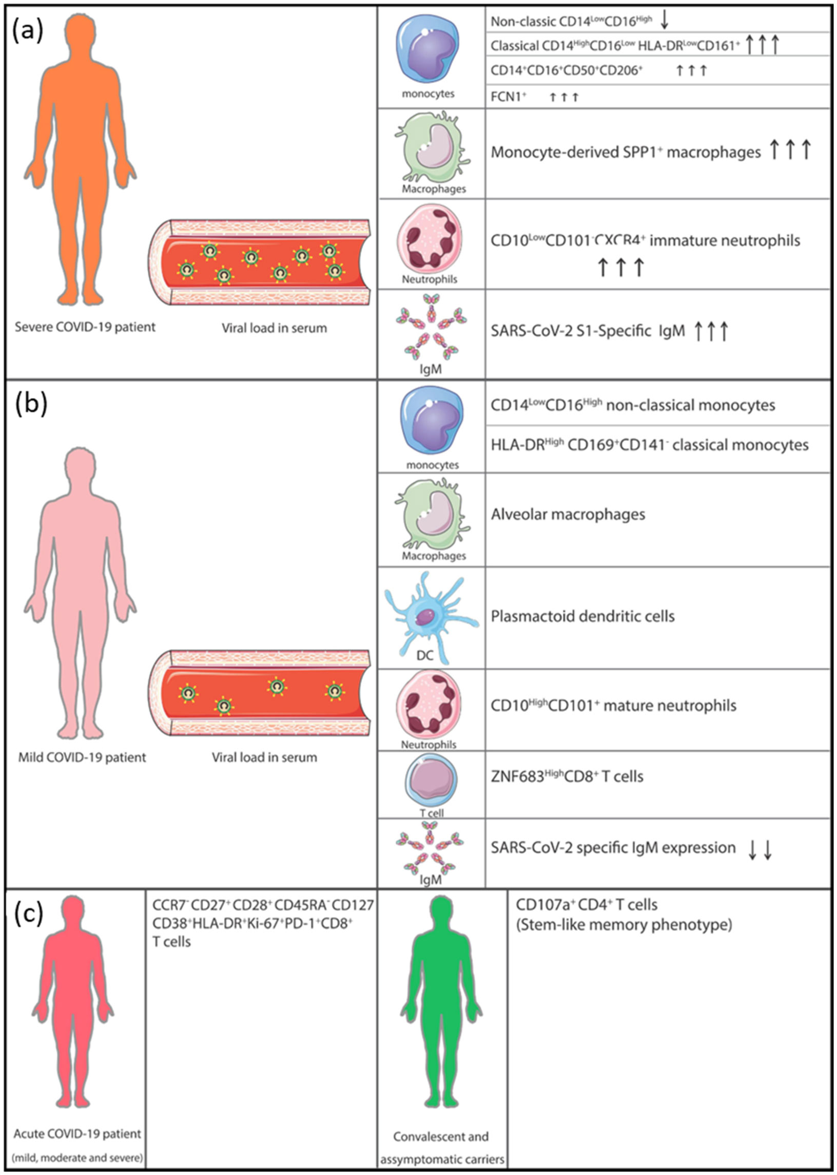
The ravaging pandemic caused by SAR-CoV-2, a member of β-coronaviruses, marks the end of the year 2019. Despite being identified and classified at the earliest stage, the virus records worldwide soaring transmissibility, morbidity, and mortality. Global data have shown the infection with SARS-CoV-2 to be severe among at least 15% of the infected; the aged and those with premorbid conditions like cancer, cardiovascular, and respiratory diseases. The highest prevalence and mortality are seen in the Americas, with African countries least affected. Severe respiratory distress and multiorgan failure are the usual findings in severe cases. A hyperinflammatory, fulminant, hypercytokinemia that is often further complicated by hypercoagulopathy and multiorgan failure has been reported extensively among severely infected patients. Scientists describe hyper-activated immune response mediated by macrophages secreting copious amounts of interleukin (IL)-6 forming the epicenter of cytokine storm (CS), thereby perpetuating signaling cascade through JAK/Kinase pathway that yields a hypercytokinemia. Researchers globally are exploring JAK/kinase inhibitors, immunomodulatory (immunosuppressive) therapy, cytokines, and cytokine receptor blockers for CS management. In which interestingly some of these agents possess antiviral activity. Here, we reviewed published studies with their respective outcome. However, a lot needs to be done to address the CS of COVID-19 to avert its fatal outcome.
Citation: Mansur Aliyu, Sayed-Hamidreza Mozhgani, Omid Kohandel Gargari, Mustapha Ahmed Yusuf, Ali Akbar Saboor-Yaraghi. The host immune responses to SARS-CoV-2 and therapeutic strategies in the treatment of COVID-19 cytokine storm[J]. AIMS Allergy and Immunology, 2021, 5(4): 240-258. doi: 10.3934/Allergy.2021018
The ravaging pandemic caused by SAR-CoV-2, a member of β-coronaviruses, marks the end of the year 2019. Despite being identified and classified at the earliest stage, the virus records worldwide soaring transmissibility, morbidity, and mortality. Global data have shown the infection with SARS-CoV-2 to be severe among at least 15% of the infected; the aged and those with premorbid conditions like cancer, cardiovascular, and respiratory diseases. The highest prevalence and mortality are seen in the Americas, with African countries least affected. Severe respiratory distress and multiorgan failure are the usual findings in severe cases. A hyperinflammatory, fulminant, hypercytokinemia that is often further complicated by hypercoagulopathy and multiorgan failure has been reported extensively among severely infected patients. Scientists describe hyper-activated immune response mediated by macrophages secreting copious amounts of interleukin (IL)-6 forming the epicenter of cytokine storm (CS), thereby perpetuating signaling cascade through JAK/Kinase pathway that yields a hypercytokinemia. Researchers globally are exploring JAK/kinase inhibitors, immunomodulatory (immunosuppressive) therapy, cytokines, and cytokine receptor blockers for CS management. In which interestingly some of these agents possess antiviral activity. Here, we reviewed published studies with their respective outcome. However, a lot needs to be done to address the CS of COVID-19 to avert its fatal outcome.
cytokine storm
noble coronavirus pneumonia disease 2019
severe acute respiratory syndrome coronavirus 2
angiotensin-converting enzyme receptor 2
transmembrane serine protease 2
numb-associated kinases
Janus kinase-signal transducer and activator of transcription
nuclear factor-kappa beta
interferon gamma
tumor necrosis factor alpha;
NLR (nod-like receptor) family pyrin domain containing 3

| [1] |
Wu F, Zhao S, Yu B, et al. (2020) A new coronavirus associated with human respiratory disease in China. Nature 579: 265-269. doi: 10.1038/s41586-020-2008-3

|
| [2] | WHO Rolling Updates on Coronavirus Disease (COVID-19). WHO, 2020 Available from: https://www.who.int/emergencies/diseases/novel-coronavirus-2019/events-as-they-happen. |
| [3] |
Li Q, Guan X, Wu P, et al. (2020) Early transmission dynamics in Wuhan, China, of novel coronavirus-infected pneumonia. N Engl J Med 382: 1199-1207. doi: 10.1056/NEJMoa2001316
|
| [4] |
Wang D, Hu B, Hu C, et al. (2020) Clinical characteristics of 138 hospitalized patients with 2019 novel coronavirus-infected pneumonia in Wuhan, China. JAMA 323: 1061-1069. doi: 10.1001/jama.2020.1585
|
| [5] |
Caly L, Druce J, Roberts J, et al. (2020) Isolation and rapid sharing of the 2019 novel coronavirus (SARS-CoV-2) from the first patient diagnosed with COVID-19 in Australia. Med J Australia 212: 459-462. doi: 10.5694/mja2.50569
|
| [6] |
Chan JFW, Kok KH, Zhu Z, et al. (2020) Genomic characterization of the 2019 novel human-pathogenic coronavirus isolated from a patient with atypical pneumonia after visiting Wuhan. Emerg Microbes Infect 9: 221-236. doi: 10.1080/22221751.2020.1719902
|
| [7] |
Ye Q, Wang B, Mao J (2020) The pathogenesis and treatment of the “Cytokine Storm” in COVID-19. J Infection 80: 607-613. doi: 10.1016/j.jinf.2020.03.037
|
| [8] |
Wang Y, Zhang L, Sang L, et al. (2020) Kinetics of viral load and antibody response in relation to COVID-19 severity. J Clin Invest 130: 5235-5244. doi: 10.1172/JCI138759
|
| [9] |
Lyons-Weiler J (2020) Pathogenic priming likely contributes to serious and critical illness and mortality in COVID-19 via autoimmunity. J Transl Autoimmun 3: 100051-100051. doi: 10.1016/j.jtauto.2020.100051
|
| [10] |
Stasi C, Fallani S, Voller F, et al. (2020) Treatment for COVID-19: An overview. Eur J Pharmacol 889: 173644. doi: 10.1016/j.ejphar.2020.173644
|
| [11] |
Wrapp D, Wang N, Corbett KS, et al. (2020) Cryo-EM structure of the 2019-nCoV spike in the prefusion conformation. Science 367: 1260-1263. doi: 10.1126/science.abb2507
|
| [12] |
Hoffmann M, Kleine-Weber H, Schroeder S, et al. (2020) SARS-CoV-2 cell entry depends on ACE2 and TMPRSS2 and is blocked by a clinically proven protease inhibitor. Cell 181: 271-280. doi: 10.1016/j.cell.2020.02.052
|
| [13] | Charoenlap S, Piromsopa K, Charoenlap C (2020) Potential role of Bacillus Calmette-Guérin (BCG) vaccination in COVID-19 pandemic mortality: Epidemiological and Immunological aspects. Asian Pac J Allergy 38: 150-161. |
| [14] |
García LF (2020) Immune response, inflammation, and the clinical spectrum of COVID-19. Front Immunol 11: 1441. doi: 10.3389/fimmu.2020.01441
|
| [15] |
Mortaz E, Tabarsi P, Varahram M, et al. (2020) The immune response and immunopathology of COVID-19. Front Immunol 11: 2037. doi: 10.3389/fimmu.2020.02037
|
| [16] |
Hadjadj J, Yatim N, Barnabei L, et al. (2020) Impaired type I interferon activity and inflammatory responses in severe COVID-19 patients. Science 369: 718-724. doi: 10.1126/science.abc6027
|
| [17] |
Bost P, Giladi A, Liu Y, et al. (2020) Host-viral infection maps reveal signatures of severe COVID-19 patients. Cell 181: 1475-1488. doi: 10.1016/j.cell.2020.05.006
|
| [18] |
Silvin A, Chapuis N, Dunsmore G, et al. (2020) Elevated calprotectin and abnormal myeloid cell subsets discriminate severe from mild COVID-19. Cell 182: 1401-1418. doi: 10.1016/j.cell.2020.08.002
|
| [19] |
Ni L, Ye F, Cheng ML, et al. (2020) Detection of SARS-CoV-2-specific humoral and cellular immunity in COVID-19 convalescent individuals. Immunity 52: 971-977. doi: 10.1016/j.immuni.2020.04.023
|
| [20] |
Sekine T, Perez-Potti A, Rivera-Ballesteros O, et al. (2020) Robust T cell immunity in convalescent individuals with asymptomatic or mild COVID-19. Cell 183: 158-168. doi: 10.1016/j.cell.2020.08.017
|
| [21] |
Kowitdamrong E, Puthanakit T, Jantarabenjakul W, et al. (2020) Antibody responses to SARS-CoV-2 in patients with differing severities of coronavirus disease 2019. PloS One 15: e0240502. doi: 10.1371/journal.pone.0240502
|
| [22] |
Lou B, Li TD, Zheng SF, et al. (2020) Serology characteristics of SARS-CoV-2 infection since exposure and post symptom onset. Eur Respir J 56: 2000763. doi: 10.1183/13993003.00763-2020
|
| [23] |
Zhao J, Yuan Q, Wang H, et al. (2020) Antibody responses to SARS-CoV-2 in patients of novel coronavirus disease 2019. Clin Infect Dis 71: 2027-2034. doi: 10.1093/cid/ciaa344
|
| [24] |
Wang F, Nie J, Wang H, et al. (2020) Characteristics of peripheral lymphocyte subset alteration in COVID-19 pneumonia. J Infect Dis 221: 1762-1769. doi: 10.1093/infdis/jiaa150
|
| [25] |
Weiskopf D, Schmitz KS, Raadsen MP, et al. (2020) Phenotype and kinetics of SARS-CoV-2-specific T cells in COVID-19 patients with acute respiratory distress syndrome. Sci Immunol 5: eabd2071. doi: 10.1126/sciimmunol.abd2071
|
| [26] |
Remy KE, Mazer M, Striker DA, et al. (2020) Severe immunosuppression and not a cytokine storm characterizes COVID-19 infections. JCI Insight 5: e140329. doi: 10.1172/jci.insight.140329
|
| [27] |
Mehta P, McAuley DF, Brown M, et al. (2020) COVID-19: consider cytokine storm syndromes and immunosuppression. Lancet 395: 1033-1034. doi: 10.1016/S0140-6736(20)30628-0
|
| [28] |
Bhaskar S, Sinha A, Banach M, et al. (2020) Cytokine storm in COVID-19-immunopathological mechanisms, clinical considerations, and therapeutic approaches: The reprogram consortium position paper. Front Immunol 11: 1648. doi: 10.3389/fimmu.2020.01648
|
| [29] |
Chen LYC, Hoiland RL, Stukas S, et al. (2020) Confronting the controversy: interleukin-6 and the COVID-19 cytokine storm syndrome. Eur Respir J 56: 2003006. doi: 10.1183/13993003.03006-2020
|
| [30] | Zhang D, Chen S (2020) Cytokine storms caused by novel coronavirus 2019 and treatment for cardiac injury. Eur Rev Med Pharmacol Sci 24: 12527-12535. |
| [31] |
Yang L, Xie X, Tu Z, et al. (2021) The signal pathways and treatment of cytokine storm in COVID-19. Signal Transduct Target Ther 6: 255. doi: 10.1038/s41392-021-00679-0
|
| [32] |
Reeh H, Rudolph N, Billing U, et al. (2019) Response to IL-6 trans- and IL-6 classic signalling is determined by the ratio of the IL-6 receptor α to gp130 expression: fusing experimental insights and dynamic modelling. Cell Commun Signal 17: 46. doi: 10.1186/s12964-019-0356-0
|
| [33] |
Mogensen TH (2019) IRF and STAT transcription factors—From basic biology to roles in infection, protective immunity, and primary immunodeficiencies. Front Immunol 9: 3047. doi: 10.3389/fimmu.2018.03047
|
| [34] |
Wang C, Xie J, Zhao L, et al. (2020) Alveolar macrophage dysfunction and cytokine storm in the pathogenesis of two severe COVID-19 patients. EBioMedicine 57: 102833. doi: 10.1016/j.ebiom.2020.102833
|
| [35] |
Caricchio R, Gallucci M, Dass C, et al. (2020) Preliminary predictive criteria for COVID-19 cytokine storm. Ann Rheum Dis 80: 88-95. doi: 10.1136/annrheumdis-2020-218323
|
| [36] | Zhang D, Guo R, Lei L, et al. (2020) COVID-19 infection induces readily detectable morphologic and inflammation-related phenotypic changes in peripheral blood monocytes. J Leukocyte Biol 2020: 1-10. |
| [37] |
Huang C, Wang Y, Li X, et al. (2020) Clinical features of patients infected with 2019 novel coronavirus in Wuhan, China. Lancet 395: 497-506. doi: 10.1016/S0140-6736(20)30183-5
|
| [38] |
Yang Y, Shen C, Li J, et al. (2020) Plasma IP-10 and MCP-3 levels are highly associated with disease severity and predict the progression of COVID-19. J Allergy Clin Immun 146: 119-127. doi: 10.1016/j.jaci.2020.04.027
|
| [39] |
Quartuccio L, Sonaglia A, McGonagle D, et al. (2020) Profiling COVID-19 pneumonia progressing into the cytokine storm syndrome: Results from a single Italian Centre study on tocilizumab versus standard of care. J Clin Virol 129: 104444. doi: 10.1016/j.jcv.2020.104444
|
| [40] |
Zachariah P, Johnson CL, Halabi KC, et al. (2020) Epidemiology, clinical features, and disease severity in patients with coronavirus disease 2019 (COVID-19) in a children's hospital in New York City, New York. JAMA Pediatr 174: e202430-e202430. doi: 10.1001/jamapediatrics.2020.2430
|
| [41] | Clinicaltrials.gov (2020) A Phase 2 Trial of Infliximab in Coronavirus Disease 2019 (COVID-19) United States National Library of Medicine, Available from: https://clinicaltrials.gov/ct2/show/NCT04425538?term=TNF-%CE%B1+inhibitors&cond=COVID&draw=2&rank=1. |
| [42] |
Patel S, Saxena B, Mehta P (2021) Recent updates in the clinical trials of therapeutic monoclonal antibodies targeting cytokine storm for the management of COVID-19. Heliyon 7: e06158. doi: 10.1016/j.heliyon.2021.e06158
|
| [43] |
Sinha P, Matthay MA, Calfee CS (2020) Is a “cytokine storm” relevant to COVID-19? JAMA Intern Med 180: 1152-1154. doi: 10.1001/jamainternmed.2020.3313
|
| [44] |
Saha A, Sharma AR, Bhattacharya M, et al. (2020) Tocilizumab: A therapeutic option for the treatment of cytokine storm syndrome in COVID-19. Arch Med Res 51: 595-597. doi: 10.1016/j.arcmed.2020.05.009
|
| [45] | Syam AF, Pitoyo CW, Suhendro S, et al. (2021) Tocilizumab as a treatment for “cytokine storm syndrome” in COVID-19: A case report. Acta Med Indones 53: 194-201. |
| [46] | Suresh K, Figart M, Formeck S, et al. (2021) Tocilizumab for the treatment of COVID-19-induced cytokine storm and acute respiratory distress syndrome: A case series from a rural level 1 Trauma Center in Western Pennsylvania. J Investig Med High Impact Case Rep 9: 1-5. |
| [47] |
Kornitzky FW, Langen HJ, Held M (2021) Treatment of a patient with a pronounced cytokine storm in severe COVID-19 pneumonia using a hemoadsorption in combination with the administration of tocilizumab. Pneumologie 75: 644-650. doi: 10.1055/a-1458-4080
|
| [48] |
Farooqi F, Dhawan N, Morgan R, et al. (2020) Treatment of severe COVID-19 with tocilizumab mitigates cytokine storm and averts mechanical ventilation during acute respiratory distress: A case report and literature review. Trop Med Infect Dis 5: 112. doi: 10.3390/tropicalmed5030112
|
| [49] |
Chitturi KR, Thacker S, Al-Saadi MA, et al. (2020) Successful treatment of acute heart failure in COVID-19-induced cytokine storm with tocilizumab: a case report. Eur Heart J Case Rep 4: 1-6. doi: 10.1093/ehjcr/ytaa188
|
| [50] |
Borku Uysal B, Ikitimur H, Yavuzer S, et al. (2020) Tocilizumab challenge: A series of cytokine storm therapy experiences in hospitalized COVID-19 pneumonia patients. J Med Virol 92: 2648-2656. doi: 10.1002/jmv.26111
|
| [51] |
Langer-Gould A, Smith JB, Gonzales EG, et al. (2020) Early identification of COVID-19 cytokine storm and treatment with anakinra or tocilizumab. Int J Infect Dis 99: 291-297. doi: 10.1016/j.ijid.2020.07.081
|
| [52] |
Ramiro S, Mostard RLM, Magro-Checa C, et al. (2020) Historically controlled comparison of glucocorticoids with or without tocilizumab versus supportive care only in patients with COVID-19-associated cytokine storm syndrome: results of the CHIC study. Ann Rheum Dis 79: 1143-1151. doi: 10.1136/annrheumdis-2020-218479
|
| [53] |
Stebbing J, Phelan A, Griffin I, et al. (2020) COVID-19: combining antiviral and anti-inflammatory treatments. Lancet Infect Dis 20: 400-402. doi: 10.1016/S1473-3099(20)30132-8
|
| [54] |
Cantini F, Niccoli L, Matarrese D, et al. (2020) Baricitinib therapy in COVID-19: A pilot study on safety and clinical impact. J Infection 81: 318-356. doi: 10.1016/j.jinf.2020.04.017
|
| [55] |
Kulkarni S, Fisk M, Kostapanos M, et al. (2020) Repurposed immunomodulatory drugs for Covid-19 in pre-ICu patients—mulTi-Arm therapeutic study in pre-ICu patients admitted with Covid-19—repurposed drugs (TACTIC-R): A structured summary of a study protocol for a randomised controlled trial. Trials 21: 626. doi: 10.1186/s13063-020-04535-4
|
| [56] |
Convertino I, Tuccori M, Ferraro S, et al. (2020) Exploring pharmacological approaches for managing cytokine storm associated with pneumonia and acute respiratory distress syndrome in COVID-19 patients. Crit Care 24: 331. doi: 10.1186/s13054-020-03020-3
|
| [57] |
Cao Y, Wei J, Zou L, et al. (2020) Ruxolitinib in treatment of severe coronavirus disease 2019 (COVID-19): A multicenter, single-blind, randomized controlled trial. J Allergy Clin Immun 146: 137-146. doi: 10.1016/j.jaci.2020.05.019
|
| [58] | Cavalli G, Dinarello CA (2015) Treating rheumatological diseases and co-morbidities with interleukin-1 blocking therapies. Rheumatology 54: 2134-2144. |
| [59] |
Cavalli G, De Luca G, Campochiaro C, et al. (2020) Interleukin-1 blockade with high-dose anakinra in patients with COVID-19, acute respiratory distress syndrome, and hyperinflammation: a retrospective cohort study. Lancet Rheumatol 2: e325-e331. doi: 10.1016/S2665-9913(20)30127-2
|
| [60] |
Schlesinger N, Firestein BL, Brunetti L (2020) Colchicine in COVID-19: an old drug, new use. Curr Pharmacol Rep 6: 137-145. doi: 10.1007/s40495-020-00225-6
|
| [61] | Clinicaltrials.gov (2020) Colchicine and COVID-19 Clinical Trials Search United States National Library of Medicine, Available from: https://clinicaltrials.gov/ct2/results?cond=COVID&term=colchicine&cntry=&state=&city=&dist=. |
| [62] |
Brunetti L, Diawara O, Tsai A, et al. (2020) Colchicine to weather the cytokine storm in hospitalized patients with COVID-19. J Clin Med 9: 2961. doi: 10.3390/jcm9092961
|
| [63] |
Jeyaraman M, John A, Koshy S, et al. (2021) Fostering mesenchymal stem cell therapy to halt cytokine storm in COVID-19. BBA-Mol Basis Diss 1867: 166014-166014. doi: 10.1016/j.bbadis.2020.166014
|
| [64] |
Farkhad NK, Reihani H, Moghadam AA, et al. (2021) Are mesenchymal stem cells able to manage cytokine storm in COVID-19 patients? A review of recent studies. Regen Ther 18: 152-160. doi: 10.1016/j.reth.2021.05.007
|
| [65] | Clinicaltrials.gov (2020) Stem Cells and COVID-19 Clinical Trials Search United States National Library of Medicine, Available from: https://clinicaltrials.gov/ct2/results?cond=COVID-19&term=stem+cell&cntry=&state=&city=&dist=. |
| [66] |
Kapugi M, Cunningham K (2019) Corticosteroids. Orthop Nurs 38: 336-339. doi: 10.1097/NOR.0000000000000595
|
| [67] |
Deng CX (2020) Glucocorticoids save lives in COVID-19 patients. Int J Biol Sci 16: 2477-2478. doi: 10.7150/ijbs.49125
|
| [68] |
Ramiro S, Mostard RLM, Magro-Checa C, et al. (2020) Historically controlled comparison of glucocorticoids with or without tocilizumab versus supportive care only in patients with COVID-19-associated cytokine storm syndrome: results of the CHIC study. Ann Rheum Dis 79: 1143-1151. doi: 10.1136/annrheumdis-2020-218479
|
| [69] |
Sharun K, Tiwari R, Dhama J, et al. (2020) Dexamethasone to combat cytokine storm in COVID-19: Clinical trials and preliminary evidence. Int J Surg 82: 179-181. doi: 10.1016/j.ijsu.2020.08.038
|
| [70] |
Ahmadikia K, Hashemi SJ, Khodavaisy S, et al. (2021) The double-edged sword of systemic corticosteroid therapy in viral pneumonia: A case report and comparative review of influenza-associated mucormycosis versus COVID-19 associated mucormycosis. Mycoses 64: 798-808. doi: 10.1111/myc.13256
|
| [71] |
Pakdel F, Ahmadikia K, Salehi M, et al. (2021) Mucormycosis in patients with COVID-19: A cross-sectional descriptive multicentre study from Iran. Mycoses 64: 1238-1252. doi: 10.1111/myc.13334
|
| [72] | Pal R, Singh B, Bhadada SK, et al. (2021) COVID-19-associated mucormycosis: An updated systematic review of literature. Mycoses In press. |
| [73] | Rodriguez-Morales AJ, Sah R, Millan-Oñate J, et al. (2021) COVID-19 associated mucormycosis: the urgent need to reconsider the indiscriminate use of immunosuppressive drugs. Ther Adv Infect Dis 8: 1-5. |
| [74] |
Tzilas V, Manali E, Papiris S, et al. (2020) Intravenous immunoglobulin for the treatment of COVID-19: a promising tool. Respiration 99: 1085. doi: 10.1159/000506650
|
| [75] |
Bongomin F, Asio LG, Ssebambulidde K, et al. (2021) Adjunctive intravenous immunoglobulins (IVIg) for moderate-severe COVID-19: emerging therapeutic roles. Curr Med Res Opin 37: 903-905. doi: 10.1080/03007995.2021.1903849
|
| [76] |
Kohler H, Kaveri S (2021) How IvIg can mitigate Covid-19 disease: A symmetrical immune network model. Monoclon Antibodies Immunodiagn Immunother 40: 17-20. doi: 10.1089/mab.2020.0041
|
| [77] |
Galeotti C, Kaveri SV, Bayry J (2020) Intravenous immunoglobulin immunotherapy for coronavirus disease-19 (COVID-19). Clin Transl Immunol 9: e1198. doi: 10.1002/cti2.1198
|
| [78] |
Calabrese LH (2020) Cytokine storm and the prospects for immunotherapy with COVID-19. Clevel Clin J Med 87: 389-393. doi: 10.3949/ccjm.87a.ccc008
|

Figures(2)

Mansur Aliyu, Sayed-Hamidreza Mozhgani, Omid Kohandel Gargari, Mustapha Ahmed Yusuf, Ali Akbar Saboor-Yaraghi. The host immune responses to SARS-CoV-2 and therapeutic strategies in the treatment of COVID-19 cytokine storm[J]. AIMS Allergy and Immunology, 2021, 5(4): 240-258. doi: 10.3934/Allergy.2021018


 DownLoad: