Loading [Contrib]/a11y/accessibility-menu.js
Research article

Sappan Lignum Extract Inhibits Restenosis in the Injured Artery through the Deactivation of Nuclear Factor-κB

  • Received: 17 February 2014 Accepted: 04 August 2014 Published: 14 August 2014
  • The aim of the study was to explore whether Sappan Lignum Extract (SLE) would inhibit vascular restenosis in injured artery and its inhibitory mechanism by using a balloon-injured rat carotid artery restenosis model. Different doses of SLE were administered to the rats by tube feeding, starting from four days before surgery and continuing twice per day for two weeks after carotid injury. Injured carotid arteries isolated from rats were embedded in paraffin block and tissue sections were stained with H&E to assess restenosis. The Effects of SLE on vascular restenosis, which are involved in smooth muscle cell cycle, NF-κB p65 expression, and Superoxide (O2-) production, was assessed by RT-PCR, western blot assay, and immunohistochemistry. The results showed that in a rat carotid model of balloon dilatation injury, SLE significantly reduced the intimal-to-medial area ratio and vascular restenosis after 14 days of the injury. Immunohistochemistry study revealed no inhibited PCNA expression caused by SLE. Rat serum containing Sappan Lignum (RSC) was found neither anti-proliferative effect in cultured vascular smooth muscle cells (VSMCs) and nor arrest cell cycle progress detected by flow cytometry. RSC remarkably decreased the expression of TNF-α mRNA and protein in cultured VSMCs. Electrophoretic mobility shift assay proved that RSC inhibited the binding of NF-κB to specific DNA sequences in TNF-α treated VSMCs. Western blot pronounced that RSC and N-acetyl-L-cysteine (NAC) reduced expression of NF-κB p65 in nuclear extracts in TNF-α treated VSMCs. RSC and NAC also attenuated superoxide anion generation in TNF-α treated VSMCs. In summary, the inhibitory effects of SLE on vascular restenosis may not be mediated through inhibiting vascular cell proliferation, but through inhibiting vascular inflammation instead, which may be attributed to the inhibition of TNF-α and subsequent deactivation of NF-κB, which was in part mediated through inhibiting superoxide anion generation.

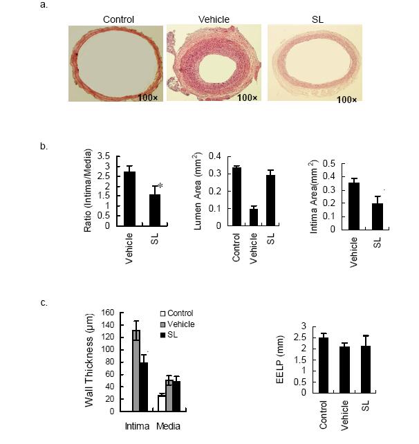
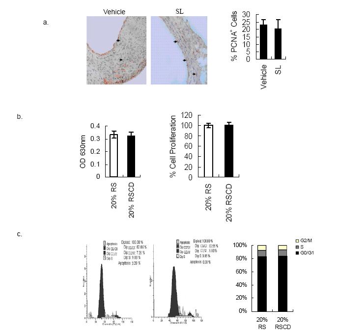
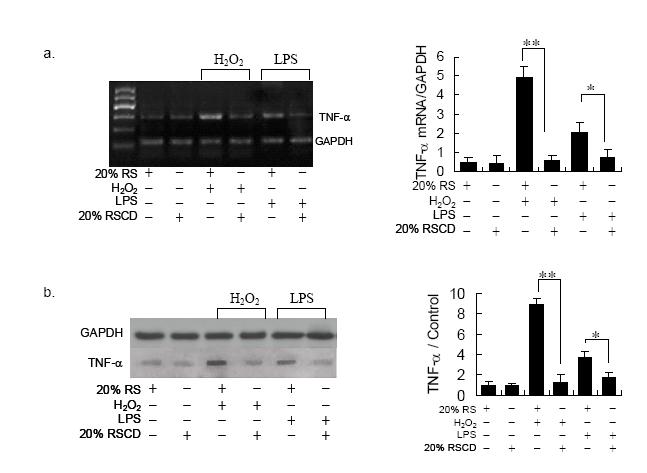
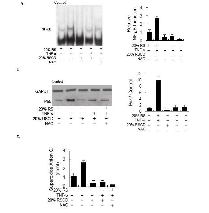
    Citation: Guang Long, Bo Lin, Lu Wang, Lingyan Wu, Tieying Yin, Donghong Yu, Guixue Wang. Sappan Lignum Extract Inhibits Restenosis in the Injured Artery through the Deactivation of Nuclear Factor-κB[J]. AIMS Bioengineering, 2014, 1(1): 25-39. doi: 10.3934/bioeng.2014.1.25

    Related Papers:

    [1] Jingwen Zhang, Lu Wang, Huolin Chen, Tieying Yin, Yanqun Teng, Kang Zhang, Donghong Yu, Guixue Wang . Effect of Caspase Inhibitor Ac-DEVD-CHO on Apoptosis of Vascular Smooth Muscle Cells Induced by Artesunate. AIMS Bioengineering, 2014, 1(1): 13-24. doi: 10.3934/bioeng.2014.1.13
    [2] Figueroa-Pizano María Dolores, Campa-Mada Alma Consuelo, Canett-Romero Rafael, Paz-Samaniego Rita, Martínez-López Ana Luisa, Carvajal-Millan Elizabeth . Influence of arabinoxylan and crosslinked arabinoxylan consumption on blood serum lipids and glucose levels of Wistar rats. AIMS Bioengineering, 2021, 8(3): 208-220. doi: 10.3934/bioeng.2021018
    [3] Takashi Nakazawa, Sohei Tasaki, Kiyohiko Nakai, Takashi Suzuki . Multicellular model of angiogenesis. AIMS Bioengineering, 2022, 9(1): 44-60. doi: 10.3934/bioeng.2022004
    [4] Ricardo I. González-Vega, Carmen L. Del-Toro-Sánchez, Ramón A. Moreno-Corral, José A. López-Elías, Aline Reyes-Díaz, Norma García-Lagunas, Elizabeth Carvajal-Millán, Diana Fimbres-Olivarría . Sulfated polysaccharide-rich extract from Navicula incerta: physicochemical characteristics, antioxidant activity, and anti-hemolytic property. AIMS Bioengineering, 2022, 9(4): 364-382. doi: 10.3934/bioeng.2022027
    [5] Júlia Novaes Matias, Gyovanna Sorrentino dos Santos Campanari, Gabriela Achete de Souza, Vinícius Marinho Lima, Ricardo José Tofano, Claudia Rucco Penteado Detregiachi, Sandra M. Barbalho . Metabolic syndrome and COVID-19. AIMS Bioengineering, 2020, 7(4): 242-253. doi: 10.3934/bioeng.2020021
    [6] Christopher N. Boyer, T. Edward Yu, Burton C. English, James A. Larson, Nicole Labbé, Lindsey M. Kline . Impact of an innovated storage technology on the quality of preprocessed switchgrass bales. AIMS Bioengineering, 2016, 3(2): 125-138. doi: 10.3934/bioeng.2016.2.125
    [7] Daniel N. Riahi, Saulo Orizaga . Modeling and computation for unsteady blood flow and solute concentration in a constricted porous artery. AIMS Bioengineering, 2023, 10(1): 67-88. doi: 10.3934/bioeng.2023007
    [8] Shaoyung Chen, Hsinyi Mao, Pinhua Tu, Naichen Cheng, Jiashing Yu . Fabrication of decellularized adipose tissue/alginate composite microspheres with pASCs encapsulation for tissue engineering. AIMS Bioengineering, 2017, 4(3): 351-365. doi: 10.3934/bioeng.2017.3.351
    [9] Jennifer L. Wood, Wuxing Liu, Caixian Tang, Ashley E. Franks . Microorganisms in heavy metal bioremediation: strategies for applying microbial-community engineering to remediate soils. AIMS Bioengineering, 2016, 3(2): 211-229. doi: 10.3934/bioeng.2016.2.211
    [10] Lindsay Arnold, Rachel Chen . One-step non-chromatography purification of a low abundant fucosylated protein from complex plant crude extract. AIMS Bioengineering, 2015, 2(3): 249-263. doi: 10.3934/bioeng.2015.3.249
  • The aim of the study was to explore whether Sappan Lignum Extract (SLE) would inhibit vascular restenosis in injured artery and its inhibitory mechanism by using a balloon-injured rat carotid artery restenosis model. Different doses of SLE were administered to the rats by tube feeding, starting from four days before surgery and continuing twice per day for two weeks after carotid injury. Injured carotid arteries isolated from rats were embedded in paraffin block and tissue sections were stained with H&E to assess restenosis. The Effects of SLE on vascular restenosis, which are involved in smooth muscle cell cycle, NF-κB p65 expression, and Superoxide (O2-) production, was assessed by RT-PCR, western blot assay, and immunohistochemistry. The results showed that in a rat carotid model of balloon dilatation injury, SLE significantly reduced the intimal-to-medial area ratio and vascular restenosis after 14 days of the injury. Immunohistochemistry study revealed no inhibited PCNA expression caused by SLE. Rat serum containing Sappan Lignum (RSC) was found neither anti-proliferative effect in cultured vascular smooth muscle cells (VSMCs) and nor arrest cell cycle progress detected by flow cytometry. RSC remarkably decreased the expression of TNF-α mRNA and protein in cultured VSMCs. Electrophoretic mobility shift assay proved that RSC inhibited the binding of NF-κB to specific DNA sequences in TNF-α treated VSMCs. Western blot pronounced that RSC and N-acetyl-L-cysteine (NAC) reduced expression of NF-κB p65 in nuclear extracts in TNF-α treated VSMCs. RSC and NAC also attenuated superoxide anion generation in TNF-α treated VSMCs. In summary, the inhibitory effects of SLE on vascular restenosis may not be mediated through inhibiting vascular cell proliferation, but through inhibiting vascular inflammation instead, which may be attributed to the inhibition of TNF-α and subsequent deactivation of NF-κB, which was in part mediated through inhibiting superoxide anion generation.


    Abbreviations list:

    SL=Sappan Lignum; RSC=Rat serum containing SL extract; TNF-α=tumor necrosis factor-α; NF-κB nuclear factor kB; PCNA=proliferating cell nuclear antigen; VSMC=vascular smooth muscle cell; TCM=traditional Chinese medicine; NAC=N-acetyl-L-cysteine.

    1. Introduction

    Recent experimental and clinical studies suggest that inhibition of inflammation can be a promising next-generation approach on prevention of vascular restenosis [1,28]. NF-kB is an important element in the activation of the inflammatory cytokines and adhesion molecule genes involved in lesion development after vascular injury [2,29]. Inhibition of NF-κB has been demonstrated to prevent neointimal formation after balloon injury in a rat carotid artery model [3,30]. Thus developing novel pharmacological methods for disruption of NF-κB signaling pathway holds great promise for the prevention of restenosis. Herbal therapies have been used for many centuries in China as treatment for individuals with cardiovascular disease as well as inflammatory disease. Many of these claims are based on anecdotes in traditional Chinese medicine (TCM). Such Chinese herbs have been used for the treatment of human diseases for thousands of years. However, over the past several years, there have been considerable interests in these agents in the treatment of human cardiovascular disease. Despite the reported benefits of these herbs in individual patients, it still remains unclear whether these agents have cardiovascular disease properties and whether there is a molecular basis to their inhibitory effects on vascular inflammation. The use of herbs in medical treatment and diseases-prevention has been dramatically rising in recent years in many countries including the United States; however, their medicinal actions have not been fully elucidated [4]. Sappan Lignum (SL) has been a commonly prescribed TCM for many anti-biotic [5] and anti-cancer [4] treatments. It has been proven to inhibit NO production [6] and has vasorelaxant activity on rat artery [7]. Although information regarding the preventive effects of SLE on restenosis is lacking,several kinds of active components in SL have been demonstrated to inhibit vascular inflammation [8,9,10,31]. Therefore it is expected that SLE plays an important role in preventing balloon injury-induced restenosis.

    TCM come mostly from crude plant extracts. Traditional pharmacological methods involving direct addition of drugs to the cell lines have been widely applied in pharmacological tests of TCM in vitro. However, recent studies have demonstrated that partial active components, which are absorbed in plasma after oral administration of TCM, are rapidly transformed into their metabolites and these sometimes are not absorbed in vivo [11,12], suggesting that validity of traditional pharmacological methods used in pharmacological tests in vitro of TCM are questionable for several reasons: if the TCM extracts are directly added to the cultured media, many active components and the transformation of theirs, which are not taken up in vivo, may generate unexpected pharmacological effects and interfere the experimental results obtained from the cell lines. Thus, pharmacological effects in vitro of some TCM using traditional pharmacological methods might be inconsistent with their pharmacological effects in vivo [13]. Moreover, since the exact components of TCM are usually unknown, equipotent dosage or concentration of extracts of TCM are rarely evaluated in vivo according to concentration in vitro. A new serum pharmacological method that involves addition of serum obtained from orally herbal-medicine treated animals has at least partially solved some of these problems. The problem of equipotent dosage or concentration of TCM is easily solved using well characterized and reproducible serum; the refined serum contains defined amounts of active components that produce pharmacological effects in vivo. The effects using the serum pharmacological method in vitro predict better than using TCM. Therefore the serum pharmacological method is now regarded as a new testing method in vitro for the TCM [14].

    The aims of this study were to determine whether SLE attenuates vascular restenosis and NF-κB associated vascular inflammation in vivo and to investigate whether SLE disrupts TNF-α induced NF-κB signaling pathway in cultured VSMCs, using the serum pharmacological method. Our results were expected to serve as a contribution to the discussion about the molecular mechanism of SLE action on cardiovascular system, especially in vascular restenosis.

    2. Materials and Methods

    2.1. Preparation of SLE and rat serum containing SL

    Sappan Lignum, which is the dried heartwood of Caesalpinia sappan L from legume family, was purchased from University Hospital, NanHua China University (HengYang, HuNan, China). These samples were authenticated by Department of Pharmacognosy, Hunan University of Chinese Medicine (Changsha, China). Voucher specimens have been deposited in Herbarium of the Medicinal Plant Garden, Department of Pharmacognosy, Hunan University of Chinese Medicine (No. 3527). The herbs (100 g) were mixed, minced with a grinder and extracted with 500 mL of distilled water under reflux for 3h by boiling the formula. The extract was filtered with 10 µ m cartridge paper. The filtrate was concentrated to about 100 mL with a rotary evaporator at 50 ℃ under vacuum. In brief, ten rats were administered a warm water extract of SL (SLE) (1000 mg· kg-1· day-1, intragastric administration) daily for 7 days, then RSC was collected from the rats. Rat serum (RS) from ten rats that were not administered SLE was also collected as a control. Rat serum containing SL (RSC) was prepared by the serum pharmacological method [14].

    2.2. Carotid artery injury model and drug treatment

    Male SD rats (400– 450 g) were fed with standard pellet feed and given water. The experimental protocol was designed in accordance with Chinese Animal Care and Use Committee standards. Animals were anesthetized with sodium pentobarbital (50 mg/kg). The right external carotid artery was inserted by a 2F coronary dilation catheter (Goodman corporation, Japan), which was advanced to the common carotid artery, inflated to a pressure of 5 atm, and rotated in a forward and retrograde direction. The catheter was then deflated, and the process was repeated three times. Treatment cohorts were divided into 3 groups (n = 6– 8/group): the control (sham-operated), the SLE (1000 mg· kg-1· day-1, intragastric administration), and the vehical. Drugs were administered daily for 14 days after injury. Two weeks postinjury, rats were euthanized by sodium pentobarbital overdose and perfused with 10 % buffered formalin.

    2.3. Histology and morphometry

    The arteries from the treated rats were fixed and embedded in paraffin. Then, the paraffin-embedded arteries were cut into 10 sections for analysis. Morphometric analysis was carried out on HE-stained arteries; images (20 ×) were transferred to a computer using a video camera for quantitative characterization. For rat balloon-injury artery, 10 sections were randomly selected for analysis. The relative parameters were determined using the Imaging software by selecting appropriate functions. The areas encompassed by the lumen surface (lumen area), internal elastic lamina– lumen surface (intima area) and External elastic lamina (EEL) perimeter were measured. For the evaluation of the degree of intimal hyperplasia, intima and media wall thickness, and the ratio of intimal area to medial area (I/M ratio) were calculated and compared using the digital imaging software (Image-pro Plus).

    2.4. Immunohistochemical analysis

    The tissue used for immunohistochemical analysis was embedded in paraffin, and cut into 5-µ m-thick slices. They were subjected to immunostaining with antibodies against rat PCNA (Sigma). For quantification of immunohistochemical images of PCNA, at least 5 representative images were selected, and the percentage of immunopositive cells per total cells in each image was calculated. The average of the 5 images was reported for each rat. Consistent positive staining involves: more than 50 % of the vascular wall was recorded as (5); positive staining of > 35 % to 50 % of the area as (4); positive staining of > 20 % to 35 % of the area as (3); positive staining of > 5 % to 20 % of the area; as (2); positive staining of 1 % to 5% of the area as (1); and staining of < 1 % of the area as (0).

    2.5. Cell culture and proliferation assay

    VSMCs were isolated from rat aorta [15]. The cells were grown in DMEM (GIBCO) containing 10 % newborn calf serum (GIBCO) at 37 °C in an atmosphere of 5 % CO2 in air. Experiments were performed on cells cultured for up to 10 passages. Cell proliferation after 24 hour incubation in medium containing 20 % rat serum was determined in 96-well plates by colorimetric assay of methylene blue incorporation [16] under 5-times repeating tests for each experiment.

    2.6. Cell cycle analysis

    The cells were harvested. Cells were fixed in 70 % ethanol for 1 h, washed with PBS, treated with 100 μg/mL RNase A for 1 h at 37 °C, and stained with 25 μg/mL propidiumiodide (PI). Flowcytometry was performed in triplicate for each experiment on a FACS Calibur system (Becton& Dickinson; San Jose, CA, USA).

    2.7. RT-PCR

    Total RNAs were extracted from VSMCs. For semiquantitative RT-PCR assay, 1 µ g of total RNAs was used to detect mRNA expression by Super-Script One-Step RT-PCR kit (Invitrogen, Carlsbad, CA). The reverse transcription reaction was performed at 25 °C for 10 minutes, 48 °C for 30 minutes, and 95 °C for 5 minutes. PCR cycles (28 cycles) were performed for 30 seconds at 94 °C, 55 °C, and 72 °C, respectively. Primers for amplification of TNF-α and GAPDH are listed as follows. The PCR product were subjected to 1 % agarose gel electrophoresis and stained with ethidium bromide. The relative levels were quantified by Matrox Inspector V. 4.1 software.

    TNF-α:

    Upstream primer: 5’- ACAACCAACTGGTGGTACCA - 3’,

    Downstream primer: 5’- AAGTACTTGGGCAGGTTGAC - 3’.

    GAPDH:

    Upstream primer: 5’- AGTTCAACGGCACCAGTCAAAG -3’,

    Downstream primer: 5’- TACTCAGCACCAGCATCACC -3’.

    2.8. Western blot

    To evaluate the presence of TNF-α and NF-κB P65 expression, cells were lysed in lysis buffer containing 20 mmol/L Tris-HCl, pH 7.4, 0.4 mol/L KCl, 2 mmol/L dithiothreitol, and 10 % glycerol. Concentrations of protein were determined with Bicinchoninic acid kit (Sigma). Extracted proteins were separated through a sodium dodecyl sulfate polyacrylamide gel, transferred to polyvinylidene difluoride membrane (Bio-Rad), and incubated with anti-TNF-α and anti-NF-κB antibodies. Protein bans were visualized with Vectastain Elite ABC kit (Vector Labratories). The density of bands was analyzed with the use of densitometry and Kodak software (Eastman Kodak).

    2.9. Nuclear extract preparation and electrophoretic mobility shift assay (EMSA)

    Cell nuclear extracts were prepared from 1 x 10⁶ cells by cell pellet homogenization in two volumes of 10 mM HEPES, pH 7.9, 10 mM KCl, 1.5 mM MgCl2, 1 mM EDTA, 0.5 mM Dithiothreitol (DTT), 0.5 mM Phenylmethyl sulfonyl fluoride (PMSF) , and 10 % glycerol. Nuclei were centrifuged at 1, 000 g for 5 min, washed, and resuspended in two volumes of the above-specified solution. KCl (3 M) was added to reach 0.39 M KCl. Nuclei were lysed at 4°C for 1 h and centrifuged at 10, 000 g for 30 min. The supernatants clarified by centrifugation were collected and stored at -80°C. Protein concentration was determined with a protein assay (Bio-Rad). The NF-κB consensus 5'-CAACGGCAGGGGAATCTCCCTCTCCTT-3'[17] oligonucleotide was end-labeled with [γ-32P] ATP (Amersham Pharmacia Biotech) using a polynucleotide kinase (Roche). End-labeled DNA fragments were incubated at room temperature for 20 min with 5 µ g of nuclear protein in the presence of 1µ g of poly (dI-dC) in 20 µ L of a buffer consisting of 10 mM Tris. HCl, pH 7.5, 50 mM NaCl, 1 mM EDTA, 1 mM DTT, and 5 % glycerol. Protein-DNA complexes were separated from free probe on a 6 % polyacrylamide gel run in 0.25 x Tris-borate buffer at 200 mV for 3 h at room temperature. The gels were dried and exposed to X-ray film (Eastman Kodak).

    2.10. Measurement of Superoxide (O2-) Products

    Superoxide products were measured by means of the SOD-inhibitable reduction of cytochrome c [18]. Briefly, being treated for 2 h, VSMC were preincubated in DMEM without phenol red for 30 minutes at 37 °C, and then cytochrome c (final concentration, 1 mg/mL) with or without SOD (final concentration, 500 U/mL) was added and kept in a CO2 incubator on the Flex Unit. At the indicated time points, the medium was removed from the cells; the absorbance was read at 550 nm against distilled water as a reference. Reduction of cytochrome c in the presence of SOD was subtracted from the values without SOD. The portion of superoxide-specific reduction of cytochrome c was between 30 % and 35 %. The optical density difference between comparable wells with or without SOD was converted to equivalent O2- production by using the molar extinction coefficient for cytochrome c [21.0×10³ (mol/L)-1· cm-1] [18]. Tests were performed in triplicates for each experiment.

    2.11. Statistical analysis

    The data are expressed as mean and SEM. Statistical analyses were performed using SPSS software. One way analysis of variance followed by paired t tests were used to determine significant differences between treatments, and significance was set at P < 0.05.

    3. Results

    3.1. Suppression of vascular restenosis by SLE after arterial injury

    On the basis of the in vitro data on the suppression of the proliferation of VSMCs by SLE, we investigated the anti-proliferative effects of SLE in vivo using a rat carotid artery balloon injury model. Administration of SLE significantly reduced the development of neointimal hyperplasia 14 days after balloon injury (Fig. 1a). Quantitative morphometric analysis showed that SLE reduced the vascular restenosis (cross-sectional areas of the lumen, neointima and the ratios between neointima and media) compared with the vehical. The ratio of intima to media and the intima area were markedly reduced, and the lumen area was increased (Fig. 1b). The intima thickness was also dramatically reduced after SLE treatment; the media thickness showed no changes (Fig. 1c). There was no remarkable change in EEL perimeter between the vehicle-treated group and SLE-treated group (Fig. 1d), which might suggest that SLE also have no effects on vascular remodelling after vascular injury.

    Figure 1. SL inhibits neointimal hyperplasia in a rat carotid artery balloon injury model. (a) Representative sections of the common carotid artery 14 days after injury. (b) Ratio of intima to media, intima area and lumen area after balloon injury (n=6 for control, n=8 for vehicle, n=9 for SL). Values are expressed as mean ± SEM. *P < 0.05.

    3.2. Effect of RSC on VSMCs proliferation

    SLE did not inhibit expression of PCNA protein in vascular restenosis compared with the vehicle (Fig. 2a, P > 0.05). In cultured VSMCs, RSC can not inhibit VSMCs growth stimulated with 20 % rat serum compared with the control group (Fig. 2b, P > 0.05). RSC can not arrest cell cycle progress in cultured VSMCs stimulated with 20% rat serum compared with the control group determined by flow cytometry (Fig. 2c, P > 0.05). It indicates that RSC can not arrest cell cycle progress in cultured VSMCs.

    Figure 2. Effects of a warm water extract of SL on VSMCs. Warm water extract of SL was administered intragastric administration to the animals at the dose of 1000 mg/kg per day. (a) The representative immunohistochemistry staining shows the effects of a warm water extract of SL on expression of PCNA. Quantification of cells stained positively for PCNA. (b) Effects of 20 % serum containing SL on VSMC growth induced by 20 % rat serum; c. Effects of 20 % serum containing SL on cell cycle progress in cultured VSMCs. RS stands for rat serum. RSC indicates serum containing SL extract. Results are mean ± SEM from five different tests. * P < 0.05.

    3.3. RSC inhibits expression of TNF-α mRNA treated with hydrogen peroxide and LPS in cultured VSMCs

    Cultured rat aortic VSMCs containing trace amounts of TNF-α mRNA was assessed by RT-PCR. Exposure of VSMCs to 1.5 mmol/mL hydrogen peroxide for 8 hours resulted in a 5 fold increased expression of TNF-α mRNA compared with the control group (Fig. 3a), and exposure of VSMCs to 10ug/mL LPS for 4 hours resulted in a 3 fold increased expression of TNF-α mRNA compared with the control group (Fig. 3a). RSC considerably attenuated the increased expression of TNF-α mRNA induced by 1.5 mmol/mL hydrogen peroxide for 8 in cultured rat aortic VSMCs (Fig. 3a). In the meantime, RSC similarly reduced the effect of 10 ug/mL LPS on expression of TNF-α mRNA (Fig. 3a).

    Figure 3. (a) Effect of 20 % rat serum containing SL on TNF-α mRNA in cultured VSMCs. Rat aortic VSMCs were cultured with hydrogen peroxide or LPS in the presence or absence of 20 % RSC. Intracellular TNF-α mRNA expression levels were determined by RT-PCR. Bands represent RT-PCR products of TNF-α protein from VSMCs. RS indicates rat serum. RSC indicates rat serum containing SL extract. (b) Effect of 20 % RSC on TNF-α protein in cultured VSMCs. Rat aortic VSMCs were cultured with hydrogen peroxide or LPS in the presence or absence of 20 % RSC. Intracellular TNF-α protein expression levels were determined by Western blot analysis. Results of TNF-α protein are expressed compared with the control for triplicate samples. RS indicates rat serum. RSC indicates rat serum containing SL extract. Results are mean ± SEM from five different tests * p < 0.05, ** p < 0.01.

    Western blot analysis demonstrated exposure of VSMCs to 1.5mmol/mL hydrogen peroxide for 8 hours resulted in a 10 fold increased expression of TNF-α protein compared with the control group, and exposure of VSMCs to 10ug/mL LPS for 4 hours resulted in a 4 fold increased expression of TNF-α protein compared with the control group. RSC greatly degraded increased expression of TNF-α protein induced by 1.5mmol/mL hydrogen peroxide for 8 hours in cultured rat aortic VSMCs (Fig. 3b). Meanwhile, RSC also inhibited expression of TNF-α protein induced by 10ug/mL LPS for 4 hours compared (Fig. 3b).

    3.4. RSC inhibits NF-kB activation treated with TNF-α in cultured VSMCs

    EMSA demonstrated that exposure of VSMCs to 20 ng/mL TNF-α for 2 hours resulted in significant enhance of the density of the shifted bands compared with the control group (Fig. 4a). RSC markedly degraded increased the density of the shifted bands treated with 20 ng/mL TNF-α for 2 hours in cultured rat aortic VSMCs compared with the TNF-α treated group (Fig. 4a). While 10 mmol/L NAC (a kind of ROS scavenger) also inhibited the shifted bands treated with 20 ng/mL TNF-α for 2 hours in cultured rat aortic VSMCs (Fig. 4a).

    Figure 4. (a) Effect of 20 % RSC on NF-κB activation in TNF-α-stimulated VSMCs. (b) Effect of 20 % RSC on NF-κB p65 expression in nuclear extracts of TNF-α-stimulated VSMCs. (c) Effect of 20 % RSC on superoxide anion generation in TNF-α-stimulated VSMCs. RS represents rat serum. RSC indicates rat serum containing SL extract. Results are mean ± SEM from five different tests. ** p < 0.01 vs. Control; ++p < 0.01 vs. treated with TNF-α.

    3.5. RSC inhibits expression of NF-kB p65 in nuclear extracts in VSMCs stimulated with TNF-α

    Western blot analysis demonstrated exposure of VSMCs to 20 ng/mL TNF-α for 2 hours resulted in striking increase of expression of NF- B p65 in nuclear extracts in VSMCs compared with the control group (Fig. 4b). RSC markedly degraded increased expression of NF- B p65 in nuclear extracts compared with the TNF-α treated group (Fig. 4b, ). And 10 mmol/L NAC also inhibited increasing expression of NF-B p65 in nuclear extracts compared with the TNF-α treated group (Fig. 4b).

    3.6. RSC inhibits superoxide anion generation in TNF-α-stimulated VSMCs

    Exposure of VSMCs to 20 ng/mL TNF-α for 2 hours resulted in significant increasing superoxide anion generation in VSMCs compared with the control group (Fig. 4c). RSC markedly degraded increasing superoxide anion generation in VSMCs compared with the TNF-α treated group (Fig. 4c). 10 mmol/L NAC also inhibited increasing superoxide anion generation compared with the TNF-α treated group (Fig. 4c).

    4. Discussions

    SL is a botanical that is used today in traditional Chinese medicine for promoting blood flow associated with cardiovascular disease. In this study,as presented in Figure 1, when applying the rat carotid model of balloon dilatation injury, potent inhibitory effects on vascular restenosis has been demonstrated using the warm water extract of SL (SLE) . To characterize the mechanism for SLE suppressing vascular restenosis, we investigated the effect of SLE on the protein expression level of PCNA. As illustrated in Figure 2a, our results revealed that there was no significant change in PCNA protein expression in vascular restenosis after SLE treatment. These results suggested that inhibitory effects of the SLE on vascular restenosis in a rat carotid model of balloon dilatation injury might not be mediated through an inhibition of vascular cell proliferation. The notion was further supported by which we did not observe any effect of rat serum containing SLE on vascular smooth muscle cells growth inhibitory or cell cycle arrest.

    NF-κB is an important element in the activation of the inflammatory cytokines and adhesion molecule genes involved in lesion development after vascular injury [2,19]. Inhibition of NF-κB has been demonstrated to prevent neointimal formation after balloon injury in a rat carotid artery model [19]. TNF-α, as a proinflammaory cytokine, which is thought to elicit NF-κB activation [20,21] has been demonstrated to be released at high levels in proliferating SMCs in the balloon-injured rabbit aorta[22], in balloon-injured rat femoral arteries[23]. Thus TNF-α-induced NF-κB signalling pathway may exert significant action in vascular restenosis. Recently, several agents that inhibit the activation of NF-κB have been identified from plants. For example, Magnolol is a NF-kB inhibitor isolated from Magnolia officinalis, a Chinese medicinal herb. Magnolol can inhibit TNF-a-induced nuclear translocation of NF-kB p65 [24]. Therefore, botanicals represent a potential source for drugs that inhibit the activation of NF-kB.

    Some studies have suggested that ROS facilitate NF-kB activation and that hydrogen peroxide or other ROS may be a second messenger in TNF and other cytokine signaling pathways upstream of NF-κB [25]. Several kinds of active components in SL have been pronounced to own antioxidant activity [26,27]. Thus we tested whether SL could block NF-κB activation induced by TNF-α through scavenging ROS. Our results proved that except inhibiting expression of TNF-α, RSC and NAC, which is a kind of ROS scavenger, markedly reduced NF-kB activation and the amount of NF-κB p65 in the nuclei. And RSC and NAC also attenuated superoxide anion generation in TNF-a treated vascular smooth muscle cells. Taken together, these data evidenced that serum containing the extract inhibits TNF-α-induced nuclear translocation of NF-κB p65, which, in part, may be attributable to the inhibition of superoxide anion generation.

    5. Conclusion

    The inhibitory effects of SLE on vascular restenosis may not be mediated through inhibiting vascular cell proliferation, but through inhibiting vascular inflammation, which may be attributed to the inhibition of TNF-α and subsequent deactivation of NF-κB, which was in part mediated through inhibiting superoxide anion generation.

    Acknowledgements

    These studies were supported by grants from the National Natural Science Foundation of China (11332003, 31370949), the National Key Technology R & D Program of China (2012BAI18B02) and the National “111 plan” Base (B06023) as well as the Public Experiment Center of State Bioindustrial Base (Chongqing), China.

    Conflict of Interests

    The authors state no conflict of interest.

    [1] Gaspardone A, Versaci F (2005) Coronary stenting and inflammation. Am J Cardiol 96:65-70.
    [2] Monaco C, Paleolog E (2004) Nuclear factor kappaB: a potential therapeutic target in atherosclerosis and thrombosis. Cardiovasc Res 61: 671-682. doi: 10.1016/j.cardiores.2003.11.038
    [3] Yoshimura S, Morishita R, Hayashi K, et al. (2001) Inhibition of intimal hyperplasia after balloon injury in rat carotid artery model using cis-element 'decoy' of nuclear factor-kappaB binding site as a novel molecular strategy. Gene Ther 8: 1635-1642. doi: 10.1038/sj.gt.3301566
    [4] Kim EC, Hwang YS, Lee HJ, et al. (2005) Caesalpinia sappan induces cell death by increasing the expression of p53 and p21WAF1/CIP1 in head and neck cancer cells. Am J Chin Med 33: 405-414. doi: 10.1142/S0192415X05003016
    [5] Xu HX, Lee SF (2004) The antibacterial principle of Caesalpina sappan. Phytother Res 18:647-651. doi: 10.1002/ptr.1524
    [6] Sasaki Y, Hosokawa T, Nagai M, et al. (2007) In vitro study for inhibition of NO production about constituents of Sappan Lignum. Biol Pharm Bull 30: 193-196. doi: 10.1248/bpb.30.193
    [7] Sasaki Y, Suzuki M, Matsumoto T, et al.(2010) Vasorelaxant activity of Sappan Lignum constituents and extracts on rat aorta and mesenteric artery. Biol Pharm Bull 33:1555-1560.
    [8] Shen J, Zhang H, Lin H, et al. (2007) Brazilein protects the brain against focal cerebral ischemia reperfusion injury correlating to inflammatory response suppression. Eur J Pharmacol 558: 88-95. doi: 10.1016/j.ejphar.2006.11.059
    [9] Oh GT, Choi JH, Hong JJ, et al. (2001) Dietary hematein ameliorates fatty streak lesions in the rabbit by the possible mechanism of reducing VCAM-1 and MCP-1 expression. Atherosclerosis 159: 17-26. doi: 10.1016/S0021-9150(01)00464-6
    [10] Washiyama M, Sasaki Y, Hosokawa T, et al. (2009) Anti-inflammatory constituents of Sappan Lignum. Biol Pharm Bull 32: 941-944. doi: 10.1248/bpb.32.941
    [11] Wang X, Sun W, Sun H, et al. (2008) Analysis of the constituents in the rat plasma after oral administration of Yin Chen Hao Tang by UPLC/Q-TOF-MS/MS. J Pharm Biomed Anal 46: 477-490. doi: 10.1016/j.jpba.2007.11.014
    [12] Yang D, Cai S, Liu H, et al. (2006) On-line identification of the constituents of Buyang Huanwu decoction in pig serum using combined HPLC-DAD-MS techniques. J Chromatogr B Analyt Technol Biomed Life Sci 831: 288-302. doi: 10.1016/j.jchromb.2005.12.032
    [13] Matsuura K, Kawakita T, Nakai S, et al. (1993) Role of B-lymphocytes in the immunopharmacological effects of a traditional Chinese medicine, xiao-chai-hu-tang (shosaiko-to). Int J Immunopharmacol 15: 237-243. doi: 10.1016/0192-0561(93)90100-D
    [14] Umeda M, Amagaya S, Ogihara Y, et al. (1988) Effects of certain herbal medicines on the biotransformation of arachidonic acid: a new pharmacological testing method using serum. J Ethnopharmacol 23: 91-98. doi: 10.1016/0378-8741(88)90117-1
    [15] Ross R, Glomset J, Kariya B, et al. (1974) A platelet-dependent serum factor that stimulates the proliferation of arterial smooth muscle cells in vitro. Proc Natl Acad Sci 71:1207-1210. doi: 10.1073/pnas.71.4.1207
    [16] Lopez-Franco O, Suzuki Y, Sanjuan G, et al. (2002) Nuclear factor-kappa B inhibitors as potential novel anti-inflammatory agents for the treatment of immune glomerulonephritis. Am J Pathol 161: 1497-1505. doi: 10.1016/S0002-9440(10)64425-2
    [17] Romano MF, Lamberti A, Tassone P, et al. (1998) Triggering of CD40 antigen inhibits fludarabine-induced apoptosis in B chronic lymphocytic leukemia cells. Blood 92:990-995.
    [18] Massey V (1959) The microestimation of succinate and the extinction coefficient of cytochrome c. Biochim Biophys Acta 34: 255-256. doi: 10.1016/0006-3002(59)90259-8
    [19] Yang TC, Zhang SW, Sun LN, et al. (2008) Ren AM. Magnolol attenuates sepsis-induced gastrointestinal dysmotility in rats by modulating inflammatory mediators. World J Gastroenterol 14: 7353-7360.
    [20] Maier W, Altwegg LA, Corti R, et al. (2005) Inflammatory markers at the site of ruptured plaque in acute myocardial infarction: locally increased interleukin-6 and serum amyloid A but decreased C-reactive protein. Circulation 111: 1355-1361. doi: 10.1161/01.CIR.0000158479.58589.0A
    [21] Wung BS, Ni CW, Wang DL,et al. (2005) ICAM-1 induction by TNFalpha and IL-6 is mediated by distinct pathways via Rac in endothelial cells. J Biomed Sci 12: 91-101. doi: 10.1007/s11373-004-8170-z
    [22] Tanaka H, Sukhova G, Schwartz D, et al. (1996) Proliferating arterial smooth muscle cells after balloon injury express TNF-alpha but not interleukin-1 or basic fibroblast growth factor. Arterioscler Thromb Vasc Biol 16: 12-18. doi: 10.1161/01.ATV.16.1.12
    [23] Jovinge S, Hultgardh-Nilsson A, Regnstrom J, et al. (1997) Tumor necrosis factor-alpha activates smooth muscle cell migration in culture and is expressed in the balloon-injured rat aorta. Arterioscler Thromb Vasc Biol 17: 490-497. doi: 10.1161/01.ATV.17.3.490
    [24] Chen YH, Lin SJ, Chen JW, et al. (2002) Magnolol attenuates VCAM-1 expression in vitro in TNF-alpha-treated human aortic endothelial cells and in vivo in the aorta of cholesterol-fed rabbits. Br J Pharmacol 135: 37-47. doi: 10.1038/sj.bjp.0704458
    [25] Shrivastava A, Aggarwal BB (1999) Antioxidants differentially regulate activation of nuclear factor-kappa B, activator protein-1, c-jun amino-terminal kinases, and apoptosis induced by tumor necrosis factor: evidence that JNK and NF-kappa B activation are not linked to apoptosis. Antioxid Redox Signal 1: 181-191. doi: 10.1089/ars.1999.1.2-181
    [26] Badami S, Moorkoth S, Rai SR, et al. (2003) Antioxidant activity of Caesalpinia sappan heartwood. Biol Pharm Bul l26: 1534-1537.
    [27] Choi BM, Lee JA, Gao SS, et al. (2007) Brazilin and the extract from Caesalpinia sappan L. protect oxidative injury through the expression of heme oxygenase-1. Biofactors 30:149-157
    [28] Lee JW, Lee BS, Lee JY, et al. (2011) The herbal extract HMC05 inhibits neointima formation in balloon-injured rat carotid arteries: possible therapeutic implications of HMC05. J Ethnopharmacol (Ireland) 133: 168-176. doi: 10.1016/j.jep.2010.09.015
    [29] Jeon J, Park KA, Lee H, et al. (2011) Water extract of Cynanchi atrati Radix regulates inflammation and apoptotic cell death through suppression of IKK-mediated NF-(ordM)B signalin. J Ethnopharmacol (Ireland) 137: 626-634. doi: 10.1016/j.jep.2011.06.022
    [30] Matsui M, Adib-Conquy M, Coste A, et al. (2012) Aqueous extract of Vitex trifolia L. (Labiatae) inhibits LPS-dependent regulation of inflammatory mediators in RAW 264.7 macrophages through inhibition of Nuclear Factor kappa B translocation and expression. J Ethnopharmacol (Ireland) 143: 24-32.
    [31] Shin W, Cuong TD, Lee JH, et al. (2011) Arginase Inhibition by Arginase Inhibition by Ethylacetate Extract of Caesalpinia sappan Lignum Contributes to Activation of Endothelial Nitric Oxide Synthase. Korean J Physiol Pharmacol 15: 123-128. doi: 10.4196/kjpp.2011.15.3.123
  • This article has been cited by:

    1. Ken Ling, Wei Zhou, Yi Guo, Guofu Hu, Jie Chu, Fen Xie, Yiqing Li, Weici Wang, H2S attenuates oxidative stress via Nrf2/NF-κB signaling to regulate restenosis after percutaneous transluminal angioplasty, 2021, 246, 1535-3702, 226, 10.1177/1535370220961038
    2. Rendan Zhang, Tudi Li, Jiao Guo, Yanqun Zhao, Yuhong Liu, Yusi Yao, Zhihuan Zeng, Fufang-Zhenzhu-Tiaozhi Capsule reduces restenosis via the downregulation of NF-kappaB and inflammatory factors in rabbits, 2018, 17, 1476-511X, 10.1186/s12944-018-0921-3
  • Reader Comments
  • © 2014 the Author(s), licensee AIMS Press. This is an open access article distributed under the terms of the Creative Commons Attribution License (http://creativecommons.org/licenses/by/4.0)
通讯作者: 陈斌, bchen63@163.com
  • 1. 

    沈阳化工大学材料科学与工程学院 沈阳 110142

  1. 本站搜索
  2. 百度学术搜索
  3. 万方数据库搜索
  4. CNKI搜索

Metrics

Article views(6205) PDF downloads(911) Cited by(2)

Article outline

Figures and Tables

Figures(4)

/

DownLoad:  Full-Size Img  PowerPoint
Return
Return

Catalog