Citation: Seidu A. Richard. High-mobility group box 1 is a promising diagnostic and therapeutic monitoring biomarker in Cancers: A review[J]. AIMS Molecular Science, 2018, 5(4): 183-241. doi: 10.3934/molsci.2018.4.183
[1] | Xin Nian, Yasuhiro Nagai, Cameron Jeffers, Kara N. Maxwell, Hongtao Zhang . Dietary influence on estrogens and cytokines in breast cancer. AIMS Molecular Science, 2017, 4(3): 252-270. doi: 10.3934/molsci.2017.3.252 |
[2] | Seidu A. Richard, Wu Min, Zhaoliang Su, Hua-Xi Xu . Epochal neuroinflammatory role of high mobility group box 1 in central nervous system diseases. AIMS Molecular Science, 2017, 4(2): 185-218. doi: 10.3934/molsci.2017.2.185 |
[3] | Jian Zou, Fulton T. Crews . Glutamate/NMDA excitotoxicity and HMGB1/TLR4 neuroimmune toxicity converge as components of neurodegeneration. AIMS Molecular Science, 2015, 2(2): 77-100. doi: 10.3934/molsci.2015.2.77 |
[4] | Tatjana Mijatovic, Dario Siniscalco, Krishnamurthy Subramanian, Eugene Bosmans, Vincent C. Lombardi, Kenny L. De Meirleir . Biomedical approach in autism spectrum disorders—the importance of assessing inflammation. AIMS Molecular Science, 2018, 5(3): 173-182. doi: 10.3934/molsci.2018.3.173 |
[5] | Sujit Nair . Current insights into the molecular systems pharmacology of lncRNA-miRNA regulatory interactions and implications in cancer translational medicine. AIMS Molecular Science, 2016, 3(2): 104-124. doi: 10.3934/molsci.2016.2.104 |
[6] | Kannan Mayuri, Sundaram Vickram, Thirunavukarasou Anand, Konda Mani Saravanan . MicroRNA-mediated regulation of BCL-2 in breast cancer. AIMS Molecular Science, 2025, 12(1): 32-48. doi: 10.3934/molsci.2025003 |
[7] | Uday Chintapula, Samir M Iqbal, Young-Tae Kim . A compendium of single cell analysis in aging and disease. AIMS Molecular Science, 2020, 7(1): 49-69. doi: 10.3934/molsci.2020004 |
[8] | Rajkumar Bavaharini, Chaitanya Sree Somala, Konda Mani Saravanan, Thirunavukarasou Anand . Neurogranin in Alzheimer's Disease: Roles in synaptic function, pathology, and potential as a diagnostic biomarker. AIMS Molecular Science, 2024, 11(4): 330-350. doi: 10.3934/molsci.2024020 |
[9] | Karen E. Livermore, Jennifer Munkley, David J. Elliott . Androgen receptor and prostate cancer. AIMS Molecular Science, 2016, 3(2): 280-299. doi: 10.3934/molsci.2016.2.280 |
[10] | Michela Illiano, Luigi Sapio, Ilaria Caiafa, Emilio Chiosi, Annamaria Spina, Silvio Naviglio . Forskolin sensitizes pancreatic cancer cells to gemcitabine via Stat3 and Erk1/2 inhibition. AIMS Molecular Science, 2017, 4(2): 224-240. doi: 10.3934/molsci.2017.2.224 |
World Cancer Report 2014 has estimated that about 5.3 million men and 4.7 million women will develop a cancer annually and 6.2 million would die from the disease [1]. Currently, clinical and radiological benchmarks are the most key indicators used for clinical decision making in patients with cancer [2,3]. Nevertheless, these measures are only suitable for solid cancers and all radiological modalities have their shortcomings. For example, sonography is sturdily reliant on the proficiency of the operating technician. Additional, modifications such as inflammation and irradiation aftermaths of the adjacent tissue can hamper the accurate findings even in computed tomography (CT) or magnetic resonance imaging (MRI) [2,4,5,6]. The most consistent diagnostic modality currently is histological as well as histoimmunochemical studies, nevertheless sample collection is invasive and not suitable for serial testing. Furthermore, the technician has an extreme probability of neglecting some parts of heterogeneous cancer cells [2,7]. Biomarkers are used in some human cancers in routine clinical diagnostics, for example, prostate specific antigen (PSA) in prostate cancer or carcinoembryonic antigen (CEA) in colorectal cancer [2,8,9,10]. Lately, a variety of auspicious molecules has immerged as a result of intense exploration in molecular structures, genomics and metabolic pathways, however only hand full of these molecules have demonstrated to be useful clinically. Molecules like human epididymis protein 4 (HE4) and progastrin associated peptide have already gone through FDA clearance and have been incorporated into clinical diagnostics [2,11,12].
High-mobility group box 1 (HMGB1), a representative of the high-mobility group gene or protein is abundant in the nuclei and cytosols of mammalian cells [13,14,15]. HMGB1 function as a DNA chaperone predominantly in the nucleus, where it stabilizes nucleosomes as well as facilitates DNA transcription, replication and recombination [13,14,16]. HMGB1 can move from the nucleus to the cytosol in certain normal or disease circumstances and then release into the extracellular milieu [13,14,16,17]. HMGB1 plays the role of a proinflammatory cytokine because of its active exudation by innate immune cells, like macrophages, neutrophils, monocytes as well as several cancer cells [18,19]. HMGB1 can correspondingly, be secreted passively when the cell is injured or dead [13,17,20]. Furthermore, HMGB1 functions as a danger-associated molecular pattern (DAMP) during blood circulation in the body. DAMP represents a collection of proteins binding to specific immune cells, and consequently facilitating phagocytation as well as presentation of pathogenic cell death products and triggering of immune responses [20,21,22,23]. Nevertheless, the binding of HMGB1 to either lipopolysaccharides (LPS), DNA, or nucleosomes has proven to augment the proficiency HMGB1 [17,21,24]. The principal mechanism via which HMGB1 function in the cell is by binding to specific receptors on dendritic or antigen-presenting cells (APCs) like the receptor for advanced glycation end products (RAGE) or the toll-like receptors (TLRs) especially TLR-4, as well as relating with bacterial LPS, and TLR-2 in juxtaposition with nucleosomes [21,24,25]. Furthermore, phagocytizes or cancer-associated elements are consequently processed intracellularly and cross-presented at the cellular surface resulting in the facilitation of cancer-specific cytotoxic T cell response [16,21,26]. Nevertheless, the secretion of DAMPs during immunogenic cell death is probably grounded on biochemical characteristics of necrosis and apoptosis. This is very fundamental for the maintenances of therapeutic response during and after chemotherapy [21,26,27]. Moreover, knockdown of HMGB1 or TLR-4 resulted in decreased anticancer immune response, both in vitro and in vivo with poor therapeutic response [7,21,26,27,28]. Therefore, this review focuses on the potential biomarker role of HMGB1 in predicting the prognosis as well as monitor therapy in various human cancers.
The conventional evaluation of the outcome of patients with cancer is typically founded on the tumor type, pathological grading as well as clinical stage. Furthermore, other factors like molecular and cellular physiognomies of the prime tumor may recuperate the assessment of cancer outcome and chaperon the choice of suitable preoperative, operative and postoperative management modalities [29,30]. Wu et al. in the meta-analysis on prognostic role of HMGB1 revealed that HMGB1 over-secretion correlated with a poorer outcome in patients with diverse kinds of cancer [29]. Studies have demonstrated that tumor cells overexpressing HMGB1 vie the extracellular matrix in erythroleukemia, neuroblastoma and colon cancer cells [29,31,32]. This incident typically happens in necrotic tumor cells or initiated by hypoxia, nutrient deprivation, lack of fundamental growth factors or administration of orthodox anticancer therapy [26,29,33]. Furthermore, the expression of HMGB1 often activates chronic inflammatory response, stimulate tumor cell survival, invasion and neoangiogenesis via the activation of intracellular signaling [29,34,35]. The inflammatory cancer microenvironment can stimulate cancer transformation as well as maintenance of cancer growth, invasion and metastasis. Extracellularly, HMGB1 perform functions similar to the prototypic DAMP such as triggering proinflammatory signaling pathways in juxtaposition with TLRs and RAGE to stimulate proinflammatory cytokine secretion. Studies have shown that inhibition of RAGE-HMGB1 interaction prevented tumor angiogenesis and growth, metastasis, migration as well as invasion of cancer cells [29,36,37]. This means that in patients with cancer, serum HMGB1 is hypothetical influential diagnostic and prognostic biomarker.
On the other hand, some studies have indicated that higher secretion of C-X-C chemokine receptor 4 (CXCR4) which is also an HMGB1's receptor could lead to poorer outcomes in different kinds of cancers [29,38]. As a prognostic biomarker, HMGB1 has hypothetical clinical usefulness such as determining the selection and stratification therapeutic modalities, evaluation of therapeutic responses and detection of cancer recurrence or metastasis. A study has shown that combination of PSA and level of serum HMGB1 is relevant in envisaging recurrence of prostate cancer [29,39]. Also, squamous cell carcinoma antigen (SCCA) and levels of HMGB1 is very useful in predicting recurrence of cervical squamous cell carcinomas [29,30]. Therefore, the combination HMGB1 and other cancer biomarkers are very crucial in determining outcomes. Since it is an effective combination tool in the above cancers, further combinations with other kinds of cancers that are not yet well researched into is warranted [29].
Gliomas epitomize 60% of major intracranial brain cancers and 80% of all cancerous categories [40,41,42,43]. Although this cancer has been reported in both man and woman, it is more common in mean than women. Glioma does not have age preference though the elderly age group are much more predisposed to this cancer [40,41]. This malignancy has an overall survival rate 5-years even with chemo-radiotherapy and surgery [40,41,42,44]. Gliomas originates from neuronal stromal cells and are categorized into four grades. These grades encompass of brain cancers arising from low (I, II) to highly brain cancers (III, IV) [40,41,45]. Clinical follow-up of patient with glioma revealed that the average existence of patient with grade III gliomas is approximately 3.5 years and beyond 10 years for anaplastic astrocytoma and anaplastic oligodendroglioma, correspondingly, while grade IV glioma, glioblastoma multiforme, has an average existence epoch of approximately one and half years [41,46]. The grading scheme of brain cancer is aligned with WHO insinuation of diverse brain cancer characteristics such as proliferative potential, mitotic activity, aplasia and so on but base on stringent histological differentiation grade [41,47]. Studies have shown that Uncharacteristic triggering of Ras arises in glioblastoma multiform and Ras is acknowledged to have nuclear factor kappa-light-chain enhancer of activated B cells (NF-κB) transcriptional augmentation endeavors [48,49,50]. Moreover, Ras and Signal transducer and Activator of transcription 3 (STAT3) controls key NF-κB stimulation in many cancers as well as in glioblastoma multiforme, hence Ras and STAT3 blockade may have curative potentials in glioblastoma multiforme [48,51]. Further studies have demonstrated that STAT3 facilitates Ras-related oncogenic revolution while oncrasin triggers programed cell death by inducing cellular foci which are able to trigger K-Ras [48,52]. Nonetheless, NF-κB-interventional blockade of c-Jun N-terminal kinase (JNK) stimulation augmented TNF-α-triggered programmed cell death and NF-κB adversely controls tumor necrosis factor alpha (TNF-α)-interventional JNK stimulation although JNK is an adverse controller of Ras-triggered carcinogenesis [20,48,53].
Studies have further demonstrated that histone deacetylase blocks the stimulation of glioma cell apoptosis in a Ras/JNK-interventional fashion and oncrasin analog intermediates programmed cell death via concurrent blockade of STAT3 and upsurge of JNK stimulation [48,52]. It has been proven that triggering glioma cell apoptosis via amplified JNK stimulation and STAT3 blockade, oncrasin introduced glioma cells to TNF-α-triggered programmed cell death in a JNK-interventional fashion. Furthermore, Oncrasin-interventional rescindment of TNFα-triggered NF-κB stimulation was concomitant with amplified JNK stimulation. Moreover, augmented stimulation of JNK after blockage of NF-κB action and vice versa, emphasized the presence of a common interface between JNK and NF-κB in glioma cells [48]. They concluded that TNFα may facilitate pro-existence benefit in glioma cells via JNK-NF-κB axis [48]. Protein Kinase B (Akt) is one of the hyperstimulated signaling pathways in human cancer and is a key kinase that have fundamental cellular roles such as cell growth, proliferation, angiogenesis, glucose metabolism, invasion, and survival, and so on [54,55]. Moreover, advanced studies have proven that Akt stimulation significance is interrelated with glioma grade [54,56]. Nevertheless, the specific functions of Akt isoforms such as Akt1, Akt2, Akt3 in cancer is still unknown. Studies have further indicated that Akt1, Akt2, and Akt3 have approximately 80% general structural characteristics and every one of them encompass three analogous domains such as pleckstrin homology, kinase, and regulatory domains [54,57]. The three isoforms have separate roles in carcinogenesis despite their similarities [54,55,58]. Research has shown that Akt2 facilitates migration, invasion, and metastasis, while Akt1 act is the opposite [54,58,59].
On the other hand, Phosphoinositide 3-kinase (PI3K) is also very crucial in the pathogenesis of Gliomas. HMGB1 facilitates Beclin-1-PI3K-III multifaceted materialization by binding to Beclin-1 via mitogen-activated protein kinase (MEK) or extracellular signal-regulated kinase (ERK) that is the MEK/ERK 1/2 pathway (Table 1) [40,60,61]. Studies have shown that the PI3K family (I, II, and III) are responsible the triggering of autophagy. While PI3K-III action is obligatory for autophagic stimulation, PI3KI on the other hand has an inverse consequence on autophagy [40,62]. Furthermore, Beclin1 muster PI3K-III to generate the Beclin-1-PI3KIII multifaceted and subsequently the production of autophagosome nucleation [40,63]. Moreover, HMGB1 curbed autophagy by inducting the MEK/ERK1/2 pathway, while the genetic suppression of PI3K-III depressed the HMGB1-stimulated phosphorylation of the MEK-ERK1/2 pathway and inhibited autophagic stimulation (Table 1). Therefore, MEK/ERK1/2 signaling led to the downstream signaling of PI3K-III in HMGB1-triggered autophagy. Nevertheless, triggering of the MEKERK1/2 signaling pathway is also connected to the HMGB1-interventional production of Beclin-1-PI3K-III multifaceted [40,60].
Studies have shown that HMGB1 is signally secreted in normal circumstances and substantiate hypothetical action of cancer suppressor gene. The proportion of apoptotic cells, comparatively amplified when an exogenous HMGB1 gene is introduced into CD133 glioma cells with an initially little secretion, which implies that HMGB1 is on the upstream of the stimulatory pathway and its activation or over-secretion can facilitate apoptosis of glioma cells [40,142]. The means via which HMGB1 cause carcinogenesis although imprecise, it is assumed that stimulation of JAK signal transducer and the trigger of STAT transcription signaling pathway, which happens via the binding of HMGB1, has extraordinary attraction of numerous receptors, such as RAGE, TLRs (TLR-2, TLR-4 and TLR-9) resulting in the development of glioma (Figure 1). Furthermore, HMGB1 expressed from dying cancer cells can TLR-2 on DC in vivo leading to T cell-interventional glioma retrogression [40].
Studies have demonstrated that management of glioma with an adenoviral vector-thymidine kinase (Ad-TK) + ganciclovir (GCV) [AdTK (+ GCV)] and adenoviruses secreting Flt3L (Ad-Flt3L) instilled right into the brain cancer microenvironment triggered a general adaptive antiglioma immune reaction [40,64]. The reaction above rigorously depends on the triggering of TLR-signaling pathway on bone marrow DCs that insinuate the local cancer environment. Furthermore, the dying glioma cells expressed HMGB1 in feedback reaction to the toxins and liquidation with Ad-TK (+ GCV) while HMGB1 in turn triggered TLR-2-interventional NF-kB signaling pathway and DC activation (Table 1). Moreover, glioma-triggered HMGB1 expression from dying cells is essential for the clonal amplification of CD8+ T cells specific for glioma antigens like the Trp2180-188 peptide (H-2Kb), and HMGB1 intervention via TLR-2 signaling pathway on cancer-invading DCs [40,64]. Therefore, endogenous TLR-2 ligands can be secreted from isogenic glioma cells (GL261 cells) treated with Ad-TK + GCV. Furthermore, HMGB1-interventional TLR-2 signaling pathway connects the Flt3L resulting in the conscription of immune cells at the brain cancer microenvironment leading to triggering of a generalize antitumor immune reaction. Therefore, endogenous TLR-2 ligands could play a part in tumor regression [40,64].
Cancer Type | Mechanism via which HMGB1 function. |
Glioma | HMGB1 facilitates Beclin-1-PI3K-III multifaceted materialization by binding to Beclin-1 via MEK/ERK 1/2 pathway [40,60,61,62]. |
HMGB1 curbed autophagy by inducting the MEK/ERK1/2 pathway, while the genetic suppression of PI3K-III depressed the HMGB1-stimulated phosphorylation of the MEK-ERK1/2 pathway and inhibited autophagic stimulation [40,60]. | |
Dying glioma cells expressed HMGB1 in feedback reaction to the toxins and liquidation with Ad-TK (+GCV) while HMGB1 in turn triggered TLR-2-interventional NF-kB signaling pathway and DC activation [40,64]. | |
Retinoblastoma | Intercommunication between HMGB1 and retinoblastoma protein stimulates HMGB1-mediated ranscriptional repression, cell growth inhibition, G1 cell cycle arrest, apoptosis induction, and cancer growth suppression [65,66]. |
HMGB1 also accelerated cancer metastasis in retinoblastoma by a number of angiogenic factors such as VEGF and cancer necrosis factor [67,68]. | |
The silencing activity of HMGB1 can apparently promote Caspase-3 activation and inhibit the cell cycle and induce apoptosis of cancer cells [67]. | |
Point mutation in the 104LFCSE108 motif of the HMGB1 protein resulted in loss of binding to retinoblastoma, backing the critical role of LXCXE (leucine-X-cysteine-X-glutamic; X = any amino acid) motif in retinoblastoma and HMGB1 interaction [66,69]. | |
Post-transcriptional regulators like microRNAs (miRNAs) have also been implicated to interact with HMGB1 during the pathogenesis of retinoblastoma [70,71]. | |
Melanoma | HMGB1 regulated cell proliferation via interrelating with Sp1 and interfering Sp1-mediated transcription of p21 which is in Coherent with the of function nuclear of HMGB1 [72]. |
UVB radiation on the skin has proven to stimulate HMGB1 secretion by epidermal keratinocytes, leading to neutrophilic inflammatory reaction that trigger angiogenesis and facilitates melanoma metastasis in mice [73,74]. | |
HMGB1 directly triggered the production of IL-10 in TAMs [73]. | |
IL-10, may be produced by melanoma cells51 and cancer-linked myeloid-derived suppressor cells may support immunoregulatory reactions by triggering the downregulation of molecules associated with antigen presentation to CD8+ T cells or by triggering regulatory T cells and/or by suppressing the production of proinflammatory cytokines such as TNFα, IFN-γ and IL-2 by T cells [73,75,76,77,78,79]. | |
HMGB1 1-IL-23-IL-17-IL-6-Stat3 axis plays a role in tumor development in murine models of melanoma [80] | |
Leukemia | HMGB1 is a direct trigger of autophagy in leukemia cells via the stimulation of PI3KC3/MEK/ERK pathway [81,82]. |
HMGB1 regulates autophagy via accumulative transcriptional activities of JNK and ERK in human myeloid leukemia cells [81,83]. | |
HMGB1 secretion by CLL cells, while blockade of the HMGB1-RAGE/TLR-9 signalling pathway inhibited NLC differentiation [84]. | |
Lymphomas | HMGB1 interacts with TLRs and RAGE on the DCs, which are associated with the cross-priming of anti-cancer T lymphocytes in vivo [85,86]. |
Gene polymorphisms in TLR-2 and TLR-4 gene sequences have been postulated as potential facilitates in follicular lymphoma and mucosa-associated lymphoid tissue lymphoma [87,88]. | |
HMGB1 may facilitate lymphoma proliferation by upregulating Survivin secretion [89,90]. | |
Breast cancer | HMGB1 is intricate in E2-mediated pro-autophagy and anti-apoptotic activities though the caveolin-1/HMGB1 pathway [91,92]. |
Caveolin-1/HMGB1 affiliation optimistically synchronizes E2-stimulated cell growth by stimulating autophagy and restraining apoptosis in BT474 cells [91,92,93]. | |
N-glycosylated HMGB1 released from breast cancer cells promoted M-MDSC differentiation from bone marrow through p38/NF-κB/Erk1/2 pathway and also participated in transformation of monocytes into MDSC-like cells [94]. | |
Nasopharyngeal carcinoma (NPC) | HMGB1 was markedly promoted by the EBV infection in NPC CNE-2 cells [95]. |
HMGB1-RAGE signalling triggers activation of key cell signalling pathways, such as MAPK, NF-κB, and Rac/Cdc42 in NPC [96,97]. | |
HMGB1 performances an anti-apoptotic factor role in cancer cells by stimulating the release of Bcl-2 and cIAP2 [97,98,99]. | |
Lung cancer | HMGB1 displays both immune activation and immune-suppressive qualities, depending on receptors, redox state and targeted cells [100,101]. |
HMGB1 has the capability of inducing apoptosis in macrophage-derived DCs in lung cancers, and hence reduction of host anticancer immunity [102]. | |
HMGB1 can stimulate tumor-infiltrating T cells to generate lymphotoxin a1b2 with the resultant recruitment of CD11b+ F4/80+ macrophages into lung cancers [100,103]. | |
Treg, a chemoattractant of HMGB1with a positive feedback mechanism releases HMGB1 receptors TLR-4 and RAGE which in turn stimulates the function of Treg [104]. | |
HMGB1 activates proinflammatory signaling pathways which also stimulate inflammatory reactions, tumor formation and metastasis [100,105]. | |
HMGB1 in necrotic cancer cell lysates amplifies ATP production by delivering a conventional connection between the inflammation and cancer energy metabolism in the lung cancer environment [100,106]. | |
HMGB1 regulates MMP-9 expression and cellular metastatic aptitude in lung cancer cells via active PI3KAkt and NF-κB pathways [107]. | |
HMGB1 and pRb can regulate Topo IIa release as well as genome stability [100,108]. | |
Esophageal squamous cell carcinoma | HMGB1 may have positively influence on the prognosis of patients with ESCC via stimulation of VEGF-C secretion which foster lymph node metastasis, as well as serve as hypothetical focus for antilymphangiogenesis therapy [109]. |
HMGB1 via its receptors can activate numerous intracellular signal transduction pathways such as Ras/MAPK, NF-kB, Rac, and Cdc42, which in turn triggered the secretion of VEGF-C [109]. | |
HMGB1 secreted from radiation-induced cancer cell death may augment engulfment of antigenic constituents by DCs via TRL-4 and mediate cross-presentation of tumor antigens into CD4 and CD8 T cells meritoriously resulting in tumor antigen-specific T-cell responses in murine model [27,86]. | |
HMGB1 and calreticulin cell surface secretions are obligatory for antigen-specific T-cell response in murine model [110,111]. | |
Gastric cancer | HMGB1 facilitates cell migration by epigenetic silencing of semaphorin 3A, reductions in Bax and p53 secretion as well as augments the secretion of Bcl-xL, Bcl-2, CyclinDl and NF-κB via the initiation of FAK/PI3K/mTOR pathway in gastric cancer [112,113]. |
HMGB1 secreted from gastric cancer cells stimulated the release of IL1-beta, IL6 and IL8 in the stromal fibroblasts through the HMGB1-TLR-2/4 signaling pathway [114]. | |
HMGB1 secretion by gastric cancer cells and concurrent nuclear translocation of NF-kB in the fibroblasts could be perceived at the deeper invasion areas [114]. | |
HMGB1, secretion of RAGE is highly associated with the invasive and metastatic activity of gastric cancer by enhancing NF-κB activity [98,107,112,115]. | |
HMGB1-mediated autophagy decreases vincristine-induced apoptosis in gastric cancer partly via upregulation of Mcl-1, a Bcl-2 family member [107]. | |
Hepatocellular carcinoma (HCC) Pancreatic cancer |
HMGB1 secreted from hypoxic tumor microenvironment binds to TLR-4 and RAGE, which in turn mediate HCC invasion and metastasis by triggering inflammasome, NF-κB, and Akt pathways [107,116]. |
P53 may positively regulator HMGB1 secretion during hepatocarcinogenesis [107,117]. | |
HMGB1 interaction with RAGE results in the triggering of the NF-kB, MAPK, and type IV collagenase (MMP-2/MMP-9) signaling pathways, all of which degrade extracellular matrix protein and influence tumor invasion and metastasis [118,119]. | |
HMGB1 or its receptor RAGE by RNAi or antisense nucleotide inhibits pancreatic cancer cell invasion and augmented chemotherapy sensitivity partly by downregulation of autophagy [107,120]. | |
HMGB1-RAGE pathway is a key controller of pancreatic cancer development and remedy [107,121]. | |
HMGB1-RAGE pathway influenced the initiation of autophagy and inhibition of HMGB1-RAGE resulted in amplified apoptosis as well as reduced autophagy in pancreatic cancer cells [122,123]. | |
RAGE and its aptitude to regulate gene secretion via NF-ĸB, basally triggered in pancreatic cancer, was anticipated as one of the likely mechanisms associate with pancreas cancer metastasis [119,122]. | |
RAGE antagonist peptide (RAP) decreased the capability of the ligands to trigger RAGE stimulation of NF-ĸB in cancer cells in vitro and in vivo [122,124]. | |
Renal cell carcinoma | HMGB1Ab triggered a substantial reduction in the proliferation and differentiation of MDSCs in bone marrow cells [125]. |
HMGB1, hence trigger cancer-specific T cell immunity, and stimulate antitumor effect through TLR-4 thereby activating DCs [27,125]. | |
DCs triggered by HMGB1 can activate the initiation and proliferation of T cells as helper T lymphocytes [125,126]. | |
HMGB1 can stimulate the JAK/STAT pathway, reduce cancer cell apoptosis, facilitates cell cycle, and trigger resistance and immune escape in cancer cells [125,127]. | |
HMGB1 could facilitate Treg cells to release IL-10 and dwindle anticancer effect of CD8+ T cells [125]. | |
HMGB1 facilitates the growth and advancement of clear cell renal cell carcinoma via ERK1/2 stimulation, which is partly mediated by RAGE [125]. | |
Prostate cancer | HMGB1 and RAGE are co-secreted in prostate cancer samples and proposed that they may have interrelated roles in the development of prostate cancer [128,129]. |
HMGB1 may regulate androgen receptor either by acting as co-activator of androgen receptor or indirectly connecting with RAGE signaling in prostate oncogenesis [128,129,130]. | |
HMGB1 can augment DNA binding activity of ETS (E-twenty-six) transcription factor in regulating peroxiredoxin-1 and -5 expressions in combating oxidative stress in prostate cancer cells [128,131]. | |
Down-regulation of HMGB1 can lead to apoptosis in LNCaP prostate cancer cells via caspase-3-dependent pathways [128]. | |
HMGB1 plays a key inflammatory role in prostate cancer. Therefore, inflammation may be one mechanism by which HMGB1 may accelerate prostate cancer [128,132]. | |
Bladder and urothelial cancer | HMGB1 can inhibit the secretion of NF-κB/p65 and VEGF-C in 5637 bladder cancer cells and HMGB1 may regulate VEGFC secretion through the NF-κB signaling pathway [133,134]. |
HMGB1 and p53 mediate bladder cancer cell survival via an equilibrium of autophagy and apoptosis [135,136]. | |
HMGB1 facilitates phosphorylation and activation of the ERK1/2 pathway in bladder cancer cell lines and HMGB1 binding to Beclin-1 base on the complex ULK1-mAtg13-FIP200 which results the establishment of the Beclin-1-PI3KC3 complex thus promoting autophagic advancement [61,135,137]. | |
Cervical cancer | HMGB1 gene is a direct target of miR-142 in cervical cancer cells [138]. |
HMGB1 suppresses the human immune function by upregulating Tregs and facilitates IL-10 production [139,140]. | |
HMGB1 inhibits the function of T cells by downregulating NF-κB signaling and polarizing Th1 cells to Th2 cells [139,140,141]. |
Retinoblastoma is a malignant cancer of maturing retina mostly seen in children. The incidence of this disorder is about 1:15,000-17,000 live births [65,143]. This cancer has genetic inclination with cells that have cancer-predisposing mutations in both copies of the RB1 gene [65,144,145]. The current treatment modalities for retinoblastoma comprises of chemotherapy, laser photocoagulation, frozen, radiotherapy as well as eyeball enucleation. The gold standard treatment modality is chemotherapy in combination with local consolidation therapy, for example photocoagulation and frozen. Nevertheless, there are still draw backs to the above treatment regime due the poor efficacy of this treatment option in patients with advanced retinoblastoma. Furthermore, chemotherapy has series adverse effects as resistance is some instances [67,146]. Currently gene therapy as a treatment modality in retinoblastoma is gaining stands due to the improvement of cancer molecular biology and genetic engineering techniques [67,147].
Wang et al. indicated that the abnormally high expression of HMGB1 in their study was closely related to retinoblastoma occurrence and development (Figure 1) [67]. Studies have shown that intercommunication between HMGB1 and retinoblastoma protein stimulates HMGB1-mediated transcriptional repression, cell growth inhibition, G1 cell cycle arrest, apoptosis induction, and cancer growth suppression (Table 1) [65,66]. Singh et al. indicated that mRNA expression of HMGB1 in retinoblastoma was also statistically significant with clinical grouping, cancer staging, poor cancer differentiation and optic nerve invasion. Furthermore, HMGB1 perhaps also accelerated cancer metastasis in retinoblastoma by a number of angiogenic factors such as vascular endothelial growth factor (VEGF) and tumor necrosis factor (Table 1). Moreover, secreted interferon itself as an angiogenic factor and perhaps promoted angiogenesis. Never the less, HMGB1 can also induce vascular endothelial cell apoptosis in retinoblastoma. Vascular endothelial cell apoptosis can reduce endothelial cells reserves and then lead to increase vascular endothelial fissure permeability resulting in augmented cancer metastasis and invasion [67,68]. Wang et al. revealed that the silencing activity of HMGB1 can apparently promote Caspase-3 activation and inhibit the cell cycle, and induce apoptosis of cancer cells (Table 1) [67].
Studies have further proven that point mutation in the 104LFCSE108 motif of the HMGB1 protein resulted in loss of binding to retinoblastoma, backing the critical role of LXCXE (leucine-X-cysteine-X-glutamic, X = any amino acid) motif in retinoblastoma and HMGB1 interaction (Table 1) [66,69]. Also, post-transcriptional regulators like microRNAs (miRNAs) have also been implicated to interact with HMGB1 during the pathogenesis of retinoblastoma (Table 1) [70,71]. They are short, ∼22 nucleotide RNA sequences that bind to complementary sequences in the 3′untranslated region (3′UTR) of multiple target mRNAs (mRNAs). These molecules are secreted in a tissue-specific and developmental stage-specific fashion and characteristically decrease mRNA stability. Modifications in the secretion of numerous miRNAs are frequently seen in human cancers and intermediate progressions in tumorigenesis and cancer therapy response and resistance [70,148,149]. Current substantiation signposted that MIR34A has a cancer suppressor role in retinoblastoma and is a hypothetical therapeutic target [70,150,151]. Studies has shown that downregulation of microRNA MIR34A-dependent HMGB1 enriched chemotherapy-induced apoptosis in the retinoblastoma cells [65,152]. This type of intercommunication between HMGB1 makes it a principal gene in the pathogenesis of retinoblastoma. Therefore, HMGB1 protein in the cell growth and radiosensitivity through retinoblastoma interaction dependent and independent mechanisms has promising novel role in retinoblastoma [66,69]. Nevertheless, the mechanisms underlying this antitumor effect are not well illustrated.
Melanoma is a fatal type of skin cancer that arises from melanocytes, the melanin-producing cells of the skin. The prevalence of this disease has melodramatically amplified in western world over the years up to nearly half a million new cases worldwide every year [153]. Malignant melanoma localized primary on skin without metastasis can be treated with surgery. Nonetheless, when it grows out of it initial boundaries, metastatic melanoma has an extremely meager prognosis [153]. Melanoma is a multifaceted cancer that demands manifold environmental and genetic modalities that function in association to drive the attainment of malignant capabilities [153]. Inflammation is frequently cancer suppressing in patients with melanoma, though new studies have implicated some inflammatory pathways that partake in melanomagenesis and tumor advancement [154,155]. The immune monikers of cancer and host regulate melanoma advancement, as well as the tumor microenvironment, cancer infiltrating lymphocyte profile, and immune features of regional lymph nodes [154,156,157]. Diverse immune cells also have essential responsibilities in the development and advancement of keratinocyte carcinomas. Studies have shown that immunosuppression and immune T-cell polarization have exhibited associations in carcinogenesis [154,158]. Furthermore, dendritic cell immunophenotype subsets have also been implicated in melanoma prognosis [154,159].
Another interesting protein or gene associated with melanoma is p21/WAF1/CIP1. It is comprehensive cyclin-dependent kinase inhibitor (CDKI) that is linked to numerous CDK multiplexes to control the advancement of cells via the cell cycle [72,160]. Several studies have shown that, in normal and tumor cell lines, the initiation of p21 secretion hypotheses led to cell cycle arrest at the G1 phase which means that p21 has an efficient inhibitory ability in the G1/S transition in CDKs such as CDK2, CDK3, CDK4, and CDK6 [72,161,162]. Furthermore, p21 has other remarkable physiologic features like apoptosis, differentiation, and senescence. Therefore, numerous cancer suppressors and oncogenes consider it a downstream target and control its secretion at transcriptional or post-translational levels [72,163]. Studies has further shown that p21 can directly be triggered by p53 resulting in its mediation in the p53-dependent cell cycle regulation of G1 phase arrest leading to the regulation of cell proliferation and cancer [72,161]. Besides p53, Sp1 is alternative key factor in the regulation of p21 [72,164,165]. Sp1 is an agent of a multigene family that binds DNA via C-terminal zinc-finger motifs [72,166]. Studies have shown that there are six Sp1 binding sites (Sp1-1 to Sp1-6) that assist in the facilitation of p21 during its transcriptional regulation activities [72,161,162]. A study by Liu et al. demonstrated in their OVA-secreting B16 melanoma murine model that intralesional Rose Bengal treatment resulted in amplification of cancer specific T cells with memory features [167]. They indicated that CD8+ T cell are key for cancer-specific response produced by intralesional Rose Bengal. Furthermore, they demonstrated that intralesional Rose Bengal therapy also amplified antigen-specific T cell proliferation and augmented tumor recession. Moreover, intralesional Rose Bengal facilitated DCs infiltrating lymph nodes draining from tumor [167].
Several studies have indicated that HMGB1 was extremely over-secreted in melanoma samples when compared with normal skin tissues (Figure 1) [73,154,167]. Li et al. demonstrated that the upsurge of HMGB1 secretion was interrelated with the advancement of melanoma as well as poorer prognosis. They further indicated that HMGB1 plays key roles in melanoma cell proliferation since HMGB1 depletion resulted in extreme inhibition of melanoma cell proliferation both in vivo and in vitro. Furthermore, decreased HMGB1 secretion resulted in distinct cell cycle arrest and mortification. Li et al. establish that HMGB1 regulated cell proliferation via interrelating with Sp1 and interfering Sp1-mediated transcription of p21 which is in Coherent with the of function nuclear of HMGB1 (Table 1) [72]. They proved that HMGB1 knockdown correlated with an obvious triggering of p21, which seemed to be accountable for the detected cell cycle arrest and mortification as these phenotypes were rescued upon depletion the secretion of p21 [72].
Huber et al. observed in their study that, cancer cells or cancer-infiltrating immune cells appear not to be the only origin of HMGB1. They argue that UVB radiation on the skin has proven to stimulate HMGB1 secretion by epidermal keratinocytes, leading to neutrophilic inflammatory reaction that trigger angiogenesis and facilitates melanoma metastasis in mice (Table 1) [73,74]. They indicated that HMGB1 directly triggered the production of IL-10 in TAMs (Table 1). They argue that blockade of IL-10 with a neutralizing antibody resulted in delayed tumor development in B16 mouse melanoma models [73]. Studies have shown that regulatory T cell-mediated/IL-10-dependent suppression of CD8+ T cells can be clogged by elimination of cancer-derived HMGB1 [73,168]. Advanced studies have indicated that IL-10, may be produced by melanoma cells51 and cancer-linked myeloid-derived suppressor cells may support immunoregulatory reactions by triggering the downregulation of molecules associated with antigen presentation to CD8+ T cells or by triggering regulatory T cells and/or by suppressing the production of proinflammatory cytokines such as TNF-α, IFN-γ and IL-2 by T cells (Table 1) [73,75,76,77,78,79]. Huber et al. concluded in their study that HMGB1, derived from hypoxic cancer cells, appreciably played a role in melanoma development by assisting the buildup of IL-10-secreting TAMs within the cancer. They stress that cancer-derived HMGB1 secreted as result of focal intra-cancer hypoxia therefore directly played a role in tumor advancement and possible epitomizes an appealing therapeutic target for tumor therapy in melanoma [73].
Leclerc et al. in their study with 40 melanoma samples observed an elevation in RAGE transcription in stage IV melanoma as compared to stage III malanoma. Similarly, they indicated that the levels of soluble RAGE (sRAGE) in both stage III and IV melanomas were considerably lesser than that of nonmalignant controls [154,169]. A Separate study with serum of human subjects with melanoma revealed lower levels of sRAGE which were autonomously and intensely linked to poor prognosis (Figure 1). On the other hand, RAGE protein secretion was extremely up-regulated in primary melanomas when compared to benign nevi [154,170]. Tang et al. demonstrated that HMGB1- RAGE pathway triggers the generation of IL-23 which facilitates the secretion of IL-17 predominantly produced by γδ T cells. IL-17 then facilitates tumor development via STAT3 activation which dependent on IL-6 stimulation. They thus concluded that HMGB1 1-IL-23-IL-17-IL-6-Stat3 axis plays a role in tumor development in murine models of melanoma (Table 1) [80]. Liu et al. demonstrated that incubation of melanoma cells with Rose Bengal resulted in necrosis and the secretion of HMGB1, which triggered DCs via up-regulation of CD40 secretion [167]. On the other hand, the blockade of HMGB1 considerably decreased the antigen presenting aptitude of DCs [167]. Immunotherapeutic strategy studies piquantly affirm that peptides derived from HMGB1 engrafted in liposomes stimulated potent antigen-specific and tumor specific immunity against B16-OVA melanoma model [167]. This could be a promising therapy in melanoma.
Osteosarcoma is the most common type of cancer in children and adolescents. It is made up of 2.4% of all childhood and adolescent cancers and about 20% of all primary bone cancers [137,171,172]. It is depicted with robust invasion and metastasis, which are the key reasons for treatment failure and mortality [68,173]. Pproliferating cell nuclear antigen (PCNA) is a nuclear protein secreted in proliferating cells and obligatory for preserving cell proliferation. It is a very key protein that is used as a biomarker to determine the proliferation of osteosarcoma cells [174,175]. On the other hand, MMP-9 is the key enzyme associated with the mortification of type IV collagen and elevated levels of MMP-9 in tissues is linked with osteosarcoma development and invasion [174,176,177]. Furthermore, P13K1AKT is a key pathway for malignant progression in diverse cancer including osteosarcoma. It is linked with the survival mediating signals that liberate Ewing sarcoma from fibroblast growth factor 2-stimulated cell death [174,178]. Studies have shown that Blockade of Ras/P13K1AKT pathways by statins decrease the secretion of TGF-β as angiogenic factors in mouse osteosarcoma [174,179]. However, grifolin or celecoxib, a cyclooxygease-2 inhibitor, stimulates apoptosis by inhibiting P13K1/AKT signalling pathway in human osteosarcoma cells [174,180], over-secretion of BMI-I facilitates cell growth and resistance to cisplatin treatment through the triggering of P13K/AKT pathway in osteosarcoma [174,181,182].
Several studies have demonstrated that HMGB 1 is extremely secreted in osteosarcoma tissues, which means that the carcinogenesis of osteosarcoma may be linked with the build-up of HMGB (Figure 1) [183,184]. Li et al. detected that the secretion of HMGBI patients was absolutely interrelated with Ennecking staging and distant metastases, but not associated with the age and gender, or the histology and location of the lesions, signifying that patients with elevated HMGB 1 secretion in osteosarcoma tissues are more liable to progression and metastasis. Further studies have implicated HMGB1 is a well-known osteoclastogenic cytokine, associated with osteoclastogenesis and contributing in focal/inflammatory osteolysis (Table 1) [174,185]. Moreover, HMGB1/RAGE signalling pathway also been implicated in bone metabolism and advance studies affirms that both osteoblasts and osteoclasts secret HMGB1, TLRs as well as RAGE [174,183]. Several studies have indicated that the secretion of RAGE and TLRs in osseous tissue offers the cellular machinery for mediating osteoblast-lineage cell and pre-osteoclast/osteoclast cell reaction with HMGB1 [107]. Studies have further indicated that PTH controls HMGB1 secretion in primary osteoblasts and osteoblast-like cells. Moreover, the RANKL/OPG/RANK signalling axis, HMGB1/RAGE may regulate cytokine complexes common to the immune and skeletal tissues. Studies have shown that the secretion of RAGE in SaOS-2 cell line is higher compared with normal fibroblasts and keratinocytes [186,187]. Martinotti et al. demonstrated that HMGB1-stimulated gene secretion in SaOS-2 appear to affirm the osteogenic effect suggested by ALP and mineralization assay. They concluded that upregulation of ITGB1 gene upon HMGB1 stimulation is obligatory to stimulate the osteomodulatory activities of SaOS-2 [186].
Li et al. demonstrated that in vitro knockdown of the endogenous HMGB 1 secretion in MG-63 osteosarcoma cells resulted in substantial blockade of the secretion of PCNA and MMP-9, P13K1AKT and decreased proliferative behaviours and invasive possibilities of the MG-63 cells. They therefore concluded that HMGBI may restrain the proliferation and metastasis of osteosarcoma cells by decreasing the secretion of PCNA and MMP-9 (Table 1). They further indicated that the cell cycle of MG-63 cells was more arrested in G0/G1 phase and with advanced apoptotic occurrence after HMGBI knockdown, while a conspicuous reduction of CyclinD 1 secretion and amplified hewed caspase-3 were seen at the same time, signifying that HMGB 1 knockdown may stimulate apoptosis and cycle arrest via caspase-3 stimulation and CyclinD 1 downregulation in osteosarcoma cells (Table 1) [174]. Meng et al. demonstrated that the secretion of HMGB1 mRNA in osteosarcoma tissues was considerably elevated than that in normal bone tissue. They further indicated that the secretion of HMGB1 mRNA in osteosarcoma tissues with lung metastasis was considerably amplified than that without lung metastasis (Figure 1). HMGB1 mRNA secretion also correlated with Enneking staging. They concluded that the over-secretion of HMGB1 partakes in the carcinogenesis, development, invasion, and metastasis of osteosarcoma. Furthermore, HMGB1 knockdown suppresses proliferation and invasion and improve apoptosis of osteosarcoma cells [68].
Huang et al. demonstrated that HMGB1 controlled autophagy by regulating the materialisation of Beclin 1-PI3KC3 complex (Table 1). They indicated that the ULK1-FIP200 complex is obligatory for the association between HMGB1 and Beclin 1, which then facilitates Beclin 1-PI3KC3 complex development [184]. Yang et al. demonstrated that the levels of miR-203 were downregulated in osteosarcoma cell lines and tissues. They indicated that compulsory over-secretion of miR-203 inhibited the osteosarcoma cell proliferation, migration as well as the inhibition of MErT. They recognized RAB22A as the direct target of miR-203 and RAB22A over-secretion blocking roles of miR-203 in osteosarcoma cell. They therefore concluded that miR-203 serves as a cancer suppressor miRNA and implicated its association with osteosarcoma progression and carcinogenesis [188]. Ke et al. demonstrated that HMGB1 markedly interrelated with MALAT1 and miR-142-3p or miR-129-5p secretion in osteosarcoma. They further indicated that MALAT1/miR-142-3p/miR-129-5p/HMGB1 axis contributed to osteosarcoma cell proliferation and cancer progression although the mechanism is unknown (Table 1) [189]. We therefore suggest further studies in this direction.
Acute leukemias are clonal malignancies of the hematopoietic system marked with the accretion of immature cell groups in the bone marrow or peripheral blood. Acute myeloid leukemia (AML) is the most common type of acute leukemia in adults with the lowest overall survival rate as compare to other leukemias [190]. Even with thorough management, about 10% of AML patients fail to respond to initial therapy and over 60% relapse with resistant disease [190,191]. Studies have shown that attaining total response to therapy relies on manifold factors such as age, the existence of cytogenetic and molecular anomalies, and functioning status of organs at time of diagnosis. Nevertheless, these factors solitarily do not completely elucidate the perceived outline of resistance [190,192].
Numerous mechanisms have been recommended by various authors to describe chemotherapy resistance in patients with AML, such as multidrug resistance and failure of present therapies to exterminate leukemia stem cells (LSCs). However, studies have also implicated autophagy as an imperative mechanism that facilitates resistance [190]. Numerous clutches of proteins that facilitates the efflux of cytostatic drugs have been reconnoitered in drug resistant acute leukemia [190,193]. One of the clutches is the ATP-binding cassette transporters also known as the ABC transporters. P-gp, encoded by the multidrug resistance gene 1 also known as MDR1 or ABCB1, is associated with resistance of numerous drugs that are currently used in AML, including anthracyclines. The secretion of P-gp has unfavorable prognostic factor hinder the accomplishment of total response and survival in adult leukemia, principally in older patients [190,194]. The breast cancer resistance protein (BCRP), encoded by the ABCG2 gene, and the multidrug resistant protein MRP3 are also associate with reduced survival rate in AML. However, inhibition of P-gp does not have any significant influence on chemotherapy outcomes [190,193].
Chronic myeloid leukemia (CML) on the other hand is another type of leukemia which also originates from the hematopoietic stem cell. This disorder is characterized with serious consequences on health as well as life expectancy [195,196]. The of CML diagnosis is based on the existence of a specific anomaly on karyotype Philadelphia chromosome which houses the BCR-ABL oncogene [195]. CML usually advances from a chronic phase to an accelerated phase or to a rapidly fatal blast crisis within 3-5-years in patients [195,197]. The current treatment option for CML comprises of imatinib, nilotinib and dasatinib [195,198]. However, the treatment modalities of leukemia are still inadequate, thus it is essential to completely understand the molecular mechanisms underlining the pathogenesis of leukemia. Some authors are of the view that molecular therapy could be combined with anti-leukemia natural compound to efficiently cure this disorder [195,199].
Studies have proven that serum levels of HMGB1 are appreciably elevated in childhood and adult lymphocytic leukemia (Figure 1) [81,200,201]. Studies has further proven that HMGB1 is a direct trigger of autophagy in leukemia cells via the stimulation of PI3KC3/MEK/ERK pathway (Table 1) [81,82]. These backups the conception that HMGB1 is a hypothetical drug target for therapeutic interventions in leukemia [81]. Zhao et al. in their experiments with mouse embryo fibroblast cells demonstrated that endogenous HMGB1 is a novel Beclin-1 binding protein active in autophagy. They indicated that it can regulate the stimulation of mitogen-actives protein kinase (MAPK), which results in ERK1/2-mediated phosphorylation of Bcl-2 and consequently alienation of the Beclin-1-Bcl-2 complex [81,83]. They concluded that HMGB1 regulates autophagy via accumulative transcriptional activities of JNK and ERK in human myeloid leukemia cells (Table 1), because: (1) cellular level: MDC is a specific marker for autophagic vacuoles, and TEM remains one of the most widely used and sensitive procedures to identify the existence of autophagic vesicles. HMGB1 triggered the development of MDC-labelled vacuoles, and more Type I autophagosomes and Type II autophagolysosomes in K562 cells gaudily founded the autophagy arising at morphology level; (2) protein level: LC3 is considered a marker for autophagy when it is proteolytically processed and conjugated to phosphatidylethanolamine (LC3-II) [62,81]. Further investigations revealed that exogenous HMGB1 amplifies the translation of LC3-I to LC3-II and triggers autophagy in K562 cells; (3) gene level: The expression of Beclin-1, VSP34 and UVRAG which are strategic genes associated with mammalian autophagy are amplified during the over-secretion of HMGB1; (4) molecular pathway: Luciferase assays document that over-secretion of HMGB1 amplifies the transcriptional activity of JNK and ERK. These means that the HMGB1 hypothetically bestows its pro-autophagic activities through the MAPK pathway [81].
Yu et al. further demonstrated that over-secretion of HMGB1 in K562 leukemia cells inhibited adriamycin (ADM)-induced down-regulation of Bcl-2 protein and the activation of caspase-3 and -9 [82]. Nevertheless, studies have further indicated that endogenous HMGB1 is a negative regulator of apoptosis in leukemia cells [82,200,201]. Over-secretion of HMGB1 by gene transfection rendered leukemia cells resistant to apoptosis; while suppression of HMGB1 secretion with RNA interference augmented the sensitivity of leukemia cells to chemotherapeutic drugs [82,200,201].
Conversely, the function of HMGB1 secretion by leukemia cells in reaction to chemotherapy was initially undetermined. Currently, Liu et al. proved that HMGB1-neutralizing antibodies augmented the cytotoxic effects of chemotherapy, while exogenous HMGB1 augmented drug resistance in leukemia cells. Jia et al. demonstrated that CLL cells secrets HMGB1 protein and DNA into the plasma, and their concentrations seem interrelated with cancer burden and adverse clinical outcome. They indicated that HMGB1 demonstrates nuclear localization as well as cytoplasmic in CLL cells prior to its secretion. They further indicated that lower nuclear and higher cytoplasmic HMGB1 secretion in CLL-LN may be linked to poor outcome in CLL patients. They stated that the CLL microenvironment comprises amplified numbers of NLCs, and NLC differentiation in vitro was linked to HMGB1 secretion by CLL cells, while blockade of the HMGB1-RAGE/TLR-9 signalling pathway inhibited NLC differentiation (Table 1) [84].
Lymphomas are a heterogeneous group of malignancies that are estimated to account for about 3-4% of cancers worldwide [202]. Lymphomas are categorized into two main groups, Non-Hodgkin's lymphoma (NHL) and Hodgkin's lymphoma (HL), based on a variety of pathologic and clinical qualities. NHL and HL are malignancies originating from cells of the lymphoid cell lines. T and B cells are usually derived from the bone marrow and migrates to the thymus or peripheral lymphoid tissues respectively [202]. They finally mature into extremely specific mediators of the adaptive immune response. NHLs are further classified into Diffuse Large B-cell lymphoma (DLBCL) and follicular lymphoma (FL) [203]. These subgroups are the most common types of aggressive and indolent NHLs, respectively [85]. During the pathogenesis of lymphoma, mutations on the p53 gene are not a very frequent occurrence in non-Hodgkin's lymphomas (NHLs), nevertheless they seem to be highly come in NHL B cell lines [204,205,206,207]. Comparatively, over-secretion of p53 protein appears to be a very common occurrence in these malignancies, predominantly in high-grade lymphomas [204,206]. The proportion of mutations in lymphoid malignancies differs in distinct histologic groups, with the highest values in adult T-cell leukemia which constitutes about 44% and Burkitt's lymphomas which also constitutes about 37% [204,205,208].
Recently, MYC has been implicated in the pathogenesis of Burkitt lymphoma (BL). MYC is a transcription factor that develops heterodimers with the related protein MAX which bind to promoter regions of target genes and modulate their secretion by the recruitment of specific coactivators and repressors [209,210,211]. Transcriptional triggering of MYC is intermediated by binding of the histone acetyltransferases CBP/p300 and TIP60/GCN5 or the transcription factor P-TEFb, inter alia [209]. MYC was originally recognised as the target oncogene dysregulated by the t(8;14) (q24;q32) translocation in BL. MYC reorganizations concerning the heavy- and light-chain immunoglobulin (IGL) loci and dissimilar non-IG genes were consequently identified in other lymphoid neoplasms usually linked to very antagonistic clinical pattern [209,211]. Furthermore, miR17-92 has also been implicated in the pathogenesis lymphomas. Studies have demonstrated that the miR17-92 polycistron at 13q31 is frequently increased in numerous subtypes of aggressive lymphomas [209,212] and its oncogenic role is mediated in part by the down-regulation of PTEN, TP53, and E2F1, promoting the stimulation of the PI3K/AKT pathway and inhibition of apoptosis, respectively [209,210]. These miRs comprises of miR15a/16-1, miR26a, miR29, and miR34, which control imperative roles in neoplastic growth such as apoptosis (miR15a/16-1 and miR34 targeting BCL2 and TP53, respectively), proliferation (miR29a targeting CDK6), or cell differentiation (miR26a targeting EZH2) [209,210].
Anaplastic lymphoma kinase (ALK) or positive large B-cell lymphoma is an aggressive cancer comprising of immunoblasts with a plasmablastic phenotype and secretion of ALK protein as a result of activating gene reorganizations with diverse associate chromosomes [209,213]. These subgroups of cancers lack mature B-cell markers and secrets BLIMP1 as well as XBP1. Contrary to other PBL types, these cancers do not have MYC translocations, but secretes high levels of MYC protein [209,214]. The mechanism triggering MYC in these cancers is unknown, nonetheless, consequence of STAT3 stimulation may lead to the triggering of MYC. Furthermore, STAT3 is a downstream effector of ALK and is phosphorylated in ALK-positive LBCL [209,215]. STAT3 also stimulates the secretion of BLIMP1 and facilitates plasma cell differentiation Comparable to PBL, the triggering of MYC by STAT3 may be a mechanism to overcome the repressing effects of BLIMP1 [209]. Studies have also demonstrated that, in lymphomas, VEGF plays a significant role in pathogenesis as a master regulator of the angiogenic switch. Sustained angiogenesis results in lymphoma advancement and metastasis. Nevertheless, in NHL for example, circulating levels of VEGF have been demonstrated to correlate with overall survival and event-free survival [87,216].
Meyer et al. demonstrated in their experiment that the secretion of HMGB1 in human NHL was amplified (Figure 1). They used real-time PCR to analyse the levels of HMGB1 secretion but did not analyse the secretion of RAGE in their experiment [87,217]. Research has demonstrated that the secretion of HMGB1 by dying lymphoma cells is obligatory to permit host DCs to process and present cancer antigens. Extracellular HMGB1 interacts with TLRs and RAGE on the DCs, which are associated with the cross-priming of anti-cancer T lymphocytes in vivo (Table 1) [85,86]. Furthermore, gene polymorphisms in TLR-2 and TLR-4 gene sequences have been postulated as potential facilitates in follicular lymphoma and mucosa-associated lymphoid tissue lymphoma (Table 1) [87,88]. Studies has revealed that an up-regulated HMGB1 signalling mediated in RAGE as well as TLR-2 and TLR-4 leading to widespread interactions between several other factors [87,88,217].
Mao et al. found out that HMGB1 secretion was amplified in T-cell lymphoma and reactive lymphoid hyperplasia as well as normal lymphoid (Figure 1). They indicated that its secretion of HMGB1 was much more elevated in T-cell lymphoma compared to reactive lymphoid hyperplasia and normal lymphoid. They also found Survivin which is an endogenous inhibitor of apoptosis and a key contribute in early event of carcinogenesis to related with the secretion of HMGB1 [89]. Studies have shown that HMGB1 and Survivin play key roles during the G2/M phase to regulate cell cycles. They also facilitate carcinogenesis by inhibiting apoptosis [89,218]. Wang et al. indicated that HMGB1 may facilitate lymphoma proliferation by upregulating Survivin secretion (Table 1) [89,90].
Mao et al. evaluated the association between HMGB1 and Survivin secretions, and found that they were positively interconnected in T-cell lymphoma, meaning that Survivin may be a downstream target of HMGB1 in T-cell lymphoma and that HMGB1 may facilitates proliferation by upregulating Survivin expression [89]. Presently, “immunogenic cell death” (ICD), a cell death modality that triggers immune response against dead cell antigens, is attracting more noticed in the field of anticancer therapy. The immunogenic physiognomies of ICD are mostly mediated by DAMPs, such as pre-mortem surface exposed calreticulin (CRT), secreted ATP, and post-mortem released HMGB1 after the introduction of certain cytotoxic agents. These danger signals are well-known by APC such as DCs preceded by the development of T cell-mediated adaptive immunity [85,110,219].
Currently, breast cancer is the second most common cancer in the world after lung cancer with females being the most predominate sex groups. The level of development of a country does not determine the occurrence of breast cancer [91]. The present all-inclusive management of breast cancer lengthens survival but distant metastasis nevertheless is an imperative risk factor hampering clinical outcomes [220,221]. Preponderance of mortality accompanying breast cancer is as a result of metastases during the advanced stages. Therefore, detection of breast cancer is very imperative so as to start treatment before the disease progress into the advance stages. The cardinal diagnostic modalities in breast cancer are the use of biomarkers [220,222,223]. Therefore, BRCA1/2, CA15.3, and CA27.29, have become the mainstream biomarkers used in the diagnosis of breast cancer [220]. Conversely, the sensitivity of BRCA1/2 and specificity of CA15.3 as well as CA27.29 alone are not adequate enough for the diagnosis of breast cancer [220,224,225]. Therefore, CA15.3 and CA27.29 are recommended not for diagnosis but rather as markers for monitoring therapy or recurrence of advanced breast cancer [220,226]. Other potential biomarkers like uPA, STAT3, PTEN, and lin28 have been investigated but have not been proven as diagnostic enough [220,227]. The process by which normal breast cells are transforms into extremely malignant derivatives is complex, encompassing numerous genetic and epigenetic vicissitudes, and the molecular mechanisms for the commencement, advancement, and metastasis of breast cancer are not entirely comprehended [220].
Studies have proven that HMGB1 is intricate in E2-mediated pro-autophagy and anti-apoptotic activities though the caveolin-1/HMGB1 pathway (Table 1) [91,92]. Additionally, E2 stimulated the release of caveolin-1 and HMGB1 as well as autophagy-like proteins such as LC3-II, Beclin-1 and Atg12/5. There was a decline in the release of Beclin-1 and LC3-II when HMGB1 was down-regulated, subsequently mitigating autophogosome establishment and stimulating apoptosis. This means that caveolin-1 or HMGB1 knockdown appreciably intimidated E2-stimulated cell growth. Further studies have indicated that caveolin-1/HMGB1 affiliation optimistically synchronizes E2-stimulated cell growth by stimulating autophagy and restraining apoptosis in BT474 cells (Table 1). When breast cancer MCF-7 cells were treated with estrogen and/or progesterone which rendered them receptor positivity, resulted in cisplatin and carboplatin sensation as well as over secretion of HMGB1 [91,93]. This led to the notion that endogenous HMGB1 proteins can enmesh in complexes with chromatin and transcription factors. Moreover, HMGB1 can ephemerally be secreted to support in accelerating ER or PR mediated transcription which can freely bind to cisplatin DNA intrastrand crosslinks. Nerve the less, HMGB1 accelerates the binding of PR by stimulating a structural modification in the target DNA [91,228].
It's imperative to note that HMGB1 and TAF(II)30 works in sequence. HMGB1 works to stimulate ER-ERE binding while TAF(II)30 works to stimulate initiate transcription [91,229]. It is further proven that estrogen stimulates over secretion of HMGB1 which safeguards cisplatin-DNA adduction from nucleotide excision repair (NER) [91,230]. Studies have demonstrated that HER2 receptor positivity is a predictive factor for the clinical outcome of breast cancer and HER2 gene augmentation or over-secretion transpires in about 15-30% of breast cancers (Figure 1) [91,231]. It is proven that this gene pungently connects to augmented tumor recurrence and a poor outcome [91]. Therefore, the monoclonal antibody trastuzumab (Herceptin) is signposted in cancers which over-secrete HER2. Besides, tumor infiltrating lymphocytes (TILs) which are usually involved in cancer cell killing signifies a good prognosis when seen in tumors (Figure 1). Also, immunogenic cell death (ICD) which usually seen in HER2-positive breast cancer is associated with HMGB1 secretion [91]. However, plasma LDH, which is a well-known marker of cell death does not have any association with HMGB1or any prognostic valve in patients with breast cancer (Figure 1). In breast cancer, macrophages have also demonstrated to be a source of HMGB1 [138,232]. Su et al. revealed that: (1) N-glycosylated HMGB1 released from breast cancer cells promoted M-myeloid-derived suppressor cells (MDSCs) differentiation from bone marrow through p38/NF-κB/Erk1/2 pathway and also participated in transformation of monocytes into MDSC-like cells (Table 1); (2) HMGB1 inhibition apparently decreased the buildup of M-MDSC in cancer-bearing mice, shelving cancer development and advancement; (3) MDSC proliferation and HMGB1 upregulation were also established in breast cancer patients [94].
Chemotherapy has demonstrated to induce the release of HMGB. The expression of HMGB1 has a strong association with high levels of sCD27 and sCD80 seen in patient with breast cancer before therapy. This finding signifies a stimulated and unbricked immune system. Furthermore, patients with upsurge in HMGB1 responded markedly with chemotherapy with a much better prognosis (Figure 1). Correspondingly, Patients with no HMGB1 upsurge had lower pretreatment values for sCD27, sCD80, and sCD273 [91,138]. However, during adjuvant chemotherapy, HMGB1 and cytoplasmic microtubule-associated protein 1 light chain 3B (MAP1LC3B/LC3B)- positive puncta is linked to lengthier survival in breast cancer patients [91,138,233]. Nerve the less, both HMGB1 and LC3B positive tumors have a better outcome in breast cancers as compare to only HMGB1 or LC3B negative tumors or both. Therefore, MAP1LC3B/LC3B and HMGB1 can directly influence the choice of the chemotherapy regime. It has been proven that pretreatment of breast cancer patients with BCF amplifies the migratory potential of cancer cells as well as intracellular and extracellular HMGB1 levels. Additionally, doxorubicin-treated cells also stimulated chemoresistance which was simultaneous to the level of secreted HMGB1. Also, recombinant HMGB1 stimulated autophagy which resulted in chemoresistance while anti-HMGB1 neutralizing antibody overturned chemoresistance. Therefore, high extracellular HMGB1 levels may be advantageous during breast cancer treatment while genetically stable cancer-associated fibroblasts may be a promising cancer therapy [91].
Studies have further demonstrated that HMGB1 may be a very useful tool in predicting the response of neoadjuvant therapy in non-metastatic breast cancer (Figure 1) [21,91]. As mention earlier, the level HMGB1 expressed by dying tumor cells may be useful in determining immunogenicity as well as effectiveness of chemotherapeutic regimens. Research has soon that HMGB1 binds to TLR-4 receptors on DCs which selectively cross-prime anti-tumor T lymphocytes in vivo. The binding of HMGB1 to TLR-4 is influenced by a TLR-4 polymorphism. Therefore, this TLR-4 polymorphism may be very useful in predicting early relapse after anthracycline-based chemotherapy in breast cancer patients [86,91]. Studies have shown that DCs signal via TLR-4 and its adaptor MyD88 to competently process and cross-present antigens from dying cancer cells during chemotherapy. Therefore, breast cancer patients with nonfunctional TLR-4 allele relapse more fleetingly after chemotherapy than patients with normal TLR-4 allele [27,91]. Furthermore, breast cancer patients with nonfunctional Asp299Gly polymorphism of TLR-4 relapsed earlier during anthracycline-based chemotherapy [28,91]. Consequently, stimulation of tumor antigen-specific T-cell immunity has led to the increase in secretion of HMGB1 and HSP70 by dying tumor cells (Figure 1). Moreover, HMGB1 and HSP70 were upregulated in the irradiated AdVEGFR2-infected 4T1 cells. In vitro, the AdVEGFR2 infected 4T1 cells exhibited augmented secretion of HMGB1 and HSP70 which stimulated tumor antigen-specific T-cell immunity. Which means that antigen-specific T-cell may be a promising vaccine breast cancer [91].
Nasopharyngeal carcinoma (NPC) is very rare in world but distinctly dominant in Asia and predominantly seen in males between the ages of 40-60 years [95,234]. The aetiology of this cancer is multifactorial. Some of the factors implicated in this cancer are, the Epstein-Barr virus (EBV) infection, host genetics, and environmental exposures [95,235]. The EBV infection plays an imperative role in the carcinogenesis of NPC because the virus has been isolated in patients with NPC tumours which means that the cancer harbours this virus. Continues interactions between EBV and host cell genes have been acknowledged to stimulate the NPC pathogenesis [95,235]. Furthermore, human leukocyte antigen (HLA) has been widely perceived to affect host reactions to EBV infection and may accelerate cancer cell evasion from the normal host immune-surveillance [95,236]. Latent membrane protein 1 (LMP1) in the deregulation has also been proposed to be a promotor of the host oncogenic signalling as well as NF-κB, Akt and JNK pathways in NPC [95,237,238].
HMGB1 over-secretion has also been known in human NPC and been shown to be connected with the malignant progression and poor outcome of NPC via immunochemical staining analysis (Figure 1) [96]. The patients with higher HMGB1 expression had poorer overall survival compared with the patients with lower HMGB1 expression. However, little is known about the mechanism underlining the HMGB1 promotion and the role of the promoted HMGB1 in NPC. Zhu and colleagues demonstrated that HMGB1 was significantly promoted in the EBV-positive NPC tissues, in contrast to the EBV-negative NPC tissues. They indicated that HMGB1 level was closely associated with the EBV-encoded LMP1 DNA in the NPC tissues. Also in vitro experiments indicated that the HMGB1 was markedly promoted by the EBV infection in NPC CNE-2 cells (Table 1). The proliferation promotion utilized by HMGB1 was RAGE-dependent. RAGE-specific siRNA transfection significantly blocked such promotion by HMGB1 in CNE-2 cells. They again stated that HMGB1 expression was significantly associated with cancer classification (T and N classifications) and clinical stage, but not with such characteristics as gender, age, pathological classification, or local relapse [95].
Furthermore, some authors have indicated that HMGB1-RAGE signalling triggers activation of key cell signalling pathways, such as MAPK, NF-κB, and Rac/Cdc42 in NPC (Table 1) [96,97], thus reprogramming cellular properties; blockade of HMGB1-RAGE signalling suppresses tumour growth and metastasis [96]. Also, HMGB1 performances an anti-apoptotic factor role in cancer cells by stimulating the release of Bcl-2 and cIAP2 (Table 1). However due to the inverse correlation between apoptosis and the metastatic possibilities of tumour cells, activation of HMGB1-RAGE signalling may similarly augment metastasis through promotion of cell survival [96,98,239]. Wu et al. further confirm that HMGB1, as a potentially oncogenic protein, might play an important role in the progression of NPC [29,96].
Lung cancer, one of the most common cancers, is a leading cause of cancer death in the world [240]. Lung cancer comprises of two main categories which are, non-small cell lung cancer and small cell lung cancer [100,107]. About 85-90% of lung cancer is non-small-cell lung cancer (NSCLC). Also, NSCLC essentially comprises adenocarcinoma (ADC) and squamous cell carcinoma (SCC) as histologic categories. The etiological factors of lung cancer include smoking, second-hand smoke, exposure to toxins and family history [107]. The over-all 5-year survival rate for NSCLC is about 18.2% notwithstanding the various management modalities such as surgery, chemotherapy, radiation and targeted therapies [240,241]. The extraordinary mortality rates of NSCLC are partly as a result of the lack of efficient predictive factors like biomarkers. The clinical pattern of NSCLC primarily reliant on its stage; there are still countless hitches in appreciably taming the survival of NSCLC due to the fact that lung cancers are detected at advanced stages with local or distant metastasis. Mutations in epidermal growth factor receptor (EGFR), K-Ras, and anaplastic lymphoma kinase (ALK) have been proposed as the initiating genetic lesions in NSCLC [107,240].
HMGB1 secretion is amplified in patients with NSCLC and linked with disease initiation, invasion, and metastasis (Figure 1) [107]. Xia et al. found out that the level of HMGB1 in patients with NSCLC with TNM stages III-IV was higher than TNM stages I-II, which means that HMGB championed the advancement of NSCLC [242]. Studies have shown that HMGB1 inhibits anti-cancer immunity, sustain inflammatory microenvironment, fulfil cancer metabolic requirements and promote angiogenesis, invasion, metastasis, genome instability and tumorigenesis on patients with lung cancer. Cancer immunity surveillance is an imperative host defense activity which leads to the inhibiting of carcinogenesis and maintenance of cellular homoeostasis. Research has demonstrated that HMGB1 displays both immune activation and immune-suppressive qualities, depending on receptors, redox state and targeted cells (Table 1) [100,101]. Furthermore, HMGB1 has the capability of inducing apoptosis in macrophage-derived DCs in lung cancers, and hence reduction of host anticancer immunity (Table 1) [100,102]. Nerve the less, HMGB1 can stimulate tumor-infiltrating T cells to generate lymphotoxin a1b2 with the resultant recruitment of CD11b+ F4/80+ macrophages into lung cancers (Table 1) [100,132]. Besides, Treg, a chemoattractant of HMGB1with a positive feedback mechanism releases HMGB1 receptors TLR-4 and RAGE which in turn stimulates the function of Treg (Table 1) [100,104]. Also, infiltrating leucocytes may express HMGB1 under injury, hypoxia or inflammatory stimuli [100,243].
In lung cancer environment, released HMGB1 activates proinflammatory signaling pathways which also stimulate inflammatory reactions, tumor formation and metastasis (Table 1) [100,105]. Studies has shown that HMGB1 participates actively during metabolism in lung cancer energy [100,106]. The secreted HMGB1 in necrotic cancer cell lysates amplifies ATP production by delivering a conventional connection between the inflammation and cancer energy metabolism in the lung cancer environment (Table 1). Furthermore, extracellular HMGB1 often amplifies mitochondrial RAGE secretion and translocation as well as augment the mitochondrial HMGB1-RAGE complex activity resulting in ATP production [100,106]. Nerve the less, loss of HMGB1 has also been implicated in the resultant upsurge in mitochondrial injury and reduction of ATP production. This means that HMGB1 has significant contribution to lung cancer progression via regulated ATP metabolism. Studies has proven that in the lung cancer environment, the release of proangiogenic growth factors like VEGF and their receptors enhance HMGB1 binding to RAGE and a resultant activation NF-κB pathway [36,100]. This means that HMGB1 contributed to angiogenesis in lung cancer. More so, reticence of RAGE-HMGB1 by antisense S-oligo deoxynucleotide or the 150-183 peptide of HMGB1 (RAGE-binding motif) subdued cancer cell growth, migration and invasion [99,100]. Studies have proven that HMGB1-RAGE signaling is cardinal in cancer invasion and metastasis. Nevertheless, restraining HMGB1-RAGE axis is an imperative approach to subdue lung cancer invasion and metastasis. Moreover, HMGB1 regulates MMP-9 expression and cellular metastatic aptitude in lung cancer cells via active PI3KAkt and NF-κB pathways (Table 1) [107].
In lung cancer however, HMGB1 modulates genome stability because HMGB1 deficiency resulted genome instability [100,244]. Also, Loss of HMGB1 led to telomere shortening although the link between HMGB1 and telomerase leading to telomere length and function still remains undetermined [100,245]. Furthermore, HMGB1 is able to bind to Topo IIa resulting in a positive feedback mechanism that comprises of induction of enzymatic activity leading to the release of more HMGB1 although this feedback can be inhibited by pRb protein. This means that the interplay between HMGB1 and pRb can regulate Topo IIa release as well as genome stability (Table 1) [100,108]. It has also been shown that in the lung cancer environment HMGB1-mediated DNA damage repair results in genome stability [100,246]. Additional, malfunctioning autophagy is concomitant to genome instability, oxidative stress, inflammation and mitochondrial injury, which then accelerates tumorigenesis in lung cancer [100,247]. Moreover, serum HMGB1 may be a biomarker for NSCLC (Figure 1) [107]. miR-218 functions as a tumor inhibitor in lung cancer partly via downregulation of HMGB1 release and consequently, metastasis [107].
The overall five-year survival rate for Esophageal squamous cell carcinoma (ESCC) is less than 15%. Lymph node metastasis is an imperative prognostic factor since metastasizes to regional or distant lymph nodes is a hallmark of esophageal cancer [109,248]. Studies have demonstrated that VEGF-C is a specific lymphatic vessel growth factor. This factor is able to stimulate the proliferation and migration of lymph vessel endothelial cells, foster lymphangiogenesis and lymph node metastasis [109,249]. Furthermore, VEGF-C augment lymph vessel permeability of tumors and thus facilitates entry of cancer cells into the lymph circulation leading distance metastasize [109,250]. Further studies have proven that VEGF-C is highly secreted in ESCC. The secreted VEGF-C accelerates lymph node metastasis as well as prognosis in patients with ESCC. However, the mechanism via which VEGF-C over-secretion occurs in ESCC is still undetermined [109,251].
ESCC is recognized to be very susceptible to radiotherapy and its amalgamation with chemotherapy has demonstrated to be clinical advantageous [111,252]. Another treatment modality that directly stimulate apoptosis or necrosis in patients with ESCC is irradiation therapy. Hyperfractionated irradiation and image-guided radiotherapy, have all proven to increase direct cytotoxic and cytostatic effects on ESCC [111,252]. Several authors have indicated that radiotherapy with/without chemotherapy may stimulate immunogenic cell death, which could initiate uptake of antigenic constituents by DCs and transfer antigenic signals to T-cell-mediated immunity, leading to the expansion of antigen-specific CTLs and development of tumor-specific monoclonal antibodies (mAb) in murine models [27,86,111]. Research has proven that the relationship between the direct consequences of irradiation and enhanced tumor-specific immunity stimulated by the irradiation could reject inoculated live tumors in murine models, where irradiation alone was not able to reject tumors [27,86,111].
Chuangui et al. found out that HMGB1 is highly secreted by ESCC and the level of HMGB1 is appreciably related to VEGF-C, MLD, metastatic lymph nodes, TNM stage and poorer clinical survival (Figure 1). In confirmation, inhibition of HMGB1 release resulted in reduction of VEGF-C secretion in vitro [109]. Therefore, HMGB1 may have positively influence on the prognosis of patients with ESCC via stimulation of VEGF-C secretion which foster lymph node metastasis, as well as serve as hypothetical focus for antilymphangiogenesis therapy (Table 1). Meta-analysis has proven that HMGB1secretion is substantial associated with prognosis of ESCC as well as act as an impartial prognostic factor for survival, which means that high secretion of HMGB1 is a substantial predictor of prognosis in patients with ESCC (Figure 1). As indicated earlier, HMGB1 significantly influence the secretion of VEGF-C, which is the precise lymphangiogenesis factor [109,253]. The mechanism of influence may be due to the interaction of HMGB1 with its receptors which led to the activation of numerous intracellular signal transduction pathways such as Ras/MAPK, NF-kB, Rac, and Cdc42, which in turn triggered the secretion of VEGF-C (Table 1) [109].
Suzuki et al. demonstrated that immunogenic cell death was stimulated by chemoradiotherapy in patients with ESCC. They indicated in their experimental findings that: (1) tumor antigen-specific T-cell responses were positive in about 38% of patients with ESCC who were put on chemoradiotherapy, (2) the serum level of HMGB1 in patients with antigen-specific T-cell responses were meaningfully elevated in contrast to patients without antigen-specific T-cell responses following chemoradiation, (3) upregulation of HMGB1 within cancer microenvironments was appreciably associated with preoperative chemoradiotherapy and the degree of HMGB1 positively associated with patients' survival, (4) both irradiation and chemotherapeutic medications could stimulate upregulation of HMGB1 and calreticulin on ESCC cell lines in vitro, and (5) HMGB1 was able to stimulate maturation of DCs in an in vitro culture system [111]. Studies has shown that between the numerous danger signals secreted by dying cells in the cancer-bearing mouse model, HMGB1, but no other familiar TLR-4 ligands, was an obligatory factor that stimulated cancer antigen-specific T-cell immunity [27,86,111]. Furthermore, HMGB1 secreted from radiation-induced cancer cell death may augment engulfment of antigenic constituents by DCs via TRL-4 and mediate cross-presentation of tumor antigens into CD4 and CD8 T cells meritoriously resulting in tumor antigen-specific T-cell responses in murine model (Table 1) [27,86]. Moreover, prompt membrane acquaintance of calreticulin stimulated by radiation may boost phagocytosis of dying tumor cells by DCs in vitro, and both HMGB1 and calreticulin cell surface secretions are obligatory for antigen-specific T-cell response in murine model (Table 1) [110,111].
Gastric cancer is one of the most fatal cancer in the world and is most often diagnosed at advanced stages. Manifold steps and factors are associated with the development of gastric cancer [254]. Chronic inflammation is the most essential factor especially in the intestinal type of gastric cancer among the numerous factors. Correa hypothesis that the development of gastric cancer advances from chronic gastritis to gastric atrophy, intestinal metaplasia, dysplasia, and finally to cancer this phenomenon is often referred to as the “gastritis-dysplasia-carcinoma” sequence [254,255]. Many cytokines and intracellular signaling often participate during each stage gastric cancer advancement [254,255]. Studies have shown that serum interleukin (IL)-6 levels in patients with gastric cancer marches with gastric cells invasion and liver metastasis. Furthermore, IL-6 stimulates gastric cancer cell invasion via c-Src/RhoA/Src pathway [114,256]. Moreover, elevated levels of serum IL-6 is an indication of poor prognosis factor of gastric cancer reoccurrence and survival [114,257]. The inflammation responses between cancer cells and cancer microenvironment is often mediated by cytokine production, which facilitates gastric cancer advancement [114].
Studies have proven that ectopic secretion of HMGB1 facilitates cell migration by epigenetic silencing of semaphorin 3A, reductions in Bax and p53 secretion as well as augments the secretion of Bcl-xL, Bcl-2, CyclinDl and NF-κB via the initiation of FAK/PI3K/mTOR pathway in numerous cancers including gastric cancer (Table 1) [112,113,258]. Furthermore, extracellular HMGB1 also augments defiance of P-gp-related drugs such as adriamycin and vincristine, whereas knockdown of HMGB1 reinstates chemosensitivity and tumor cell death in gastric cancer [112,152,259]. Zhang et al. demonstrated that HMGB1 secretion in gastric cancer tissues and serum was significantly amplified compared to the controls and healthy serum (Figure 1). They further indicated that gastric carcinoma cells exhibited an amplified HMGB1 levels in the nuclei and cytoplasm, while GES-1 cells exhibited a lesser HMGB1 levels in nuclei [112].
Numerous authors have indicated that over-secretion of HMGB1 performances a significant role in tumorigenesis, advancement, and invasion during gastric cancer (Figure 1) [98,254,260]. Chung et al. discovered that serum levels of HMGB1 were associated with depth of invasion (T stage), lymph node metastasis (N stage), cancer size, and poor prognosis. They further indicated that serum HMGB1 levels did not have any correlation with the patient's gender, age, and lymphovascular or any perineural invasion. Moreover, HMGB1 levels did not change when they compared gastric cancer with only local invasion to gastric cancers with distance metastatic. Several authors have indicated that for metastasis to occur, tumor cells must pass through a multi-step process comprising of a series of sequential and selective events [254,261]. Abe et al. demonstrated that HMGB1 secreted from gastric cancer cells stimulated the release of IL1-beta, IL6 and IL8 in the stromal fibroblasts through the HMGB1-TLR-2/4 signaling pathway (Table 1). They indicated that HMGB1 secretion by gastric cancer cells and concurrent nuclear translocation of NF-kB in the fibroblasts could be perceived at the deeper invasion areas (Table 1) [114].
Zhang et al. demonstrated that BGC-823 cells incubated with HMGB1 had amplified ERK1/2 phosphorylation, whereas the levels of JNK, p38 or Akt were not affected. They further indicated that blocking RAGE-HMGB1 interaction with antibody or siRNA suppressed the ERK1/2 activation and gastric cancer cell growth, meaning RAGE-mediation in ERK1/2 signaling was essential for tumor advancement [115]. Also, many authors have indicated that mRNA and protein expression levels of HMGB1 are amplified and correlates with inflammatory status in gastric cancer (Figure 1) [107,254]. Serum HMGB1 is a potential early diagnostic marker for gastric cancer [107,254]. In addition to HMGB1, secretion of RAGE is highly associated with the invasive and metastatic activity of gastric cancer by enhancing NF-κB activity (Table 1) [98,107,112,115]. During chemotherapy, HMGB1-mediated autophagy decreases vincristine-induced apoptosis in gastric cancer partly via upregulation of Mcl-1, a Bcl-2 family member (Table 1) [107]. However, over-secretion of HMGB1 may mark good prognosis of gastric cancer after anticancer therapy [107]. Gefitinib augmented autophagy and cytoplasmic HMGB1 secretion from the BGC-823 cells. Extracellular HMGB1 in autophagic cell supernatant facilitated proliferation that was abolished by glycyrrhizic acid, an HMGB1 inhibitor. Which means that HMGB1 contributed to the pathogenesis of gastric cancer [107].
Hepatocellular carcinoma (HCC) is very common cancer in both sexes, and one of principal cause of cancer-related mortality globally [118,262]. HCC frequently occurs in Asia and Africa; however, its occurrence is currently on the rise in Western countries [118,263]. This disease is characterized by distinct inflammation-related carcinoma with widespread inflammation and fibrosis. HCC is often discovered in late stages because of absence of efficient biomarkers for diagnosis and prognosis. Furthermore, it is often noticed at critical stages when curative therapy approaches like resection, liver transplantation, radio frequency ablation, and transarterial chemoembolization don't yield suitable clinical outcomes. HCC patients are usually confronted with high recurrence and metastasis with a 5-year overall survival rate of 25-39% even after [118,264,265]. Currently, serum α-fetoprotein (AFP) is generally used as a risk evaluation factor in patients with cirrhosis and a screening tool for prompt recognition of HCC as well as a prognostic factor for prediction of cancer recurrence. AFP is the only HCC biomarker that has been evaluated systematically up to phase 5 of biomarker development [118,266]. However, its sensitivity and specificity differ drastically oscillating from 40-65% and 76-96%, respectively [118,267]. Other possible biomarkers for initial diagnosis of HCC, includes; circulating AFP isoform AFP-L3, des-gamma-carboxy prothrombin (DCP), Golgi protein-73 (GP73) and circulating miRNA [118,268,269,270,271]. Nonetheless, most of them are only in phase 1 or 2 stages requires further evaluation to determine whether these biomarkers can be translated from experimental setting to clinical practice. Although VEGF, CDK, b-catenin/Wnt pathway and microRNAs have also been proposed as potential biomarkers in HCC, their use in clinical practice is still a matter of debatable [118,272].
Several studies have indicated that serum HMGB1 levels are amplified in patients with HCC and secretion of HMGB1 in the liver meticulously correlates with pathological grade, distant metastases, and drug resistance of liver cancer (Figure 1) [107,273,274]. Furthermore, HMGB1 secreted from hypoxic tumor microenvironment binds to TLR-4 and RAGE, which in turn mediate HCC invasion and metastasis by triggering inflammasome, NF-κB, and Akt pathways (Table 1) [107,116]. Nonetheless, p53 may positively regulator HMGB1 secretion during hepatocarcinogenesis (Table 1) [107,117]. Research has proven that serum HMGB1 levels correlates with the TNM staging (I, II, III, IV) in patients with HCC. The differences are very significant between stage I and II, stage II and III, as well as stage III and IV groups. Higher HMGB1 secretion was noticed in advanced stages of the disease. Which confirms that HMGB1 contributes significantly to the invasion, metastasis and progression of HCC (Figure 1) [118].
Research has further indicated that HMGB1 was co-exist with RAGE, which affirm their hypothetical influence to cellular migration and tumor invasion. The interaction of HMGB1 with RAGE results in the triggering of the NF-kB, MAPK, and type IV collagenase (MMP-2/MMP-9) signaling pathways, all of which degrade extracellular matrix protein and influence tumor invasion and metastasis (Table 1) [118,119]. Thus, the over-secretion of HMGB1 may have dual roles such a potential biomarker for early diagnosis and a predictor of prognosis in HCC patients (Figure 1). Nevertheless, some authors have conflicting arguments against the relationship between HMGB1 secretion and the prognosis of patients with HCC [118,275]. In likened to low HMGB1 secretion, HMGB1 high secretion correlates with more than 1.3 folds' risk of death. HMGB1 influence the outcome of HCC via complex pathways. HMGB1 has the following capabilities to stimulate tumorigenesis: (1) tumor cells and tumor-infiltrating leukocytes express HMGB1 in tumor microenvironment which triggers NK-kB and inflammatory pathways, facilitating tumor growth, invasion and metastasis; (2) HMGB1 also has the capability to amplify the generation of ATP which offer more energy for cancer development; (3) HMGB1 facilitates angiogenesis for cancer development and metastasis; (4) HMGB1 restrains antitumor immunity for cancer survival [118]. All these behaviors play very critical roles in the poor prognosis of HCC patients with high secretion of HMGB1. Thus, HMGB1 over-secretion predicts a poor prognosis in patients with HCC.
Studies has further demonstrated that serum HMGB1 levels were drastically higher in HCC patients with Hepatitis C viral (HCV) or Hepatitis B Viral (HBV) infection [107,276]. Furthermore, HMGB1 is translocated from the nucleus to the cytoplasm during infection and consequently secreted into the extracellular milieu during HCV or HBV infection [118]. Moreover, therapeutic targeting of HMGB1 with RNAi, ethyl pyruvate, and N-acetylcysteine inhibits HMGB1 secretion and movements, which in turn suppresses cancer growth in liver metastasis models of colon cancer [107,116]. These findings indicate that HMGB1 as very key influence roles in the pathogenesis and treatment of HCC.
Pancreatic cancer one of the leading cause of death from cancer in the whole world with a 5-year relative survival rate of less than 7% [122]. Pancreatic ductal adenocarcinoma (PDAC), the most frequent form of pancreatic cancer, advances from non-invasive pancreatic lesions called pancreatic intraepithelial cancers [107]. Research have proven that mutations of the K-Ras gene is observed about 90% of pancreatic carcinomas and are propositioned to be the instigating genetic lesion in PDAC [107]. More to the point, PDAC often exhibitions reactivation of embryonic signaling pathways in the initial stage [107]. The functional role of autophagy in pancreatic cancer is multifarious since it is plays a paradoxical role of tumor suppression as well as amplified [122,277]. Studies have demonstrated that pancreatic cancer cells performance fundamental autophagic role under basal conditions. Furthermore, when autophagy was inhibited in pancreatic cancer cells, the number of reactive oxygen species (ROS) amplified, resulting in DNA damage and a decrease in mitochondrial oxidative phosphorylation. This led to substantial development suppression of pancreatic cancer cells in vitro resulting in tumor regression [122,278]. Also, in a hypoxic microenvironment, hypoxia-induced autophagy mediates survival of pancreatic tumor-initiating cells. Additionally, physiological enrichment of autophagy made pancreatic cancer stem-like cells resistant to stimulation of apoptosis by hypoxia and starvation, while inhibition of autophagy triggered death of pancreatic cancer stem-like cells and inhibits self-renewal potential [122,279].
Studies have proven that Serum HMGB1 was elevated in pancreatic cancer patients with or without chemotherapy (Figure 1) [33,107,280]. In pancreatic cancer environment, HMGB1 release from necrotic or inflammatory cells facilitates ATP generation and pancreatic cancer development via RAGE [107]. Further studies have demonstrated that knockdown of HMGB1 or its receptor RAGE by RNAi or antisense nucleotide inhibits pancreatic cancer cell invasion and augmented chemotherapy sensitivity partly by downregulation of autophagy (Table 1) [107,120]. Also, absence of RAGE led to the inhibition of oncogenic K-Ras driven pancreatic carcinogenesis [107,121]. This means that HMGB1-RAGE pathway is a key controller of pancreatic cancer development and remedy (Table 1). Moreover, the ROS initiated the translocation and secretion of HMGB1 in pancreatic cancer cells.
The intramolecular disulfide bridge in HMGB1 is required to bind Beclin1 and sustain autophagy. Therefore, disulfide HMGB1 binding to RAGE, stimulates Beclin 1-dependent autophagy and facilitated cancer cell resistance to chemotherapeutic agents or ionizing radiation. On the other hand, oxidized HMGB1 amplifies the cytotoxicity of these agents and stimulates apoptosis through the mitochondrial pathway [122,281]. Furthermore, the HMGB1-RAGE pathway influenced the initiation of autophagy and inhibition of HMGB1-RAGE resulted in amplified apoptosis as well as reduced autophagy in pancreatic cancer cells (Table 1). RAGE inhibits apoptosis via p53 in reaction to chemotherapy. Studies have shown that p53 has twofold roles. Nuclear p53 stimulates autophagy in a transcription-dependent fashion, while cytoplasmic p53 inhibits the formation of autophagosomes in a transcription-independent manner [122,123]. On the contrary, p53-deficient mice produced blatantly fewer autophagosomes [122,282]. Moreover, RAGE maintains autophagy by connecting with definite molecules associated with the development of autophagosome as it reduces phosphorylation of the mammalian target of rapamycin (mTOR) or amplified Beclin 1-Vps34 interaction [122,123]. These findings can be inferred to HMGB1 because endogenous HMGB1 is the key pro-autophagic protein that boosts cell survival and restricts apoptosis [122].
Many authors have implicated HMGB1 as a gene responsible for the metastatic phenotype of pancreatic cancer. They found out that up-regulation of this gene usually leads to over-secretion of HMGB1 in cell lines derived from the metastatic lesions in patients with pancreatic cancer [122,283]. Thus, HMGB1 has been meticulously linked to carcinogenesis in the pancreas and may be a promising diagnostic marker for pancreatic cancer (Figure 1) [122,284]. The pivotal invasive role of HMGB1 in pancreatic cancer was further established when transfecting antisense-HMGB1 secretion vector into a human pancreatic cancer cell line. The antisense-HMGB1 inhibited the secretion of HMGB1, and that of MMP-2 and MMP-9 mRNA release, which resulted in substantial decrease in cell migration [122,285]. Besides, robust secretion of HMGB1 receptor, RAGE, was revealed in human pancreatic carcinoma cells with extreme metastatic capability. On the other hand, low levels of RAGE were detected in cells with low capability. Correspondingly, secretion of MMP-9 exhibited approximately the same propensity. Therefore, RAGE and MMP-9 are secreted simultaneously with the metastatic capacity of the human pancreatic cancer cells (Figure 1) [119,122], though the association between them has not yet been well establish. Furthermore, RAGE and its aptitude to regulate gene secretion via NF-ĸB, basally triggered in pancreatic cancer, was anticipated as one of the likely mechanisms associate with pancreas cancer metastasis (Table 1) [119,122]. Nevertheless, systemic in vivo injection of small RAGE antagonist peptide (RAP) blocking receptor stimulation by numerous ligands such as S100P, S100A4 and HMGB-1 decreased the development and metastasis of pancreatic tumors. Besides, RAP inhibited the interaction of S100P, S100A4 and HMGB-1 with RAGE at micromolar concentrations. Lastly, RAP similarly decreased the capability of the ligands to trigger RAGE stimulation of NF-ĸB in cancer cells in vitro and in vivo (Table 1) [122,124].
Thus, targeting RAGE may an epitomize potential tool for impending treatment of pancreatic cancer. Studies have proven that amplified amounts of HMGB1, and low serum levels of sRAGE, are linked with an insufficient reaction to therapy and poorer outcomes (Figure 1). HMGB1 might, therefore, be considered as a potential diagnostic and prognostic/predictive biomarker for pancreatic cancer, though cancer antigen (CA) 19-9 which is the only recognized serum biomarker for pancreatic cancer to date and cytokeratin-19 fragments (CYFRA 21-1) are established to be outstandingly predictive biomarkers in patient with pancreatic cancer [122,280,286]. Some authors are of the view that even if immunogenic biomarkers are not as influential as these molecules, they should still be taken into account with regard to chemotherapy reaction assessment and patient outcome [122,280]. However, HMGB1 has its restrictions. Serum HMGB1 can be higher in other cancers or in pancreatic inflammatory diseases, such as acute pancreatitis though in these inflammatory conditions, levels of HMGB1 are characteristically lower [122,287]. To resolve the preceding drawback, HMGB1 can be quantified together with CA 19-9, to make it more specificity and sensitivity [122,288,289].
Colorectal cancer is one of the leading causes of cancer-related deaths worldwide and constitutes about 10% of all cancer deaths [290,291]. The prognosis of patients with colorectal cancer are currently not encouraging due to the high occurrence rate and lack of efficient diagnostic biomarkers as well as standard treatment modalities [290,292]. The tumor, node, and metastasis (TNM) staging system is currently the most effective tool in prediction the prognosis of colorectal cancer [290,293]. Colorectal cancers are depicted with infiltration into multiple stromal cells. Therefore, tumor infiltrating lymphocytes (TILs) usually serves as prognostic and predictive factors [294,295,296]. These TILs comprise of natural killer (NK) cells, CD8+ T cells, and CD4+ T cells with subgroup such as Th1, Th2, Th17, and Treg cells. Though the functions of TILs in tumor development is debatable, CD45RO+ T cells have been implicated as the key anti-cancer initiator in early colorectal cancers [294]. Categorization sampling has proven that markers of T-cell migration, activation, and differentiation are elevated in tumors without physical indicates of early metastatic invasion. Studies have shown that these tumors have amplified quantities of CD8+ T cells, alternating from early memory such as CD45RO+, CCR7−, CD28+, and CD27+ to effector memory such as CD45RO+, CCR7−, CD28−, and CD27− T cells [294].
The existence of high levels of infiltrating memory CD45RO+ T cells is associated with the lack of physical indicators of early metastatic invasion, a less progressive pathologic stage, and improved survival, which has been established in numerous successions of patients [294,297]. These relations display the existence of protective immune responses in a subset of colorectal cancer patients. Studies have demonstrated that activated tissue-resident memory T cells, with powerful lytic capabilities and the secretion of perforin as well as granzyme B have the abilities of offering instant effector role at the site of cancer cells and can also produce an efficient secondary immune response [294,298]. Research has further demonstrated that radiotherapy and some chemotherapeutics have the capabilities of trigging immunogenic death in cancer cells and consequently stimulation of memory T cells. Moreover, clinical trials have proven that the efficacy of chemotherapy against colon cancer can be enhanced when combined with cytokines. These findings therefore mean that activation of memory T cells that have infiltrated into tumor tissues may have therapeutic potentials [26,101,294].
Studies has further proven that in the colorectal cancer environment, dying tumor cells after radiotherapy can trigger the proliferation of residual living tumor cells, which may result in tumor resistance as well as recurrence [299,300]. Interestingly, the mechanism above has been proven to be linked with the activation of caspase-3 and caspase-7. These triggered caspases have the ability to extra trigger downstream effectors. Cytosolic calcium-independent phospholipase A2 (iPLA2) is one of these crucial effectors that facilities prostaglandin E2 (PGE2) secretion. PGE2 subsequently trigger the proliferation of living tumor cells in vitro and in vivo [299,300,301]. Studies have further proven that chemotherapy meritoriously stimulates apoptosis and PGE2 secretion, which illogically facilitates neighboring cancer stem cell recurrence resulting in resistance to chemotherapy [299,302]. Gene mutations such as APC, K-Ras, and p53, as well as chromosomal instability, DNA-repair defects, and aberrant DNA methylation have been documented at the molecular level as genetic bases in colorectal cancer and nuclear HMGB1 may be an imperative regulatory factor at these molecular levels [107].
HMGB1 secretion often amplified in patients with colorectal cancer. The level of secreted HMGB1 usually matches with cancer development as well as poor prognosis (Figure 1) [107]. Also, anti-HMGB1 autoantibody is elevated in serum from patients with colorectal cancer, though the implication of this elevation is not well understood [107]. Studies has demonstrated that E-selectin facilitates HMGB1 secretion in metastatic colorectal carcinoma cells, which in turn augments E-selectin secretion by endothelial cells, signifying a novel mechanism controlling HMGB1 secretion and cancer metastasis (Table 1). Furthermore, extracellular HMGB1 triggered macrophage apoptosis and restrained macrophage infiltration into colon cancer boosting host immunity against cancer [97,107]. Moreover, phosphorylated HMGB1 secreted from colon cancer cells facilitated cancer cell migration by RAGE (Table 1) [107].
Zhang et al. demonstrated that positive RAGE secretion is interrelated with higher TNM staging and lymph node metastasis in colorectal cancer patients. They argue that positive association between RAGE secretion and TNM stage implies that RAGE is a potential influential factor in colorectal cancer prognosis (Table 1) [299]. Zhang et al. further confirmed the roles of CC3 in the proliferation of tumor cells as well as its association with poor prognosis in colorectal cancer patients [299]. Also, the onus of proliferative marker Ki67 in colorectal cancer patients was also established as well as it's interrelated with PCNA secretion in tumor cells in vitro [299,303]. Studies has proven that knockdown of HMGB1 amplified the sensitivity of chemotherapy in clone cancer cells partly by regulating p53-mediated autophagy and apoptosis [107,304]. Additionally, HMGB1 secreted from chemotherapeutic agent such as oxaliplatin triggered cell death and amplified antitumor immunity in colon cancer cells [107]. Peng et al. demonstrated that co-secretion of nuclear and cytoplasmic HMGB1 is inversely correlated to the infiltration of CD45RO+ cells and CD3+ cells. They indicated further that the consequence of the configuration of co-secretion of HMGB1 on survival is linked to the local modulation of the immunologic response by HMGB1. They stressed that the co-secretion of HMGB1 in the nuclear and cytoplasm is related to poorer prognoses and lower infiltration of CD45RO+ cells, signifying that extemporaneous secretion and damage-induced secretion of HMGB1 functions inversely in the advancement of colon cancer (Figure 1) [294]. On the other hand, studies have proven that HMGB1 secreted by colon cancers facilitates the angiogenesis switch and accelerates cancer cell invasiveness [294,305]. Another possible mechanism via which HMGB1 facilitates the angiogenesis switch and accelerates cancer cell invasiveness is that extracellular HMGB1 activates the inflammatory cascade, modulates the local immunologic microenvironment towards acceptance by polarizing the response of helper T cells and inappropriately triggering macrophages and DCs (Table 1) [294,306]. Furthermore, the higher level of HMGB1 perceived in DCs was already present in metastatic lymph nodes of colon cancer patients. Therefore, HMGB1 secretion in colon cancer cells may facilitates efficient angiogenesis and immune escape [102,294].
Renal cell carcinoma is the most common malignant renal cancer, and it has capricious biological physiognomies as well as more susceptible to metastasis and recurrence [307]. The incidence of renal carcinoma is on the rise and contributes to morbidity and mortality each year. The principal histological subtypes are clear cell renal carcinoma which constitutes about 75%, papillary renal cell carcinoma which also made up about 15% and chromophobe renal cell carcinoma which constitutes about 5% [308,309]. Myeloid-derived suppressor cells (MDSCs) are a faction of heterogeneous cells originating from bone marrow with inhibitory effect on immune cell responses. Studies have shown that MDSCs play critical role in the growth of renal cell carcinoma [125,310]. MDSCs have been implicated in renal cell cancer immune escape and facilitated cancer development [125,311]. Furthermore, VEGF, granulocytecolony stimulating factor, granulocyte-macrophage colony stimulating factor, IL-6, TNF-α, have also proven to facilitate the proliferation and differentiation of bone marrow stromal cells into MDSCs by NF-κB and JAK/STAT signal pathway during renal cell cancer [125,312]. Other mediators like interferon gamma (IFN-γ) and tumor growth factor beta (TGF-β) produced by cancer stromal cells and activated T cells can also directly activate MDSCs, which aid cancer escape from immune surveillance and attack [125,313].
Studies have shown that MDSCs can inhibit the viability of NK cell, increase the secretion and activity of inducible nitric oxide synthase and arginase1, boost the secretion of suppressive cytokines like TGF-β, and stimulate the production of cancer antigen-specific cells, thereby directly or indirectly influencing the proliferation and activation of T cells and inhibiting antitumor immunity [125,314,315,316,317]. Moreover, MDSCs can interact with immunosuppressive M2 cancer-associated macrophage via TGF-β and IL-10, which increase suppressive immune microenvironment [125,318]. MDSCs have been established as the essential cell subset triggering cancer immune escape. Thus, dissipating immunosuppressive MDSCs, eliminating cancer immune tolerance status and organising systemic immune killing role can offer perhaps favourable ideas for cancer immunotherapy [125].
Li et al. demonstrated that, by down-regulating HMGB1 release in Renca-bearing mice. The indicated that HMGB1Ab triggered a substantial reduction in the proliferation and differentiation of MDSCs in bone marrow cells (Table 1). They also observed that cancer growth in their experimental mice was inhibited. Therefore, they concluded that HMGB1 could potentially regulate the amplification and participation of MDSCs in cancer immune escape (Figure 1) [125]. It is evidence that in the renal cell cancer environment, dead cancer cells could secret HMGB1, hence trigger cancer-specific T cell immunity, and stimulate antitumor effect through TLR-4 thereby activating DCs (Table 1) [27,125]. Furthermore, HMGB1 can also stimulate the maturation of immature DCs and initiate adaptive immune response as an in vitro signal [125,319]. Moreover, DCs triggered by HMGB1 can activate the initiation and proliferation of T cells as helper T lymphocytes (Table 1) [125,126].
On the other hand, Studies have confirmed that highly secreted HMGB1 did not mediate the proliferation of cancer cells via direct inhibition of adaptive immune response. Li et al. explain further that HMGB1Ab could stimulate the differentiation of mature DCs, restrain the differentiation of MDSCs, and indirectly result in the slight upsurge in secretion of CD3 and B220 in vitro [125]. Studies has further indicated that, in renal cell cancer environment, HMGB1 can stimulate the JAK/STAT pathway, reduce cancer cell apoptosis, facilitates cell cycle, and trigger resistance and immune escape in cancer cells (Table 1) [125,127]. Also, Li et al. demonstrated that the upsurge in secretion of HMGB1 could facilitate Treg cells to release IL-10 and dwindle anticancer effect of CD8+ T cells (Table 1) [22,125]. They further indicated that HMGB1 facilitates the growth and advancement of clear cell renal cell carcinoma via ERK1/2 stimulation, which is partly mediated by RAGE (Table 1) [125]. Lin et al. indicated that the stimulating outcomes of low dose HMGB1Ab on cancer reduction and host survival was meagre as compared to high dose which could be due to the fact the cancer cells and activated immune cells, such as macrophages, DCs, and NK cells, could actively secrete HMGB1 [308,320].
Prostate cancer is one of the most common cancers in males worldwide. It was initial prevalent in developing countries but now very frequent in many Western countries [321]. Currently, the upsurge in public sentience of prostate cancer and the approve of an outstanding tumor marker, PSA, have contributed to the recognition of prostate cancer at early stages [321]. Also, early recognition and enhanced surgical procedures have made prostate cancer hypothetically treatable by surgery alone [321]. Risk factors for Prostate cancer comprise of genetic factors, hormonal changes, chronic inflammation, and dietary differences [128,322,323]. Furthermore, investigations revealed that androgen receptor is a key gene prerequisite for prostate cancer survival and prostate cancer advancement [128,324]. In addition, the triggering of androgen receptor is also known to play a major role in the development of androgen-independent prostate cancer [128,324]. Nevertheless, activation or secretion of androgen receptor appear to be regulated by many signaling pathways [128,325]. Studies have demonstrated that targeting RAGEs downregulated the secretion of PSA, the downstream target gene of androgen receptor, signifying that RAGE may have a role in the regulation of androgen receptor in prostate cancer cells [128,130].
Studies have shown that HSP70 is stimulated after stress and, when it is localized intracellular, juxtaposes apoptotic cell death and inhibits the aggregation of cellular proteins. When it is secreted by dying necrotic cells, HSP70 plays concurrent roles as a source of antigen due to its ability to chaperone intracellular peptides and as a DCs maturation and migratory signal (DC), because of its ability to interact with the receptors CD40, CD14, CD91, TLR-2 or 4 and the lectin-like oxidized low density lipoprotein receptor 1 [326,327]. Furthermore, during the maturation step in tumor development, HSP70 plays a crucial role of initiating DCs cross-presentation of TAA peptides to class I-restricted CD8+ T cells [326,327]. These leads to a chain reaction by cancer cells to escape immunosurveillance [326]. Prostate cancer can directly inhibit cytotoxic T lymphocytes or escape their invasion by down-regulating or mispresenting the MHC I-antigen complexes on the membrane [326,328,329]. Further studies have proven that LNCaP prostate cancer cells inhibited the in vitro maturation of DCs and the initiation of a MHC I-unrestricted Th1 response, but when necrotic death was induced, they permitted normal DC maturation and TAA cross-presentation [326].
Numerous studies have demonstrated high elevation of HMGB1 in prostate cancer cells (Figure 1) [128,129]. He and associates with their transgenic adenocarcinoma mouse prostate (TRAMP) model revealed that HMGB1 facilitated invasiveness of prostate cancer in experimental models. They further indicated that the level HMGB1 in the serum during cancer development correlating with gravity of the clinical manifestations of prostate cancer [103,128]. On the contrary, Mengus and colleagues demonstrated that they were no significate differences in the circulating levels of cytokines in early stage of prostate cancer (1-2c) and HMGB1 levels when compared to control benign hyperplastic prostate (BPH) samples [128,276]. Studies has further proven that there is a strong link between apoptotic cell death is and membrane translocation of calreticulin, which improves the uptake of the cancer by DCs, and necrotic cell death which depicted with the secretion of DAMP molecules such as HMGB1 and HSP70 [23,326]. The secreted HMGB1 therefore amplifies the effectiveness of the DC antigen cross-presentation [23,326]. On the other hand, the HSP70 secreted by necrotic cells transports the TAAs to DC and serve as a DC maturating factor [326]. Studies has further confirm that maturation of DC and DC antigen cross presentation are linked to secretion of both HMGB1 and HSP70 during LNCaP necrotic cells, however there is no evidence that tumor uptake is connected with this kind of cell death [128,326]. Studies have proven that HMGB1 transactivate sex steroid hormone receptors like androgen receptor, mineralocorticoid receptor, progesterone receptor, and glucocorticoid receptor [326,330]. A study indicated that, in prostate cancer environment, transactivation of androgen receptor by HMGB1 may have clinical significance [326,330].
Researchers have demonstrated that HMGB1 and RAGE are co-secreted in prostate cancer samples and proposed that they may have interrelated roles in the development of prostate cancer (Table 1) [128,129]. It is further shown that the interact of HMGB1with RAGE receptor in prostate cancer cells occur in extracellular milieu. Further investigations have proven that HMGB1 may regulate androgen receptor either by acting as co-activator of androgen receptor or indirectly connecting with RAGE signaling in prostate oncogenesis (Table 1). Additionally, silencing RAGE secretion using RNAi technique abolished the cell proliferative effects of extracellular recombinant HMGB1 on prostate cancer cells [128,130]. Furthermore, HMGB1 can augment DNA binding activity of ETS (E-twenty-six) transcription factor in regulating peroxiredoxin-1 and -5 expression in combating oxidative stress in prostate cancer cells (Table 1) [128,131]. Also, HMGB1 directly interact with ETS to augment its target gene transcriptional activity resulting in prostate cancer disease progression, because ETS is recognized as key factor associated with prostate cancer advancement, androgen independence, and metastasis [128,331]. Moreover, HMGB1 can promote gene recombination and with the advent of frequent gene rearrangements of ETS derived transcription factors in prostate cancer, the likelihood of ETS gene recombination driven by HMGB1 may support facilitate aggressive prostate cancer [128,331]. Gnanasekar et al. again demonstrated that down-regulation of HMGB1 can lead to apoptosis in LNCaP prostate cancer cells via caspase-3-dependent pathways (Table 1) [128].
He et al. in their TRAMP animal model of prostate cancer demonstrated that HMGB1 plays a key inflammatory role in prostate cancer (Table 1) [128,132]. They indicated that targeting HMGB1 disrupts cancer development by inhibiting the activation of T-cells and reducing the infiltration of macrophages, which are key inflammatory cells that facilitates prostate cancer [128]. Therefore, inflammation may be one mechanism by which HMGB1 may accelerate prostate cancer. Gnanasekar et al. demonstrated that HMGB1 is a target inflammatory gene for 18-alpha glycyrrhetinic acid in prostate cancer cells. They therefore support the notion that HMGB1 is an inflammation related gene which plays a multistep role in prostate cancer development [128,332]. The two studies above suggest that HMGB1 may a biomarker use in defining the advanced stages of prostate cancer (Figure 1). Studies has proven that androgen deprivation led to significate release of HMGB1 in prostatic stromal cells and correlated with metastatic prostate cancer [128,333]. This means that androgen deprivation therapy may upregulate the secretion of HMGB1 resulting in either hormone resistance or metastatic disease. Researchers have demonstrated that antisense targeting of HMGB1 in PC-3 cells appreciably inhibited the invasive potential of these cells in vitro [128,333]. Furthermore, targeting HMGB1 by RNAi has also proven to inhibit osseous metastasis of prostate cancer cells in an experimental metastases model [128,334]. Therefore, antisense and RNAi strategies demonstrate promising approaches to target HMGB1 secretion thereby achieving therapeutic effects against prostate cancer. Preliminary investigation has shown that the potentials of HMGB1 as a vaccine for prostate cancer is realistic. Studies have established that HMGB1 antigenic peptides can act as an adjuvant for subunit cancer vaccines [128,335]. Therefore, these HMGB1 derived peptides could also be tested as adjuvant for augmenting the efficacy of prostate cancer vaccines [128].
Bladder carcinoma is one of the most frequent cancer of the urinary tract. It is the seventh most frequent malignancy in males and the 17th in females worldwide [133,336]. Notwithstanding the improvement of surgical procedures and instruments, patients with non-muscle invasive bladder cancer still experience a high risk of recurrence, and one third of these patients will progress to muscle-invasive bladder cancer [133,337]. The prognosis of muscle-invasive bladder cancer is poor, with an overall survival rate of 48-67% within 5 years [133,338]. Bladder urothelial carcinomas constitute approximately 90% of bladder carcinoma that originates from an epithelial source. Studies have implicated long non-coding RNAs (lncRNAs) in the pathogenesis of bladder cancer. LncRNAs are non-proteincoding transcripts longer than 200 nucleotides, which exercise their physiological and pathological functions via interactions with genomic DNA, miRNAs, mRNAs and proteins [339,340]. Further studies have demonstrated that lncRNAs are key molecules associated with normal growth as well as carcinogenesis [339,341]. Thus, abnormal secretion of lncRNAs may act as oncogenes and cancer suppressors, meticulously linked with carcinogenesis, metastasis, prognosis or diagnosis [339,342]. Moreover, several studies have indicated that lncRNAs are associated with radiotherapy resistance of bladder cancers as well as other cancers [339,343]. On the other hand, aberrant secretion of taurine up-regulated 1(TUG1) has also been detected in bladder cancer cells. Studies have indicated that TUG1 secretion is extraordinarily amplified in high-grade methyl isobutyl carbinol (MIBC) cancer tissues. Moreover, silencing TUG1 secretion suppressed proliferation and migration in high-grade MIBC [339,344]. TUG1 was upregulated in bladder cancer tissues and cell lines, and facilitates cancer cell invasion and radioresistance via the stimulation of epithelial-to-mesenchymal transition (EMT) [339,345].
Several studies have proven that HMGB1 is over-secreted in bladder carcinoma tissues, compared with normal tissues (Figure 1). Also, the over-secretion of HMGB1 also correlates with cancer grade and stage, which means that HMGB1 protein play a key role in the development of bladder carcinoma [339,346]. Furthermore, HMGB1 over-secretion has proven to regulate tumor growth, metastasis and survival in bladder cancer [339,347]. A study has shown that both mRNA and protein levels of HMGB1 were significantly higher in bladder urothelial carcinomas tissues and cell lines than in non-cancer cells. This study further indicated that HMGB1 protein secretion is inversely correlated with disease-free survival and overall survival. The patients with higher secretion of HMGB1 had a shorter survival. In multivariate analyses, high secretion of HMGB1 was a significant predictor of poor prognosis for patients with bladder carcinoma [347]. Huang et al. demonstrated that Specific lentivirus-mediated knockdown of HMGB1 meaningfully contributed to the proliferation, cell cycle, cells apoptosis and invasive ability of bladder carcinoma cells. They indicated that injecting lentivirus delivering shRNA against HMGB1 reduced bladder carcinoma cells proliferation and made bladder cancer cells nearly lost their carcinogenicity in nude mice models. Also, down-regulation of HMGB1 by shRNA plasmids can specifically and effectively inhibit the proliferation, migration, and invasion of cells, and induce apoptosis and G0/G1 arrest in bladder urothelial carcinomas cells [133]. Studies have shown that downregulation of HMGB1 can inhibit the secretion of NF-κB/p65 and VEGF-C in 5637 bladder cancer cells and HMGB1 may regulate VEGFC secretion through the NF-κB signaling pathway (Table 1). Furthermore, action of HMGB1 in the advancement of bladder urothelial carcinomas cells may regulated by VEGF-C via the NF-κB signaling pathway. Therefore, due to the high efficiency and specificity in knockdown gene secretion, silencing HMGB1 gene secretion by specific shRNA plasmids could be a potential therapeutic strategy against bladder urothelial carcinomas by focusing on extracellular HMGB1 protein [133,134].
Jiang et al. demonstrated that TUG1 knockdown augmented radiosensitivity of bladder cancer cells in vivo and in vitro by suppressing the secretion of HMGB1. Moreover, HMGB1 knockdown significantly induced the bladder cancer cells to radiotherapy [135,339]. A study has proven that TUG1 facilitated cancer cell invasion and radioresistance via triggering EMT in bladder cancer [339,345]. Radiation-induced DNA damage primarily comprises of DNA lesions like double stranded breaks (DSBs) which can be repaired either by homologous recombination (HR) or nonhomologous end joining (NHEJ). The role of HMGB1 in targeting DNA-PKcs to DNA break ends for improving ligation during NHEJ has been demonstrated in several studies [135,348]. Also, the extent of DSB damage during post-radiation in HMGB1 knockdown cell lines, is determine by the level of histone H2AX phosphorylation (gH2AX). It has proven that gH2AX is a key damage identification step during DSB repair which decides whether the cell will undergo DNA repair, cell-cycle arrest, or apoptosis [135,349]. Shrivastava et al. demonstrated that loss of HMGB1 amasses gH2AX in bladder cancer cells during post-radiation, implies an inhibition in the DSB repair pathway of these cells [135].
Studies has proven that HMGB1 and p53 mediate bladder cancer cell survival via an equilibrium of autophagy and apoptosis (Table 1) [135,136]. In the bladder cancer, HMGB1 translocate from the nucleus to the cytoplasm and trigger autophagy by binding to Beclin-1. Current studies have indicated that during autophagy, HMGB1 facilitates phosphorylation and activation of the ERK1/2 pathway in bladder cancer cell lines and HMGB1 binding to Beclin-1 base on the complex ULK1-mAtg13-FIP200 which results the establishment of the Beclin-1-PI3KC3 complex thus promoting autophagic advancement (Table 1) [61,135,137]. Further studies have shown that HMGB1-mediated autophagy is triggered during radiation in bladder cancer cells and loss of HMGB1 results in mitigation of this process. Although his process is not well understood, investigating the BCL-2/Beclin-1 interaction as well as looking at upstream signalling pathways like MEK/ERK and MAPK would be imperative in detecting major thespians in this process [61,135,350]. In the bladder cancer environment, substantial interactions have been recognized between the autophagy and NF-kB pathways [135,351]. Studies has shown that assessing the mechanism via which HMGB1 activates autophagy through NF-kB pathways in bladder cancer could throw more light on cancer cell metabolism and offer prospect on the improvement of radiosensitization modalities. Further studies have demonstrated that the Bcl-2 and Bax are associated with apoptosis in HMGB1 knockdown/out bladder urothelial carcinomas cells when they assessed Bcl-2 and Bax after HMGB1 knockdown/out and discovered that Bax amplified sharply and Bcl-2 acted in the opposite way when compared with those of controls (Figure 1). Initial studies indicated that the major determinants of cell survival are the balance between the antiapoptotic (Bcl-2, Bcl-XL, and Mcl-1) and proapoptotic members (Bid, Bax, and Bad) of the Bcl-2 family [346]. In the Bcl-2 protein family, proapoptotic member Bax and antiapoptotic member Bcl-2 are the active effectors and regulators, and the ratio between Bcl-2 and Bax affects apoptosis induction [346].
Ovarian cancer is one of the most common gynaecologic malignancies with a very highest mortality rate [352]. Notwithstanding innovations in diagnosis and therapy, the overall 5-year survival rate for ovarian cancer is still about 44% and cancer metastasis is responsible for the poor prognosis and death [353]. Clinically, lymph node is a common channel via which ovarian cancer cells metastasis and invade. Moreover, because of the silent clinical nature of this cancer, women are often diagnosed in the advanced stages of the disease. Actually, 75% of case are seen at the advanced stages (III or IV) of cancer, and though more than 80% of these case benefit from first-line therapy, cancer recurrences occur in almost all cases in a median time of 15 months from diagnosis [352]. Studies have demonstrated that in patients with ovarian cancer the secretion of NAC1 is considerably higher in post-treatment cancer recurrences than in untreated specimens, and the upregulation of NAC1 in ovarian cancers and other cancer contributes to cancer growth and survival, as well as the resistance of cancer cells to chemotherapeutic drug paclitaxel [354,355]. Moreover, NACC1 that encodes NAC1 is increased in many ovarian high-grade serous carcinomas. Studies have indicated that Cyclin D1, a member of G1 cyclins, controls the cell-cycle transit from the G1 to S phase in the ovarian cancer environment [354,355,356,357]. Further investigations revealed that, PCNA, a nuclear protein synthesized in the late G1 and S phases of the cell cycle is commonly used to evaluate the variations of ovarian cancer cell growth as well as other cancers [356,358]. Also, TAMs which are mostly perceived to be polarized to M2 macrophages by cancer derived factors like IL-6, leukemia inhibitory factor, and MCSF in the ascites of advanced ovarian cancer patients, could in turn facilitate cancer metastasis and advancement by refashioning the ovarian cancer microenvironment [353,359].
Several studies have indicated that the levels of serum HMGB1 in ovarian cancer are much higher than that of healthy control and benign ovarian cancer (Figure 1) [353,354,356]. Moreover, high levels of serum of HMGB1 are related to the stage and lymph node metastasis in ovarian cancer. This means that HMGB1 may play a key role in the development of ovarian cancer. Zhang et al. demonstrated that HMGB1 and tumor-associated macrophages (TAMs) were over-secreted in ovarian cancer specimens and were linked to lymph node metastasis. They further indicated that HMGB1 as well as TAMs isolated from ascites of ovarian cancer patients could facilitates lymphangiogenesis by triggering LEC proliferation, migration, and tube formation (Table 1) [353].
Furthermore, the ovarian cancer microenvironment is relatively complex because it comprises of cancer cells as well as non-cancer cells such as endothelial cells (ECs), cancer-associated fibroblasts (CAFs), TAMs, and non-cellular components like pro-cancer mediators [353,360]. Studies have proven that knockdown of HMGB1 can reduce the secretion of Cyclin D1 and PCNA, which means that HMGB1 can play an effect role in ovarian cancer proliferation via controlling the secretion of the cell cycle proteins (Table 1) [354,356]. Studies have established that regulation of autophagy by NAC1 is mediated by its influence on HMGB1, as the functional status of NAC1 imitates the secretion and translocation of this key autophagy regulator which is known to trigger autophagy by disrupting the interaction between beclin-1 and bcl-2 [107,354,356]. Knockdown of HMGB1 triggers ovarian cancer cell apoptosis. Apoptosis is controlled partly by Bcl-2 family such as apoptosis inhibiting genes (Bcl-2, Bcl-xL, Mcl-1, A1, Bcl-w) and apoptosis accelerating genes (Bax, Bak, Bcl-xS, Bim). Therefore, HMGB1 may serve as a potential therapeutic target for the treatment of ovarian cancer in the future [356,361].
Cervical cancer is one of the most common gynecologic malignancies and the third most common cause of cancer-related death among females worldwide. The etiology of this cancer is as a result of continues infection of the cervical epithelium with high-risk human papillomavirus (hrHPV) via recognized stages of cervical intraepithelial neoplasia (CIN1-3) [362]. Irrespective of the level of development of a country, the prognosis for women with diagnosed invasive cervical cancer remains poor in all countries around the world [362]. Studies have established that the suppression of apoptosis by hrHPV oncoproteins may participate in decreasing the response of cervical cancers to chemoradiation [362]. Studies have demonstrated that hepsin protein, a type II transmembrane serine protease participates in the breakdown of cell membrane matrix formation leading to anomalous signal transduction between cells. Further studies have implicated an association between the high levels of hepsin and the development and metastasis of malignant tumors principally cervical cancer [363,364]. Research has shown that hepsin ability to breakdown extracellular basement membrane and guide cancer cell invasion and metastasis is via triggering pathways that involves plasminogen activator (pro-uPA) of the precursor urokinase type [363,365]. Moreover, hepsin may stimulate the double chain hepatocyte growth factor, resulting in the degradation of the cell basement membrane leading to a weaker intercellular adhesion force, that promotes cellular movement, invasion and metastasis [363,366]. Additionally, hepsin gene interact with cancer cells via the HGF/Met pathway, decreasing the adhesion between cells, and thus facilitating invasion and metastasis of malignant cancer cells like ovarian cancer cells [363,367].
Studies have proven that during the development of ovarian cancer, factors such as hrHPV infection and transformation, inflammatory cytokine production (e.g. type 1 IFNs, TNF-α, IL-1β, and different chemokines) leads to suppressing of keratinocytes [362,368]. Also, the HPV oncoproteins also inhibit inflammatory cytokine secretion in reaction to robust triggers like dsRNA [362,368]. Moreover, the condition is even serious in cervical cancer, in which the cancer cells usually exhibit very little secretion of inflammatory cytokines and chemokines [362,369]. A significant example is IL-1β secretion, which slowly reduces during cervical carcinogenesis [362,368]. On the other hand, the alarmin IL-1α is stored intracellularly in keratinocytes and is secreted during cervical carcinogenesis [362,370]. Furthermore, huge efforts have been staunch to the revelation of the cellular mechanisms intertwined in immunogenic cell death (ICD) to ascertain the molecular pathways in dying cells. these pathways comprise of: (i) endoplasmic reticulum (ER) stress elicited, caspase-dependent pre-apoptotic co-exposure or the ER chaperones calreticulin and ERp57 on the outer leaflet of the plasma membrane; (ii) autophagy-dependent pre-apoptotic secretion of ATP; (iii) post-apoptotic release of the non-histone chromatin binding protein HMGB1; and (iv) cell surface exposure or release of HSP70 and HSP90. Therefore, ICD can be triggered in patients with HPV-associated cervical cancer treated with doxorubicin [86,371,372,373,374].
Several studies have indicated that HMGB1 is over-secreted in cervical cancer tissues (Figure 1) [138,375]. Jiang et al. demonstrated HMGB1 gene is a direct target of miR-142 in cervical cancer cells and confirmed that the inhibitory effect of miR-142 on cervical cancer cells was mediated by the regulating the secretion of HMGB1 (Table 1). They further demonstrated that an inverse relationship exists between the miR-142 secretion and HMGB1 mRNA in cervical cancer tissues. They indicated that HMGB1 is also associated in cancer cell growth, invasiveness and apoptosis of cervical cancer [375]. Cheng et al. demonstrated that the levels of secretion of hepsin and HMGB1 were related to the depth of invasion, the manifestation of lymph node metastasis of cervical cancer and the prognosis for patients (Figure 1). This means that both proteins may play a synergistic role in the genesis and development of cervical cancer. Several studies have proved that high levels of HMGB1 secretion may beneficial as an early prognostic marker in recurrent cervical cancer patients [139,376].
Furthermore, studies have established that patients with elevated HMGB1 secretion have a higher rate of hrHPV infection recurrence than those with weak HMGB1 secretion [139,140]. The signaling mechanisms via which HMGB1 to trigger the immune tolerance to hrHPV infection are: (i) the secreted HMGB1 suppresses the human immune function by upregulating Tregs and facilitates IL-10 production, (ii) HMGB1 inhibits the function of T cells by downregulating NF-κB signaling and polarizing Th1 cells to Th2 cells (Table 1). Thus, HMGB1 is becoming a valuable biomarker for the assessment of hrHPV infection persistency and cervical cancer advancement [139,141]. Du et al. did not discover any substantial association between RAGE secretion and size, histological grade, clinical stage and metastasis of cervical cancer, but they however noticed in their study that RAGE over-secretion in the cervical cancer metastatic group was appreciably associated with HMGB1 using statistical analysis. Their findings mean that the over-secretion of HMGB1 may cause modulation of the transcriptional secretion of numerous clutches of genes described to partake in different biological processes of cervical cancer development, proliferation and metastasis. Therefore, RAGE has a role in cervical cancer metastasis. Over-secretion of RAGE may associate with HMGB1 to facilitate cervical cancer metastasis [377].
Intracellular and extracellular HMGB1 plays meaningfully roles in numerous cancers as outlined above. During the pathogenesis of cancer, HMGB1 has proven to be involved in several carcinogenic as well as anticancer activities. HMGB1 has been implicated in cancer staging in almost all the cancers discussed above. This implies that HMGB1 is a promising biomarker that can also assist in cancer staging besides TNM cancer staging. HMGB1 partakes in apoptosis, autophagy, growth as well as anti-growth signaling pathways, cancer immunity, angiogenesis, lymphagiogenesis, tissue invasion and metastasis during carcinogenesis. In lined with the role of HMGB1 in the pathological process of numerous cancers above, we proposed HMGB1 could become a fundamental biomarker in diagnosis as well as determining the prognosis and therapeutic target in cancer although further studies are required to arrive at conclusions.
The authors declare no conflict of interest.
[1] | Mcguire S (2016) World cancer report 2014. Geneva, Switzerland: World Health Organization, international agency for research on cancer, WHO Press, 2015. Adv Nutr 7: 418–419. |
[2] |
Pilzweger C, Holdenrieder S (2015) Circulating HMGB1 and RAGE as clinical biomarkers in malignant and autoimmune diseases. Diagnostics 5: 219–253. doi: 10.3390/diagnostics5020219
![]() |
[3] |
Therasse P, Arbuck SG, Eisenhauer EA, et al. (2000) New guidelines to evaluate the response to treatment in solid tumors. J Natl Cancer I 92: 205–216. doi: 10.1093/jnci/92.3.205
![]() |
[4] | Brambs HJ, Claussen C (1993) Pancreatic and ampullary carcinoma. Ultrasound, computed tomography, magnetic resonance imaging and angiography. Endoscopy 25: 58–68. |
[5] |
Gress F, Gottlieb K, Sherman S, et al. (2001) Endoscopic ultrasonography-guided fine-needle aspiration biopsy of suspected pancreatic cancer. Ann Intern Med 134: 459–464. doi: 10.7326/0003-4819-134-6-200103200-00010
![]() |
[6] |
Yang X, Liu J, Ren Y, et al. (2017) Isolated intracranial Rosai-Dorfman disease mimicking petroclival meningioma in a child: Case report and review of the literature. Medicine 96: e8754. doi: 10.1097/MD.0000000000008754
![]() |
[7] |
Gerlinger M, Rowan AJ, Horswell S, et al. (2012) Intratumor heterogeneity and branched evolution revealed by multiregion sequencing. New Eng J Med 366: 883–892. doi: 10.1056/NEJMoa1113205
![]() |
[8] |
Sturgeon C, Hill R, Hortin GL, et al. (2010) Taking a new biomarker into routine use-a perspective from the routine clinical biochemistry laboratory. Proteomics Clin Appl 4: 892–903. doi: 10.1002/prca.201000073
![]() |
[9] |
Haas M, Heinemann V, Kullmann F, et al. (2013) Prognostic value of CA 19–9, CEA, CRP, LDH and bilirubin levels in locally advanced and metastatic pancreatic cancer: Results from a multicenter, pooled analysis of patients receiving palliative chemotherapy. J Cancer Res Clin Oncol 139: 681–689. doi: 10.1007/s00432-012-1371-3
![]() |
[10] |
Behesnilian AS, Reiter RE (2015) Risk stratification of prostate cancer in the modern era. Curr Opin Urol 25: 246. doi: 10.1097/MOU.0000000000000164
![]() |
[11] |
Korse CM, Holdenrieder S, Zhi XY, et al. (2015) Multicenter evaluation of a new progastrin-releasing peptide (ProGRP) immunoassay across Europe and China. Clin Chim Acta 438: 388–395. doi: 10.1016/j.cca.2014.09.015
![]() |
[12] |
Molina R, Escudero JM, Augé JM, et al. (2011) HE4 a novel tumour marker for ovarian cancer: Comparison with CA 125 and ROMA algorithm in patients with gynaecological diseases. Tumor Biol 32: 1087. doi: 10.1007/s13277-011-0204-3
![]() |
[13] | Ying S, Jiang Z, He X, et al. (2017) Serum HMGB1 as a potential biomarker for patients with asbestos-related diseases. Dis Markers 2017: 5756102. |
[14] | Naglova H, Bucova M (2012) HMGB1 and its physiological and pathological roles. Bratisl Lek Listy 113: 163–171. |
[15] | Richard SA, Jiang Y, Xiang LH, et al. (2017) Post-translational modifications of high mobility group box 1 and cancer. Am J Transl Res 9: 5181–5196. |
[16] | Sims GP, Rowe DC, Rietdijk ST, et al. (2009) HMGB1 and RAGE in inflammation and cancer. Annu Rev Immunol 28: 367–388. |
[17] |
Richard SA, Min W, Su Z, et al. (2017) High Mobility Group Box 1 and Traumatic Brain Injury. J Behav Brain Sci 7: 50. doi: 10.4236/jbbs.2017.72006
![]() |
[18] | Richard SA, Sackey M, Su Z, et al. (2017) Pivotal neuroinflammatory and therapeutic role of high mobility group box 1 in ischemic stroke. Biosci Rep, 37. |
[19] | Richard SA, Xiang L, Yun J, et al. (2017) Carcinogenic and therapeutic role of High-Mobility Group Box 1 in Cancer: Is it a cancer facilitator, a cancer inhibitor or both? World Cancer Res J 4: e919. |
[20] |
Richard SA, Min W, Su Z, et al. (2017) Epochal neuroinflammatory role of high mobility group box 1 in central nervous system diseases. AIMS Mol Sci 4: 185–218. doi: 10.3934/molsci.2017.2.185
![]() |
[21] |
Stoetzer OJ, Fersching DM, Salat C, et al. (2013) Circulating immunogenic cell death biomarkers HMGB1 and RAGE in breast cancer patients during neoadjuvant chemotherapy. Tumor Biol 34: 81–90. doi: 10.1007/s13277-012-0513-1
![]() |
[22] |
Lotze MT, Tracey KJ (2005) High-mobility group box 1 protein (HMGB1): Nuclear weapon in the immune arsenal. Nat Rev Immunol 5: 331. doi: 10.1038/nri1594
![]() |
[23] |
Tesniere A, Panaretakis T, Kepp O, et al. (2008) Molecular characteristics of immunogenic cancer cell death. Cell Death Differ 15: 3. doi: 10.1038/sj.cdd.4402269
![]() |
[24] |
Bianchi ME (2009) HMGB1 loves company. J Leukocyte Biol 86: 573–576. doi: 10.1189/jlb.1008585
![]() |
[25] |
Park JS, Gamboni-Robertson F, He Q, et al. (2006) High mobility group box 1 protein interacts with multiple Toll-like receptors. Am J Physiol-cell Ph 290: C917–C924. doi: 10.1152/ajpcell.00401.2005
![]() |
[26] |
Kepp O, Tesniere A, Schlemmer F, et al. (2009) Immunogenic cell death modalities and their impact on cancer treatment. Apoptosis 14: 364–375. doi: 10.1007/s10495-008-0303-9
![]() |
[27] |
Apetoh L, Ghiringhelli F, Tesniere A, et al. (2007) Toll-like receptor 4-dependent contribution of the immune system to anticancer chemotherapy and radiotherapy. Nat Med 13: 1050–1059. doi: 10.1038/nm1622
![]() |
[28] |
Apetoh L, Tesniere A, Ghiringhelli F, et al. (2008) Molecular interactions between dying tumor cells and the innate immune system determine the efficacy of conventional anticancer therapies. Cancer Res 68: 4026–4030. doi: 10.1158/0008-5472.CAN-08-0427
![]() |
[29] | Wu T, Zhang W, Yang G, et al. (2016) HMGB1 overexpression as a prognostic factor for survival in cancer: A meta-analysis and systematic review. Oncotarget 7: 50417. |
[30] |
Johann DJ, Mcguigan MD, Patel AR, et al. (2004) Clinical proteomics and biomarker discovery. Ann Ny Acad Sci 1022: 295–305. doi: 10.1196/annals.1318.045
![]() |
[31] |
Passalacqua M, Zicca A, Sparatore B, et al. (1997) Secretion and binding of HMG1 protein to the external surface of the membrane are required for murine erythroleukemia cell differentiation. Febs Lett 400: 275–279. doi: 10.1016/S0014-5793(96)01402-0
![]() |
[32] |
Wähämaa H, Vallerskog T, Qin S, et al. (2007) HMGB1-secreting capacity of multiple cell lineages revealed by a novel HMGB1 ELISPOT assay. J Leukocyte Biol 81: 129–136. doi: 10.1189/jlb.0506349
![]() |
[33] |
Dong XDE, Ito N, Lotze MT, et al. (2007) High mobility group box I (HMGB1) release from tumor cells after treatment: implications for development of targeted chemoimmunotherapy. J Immunother 30: 596–606. doi: 10.1097/CJI.0b013e31804efc76
![]() |
[34] |
Schiraldi M, Raucci A, Muñoz LM, et al. (2012) HMGB1 promotes recruitment of inflammatory cells to damaged tissues by forming a complex with CXCL12 and signaling via CXCR4. J Exp Med 209: 551–563. doi: 10.1084/jem.20111739
![]() |
[35] |
van Beijnum JR, Dings RP, van der Linden E, et al. (2006) Gene expression of tumor angiogenesis dissected: Specific targeting of colon cancer angiogenic vasculature. Blood 108: 2339–2348. doi: 10.1182/blood-2006-02-004291
![]() |
[36] |
van Beijnum J, Nowak-Sliwinska P, van den Boezem E, et al. (2013) Tumor angiogenesis is enforced by autocrine regulation of high-mobility group box 1. Oncogene 32: 363–374. doi: 10.1038/onc.2012.49
![]() |
[37] |
Taguchi A, Blood DC, del Toro G, et al. (2000) Blockade of RAGE-amphoterin signalling suppresses tumour growth and metastases. Nature 405: 354–360. doi: 10.1038/35012626
![]() |
[38] | Zhao H, Guo L, Zhao H, et al. (2015) CXCR4 over-expression and survival in cancer: A system review and meta-analysis. Oncotarget 6: 5022. |
[39] | Ueda M, Takahashi Y, Shinden Y, et al. (2014) Prognostic significance of high mobility group box 1 (HMGB1) expression in patients with colorectal cancer. Anticancer Res 34: 5357–5362. |
[40] | Seidu RA, Wu M, Su Z, et al. (2017) Paradoxical Role of High Mobility Group Box 1 in Glioma: A Suppressor or a Promoter? Oncol Rev 11: 325. |
[41] |
Angelopoulou E, Piperi C, Adamopoulos C, et al. (2016) Pivotal role of high-mobility group box 1 (HMGB1) signaling pathways in glioma development and progression. J Mol Med 94: 867–874. doi: 10.1007/s00109-016-1435-y
![]() |
[42] | Zhang J, Liu C, Hou R (2014) Knockdown of HMGB1 improves apoptosis and suppresses proliferation and invasion of glioma cells. Chin J Cancer Res 26: 658–668. |
[43] | Seidu A, Richard YY, Hao Li, et al. (2018) Glioblastoma multiforme subterfuge as acute cerebral hemorrhage: A case report and literature review. Neurol Int 10: 7558. |
[44] | Richard SA, Liang RF, Lei CF, et al. (2017) Diffuse leptomeningeal tuberculoma masquerading as leptomeningeal gliomatosis: A case report and review of literature. Infect Dis Trop Med 3: e406. |
[45] |
Wang X, Zhou S, Fu X, et al. (2015) Clinical and prognostic significance of high-mobility group box-1 in human gliomas. Exp Ther Med 9: 513–518. doi: 10.3892/etm.2014.2089
![]() |
[46] |
Stupp R, Brada M, van den Bent M, et al. (2014) High-grade glioma: ESMO Clinical Practice Guidelines for diagnosis, treatment and follow-up. Ann Oncol 25: iii93–iii101. doi: 10.1093/annonc/mdu050
![]() |
[47] |
Louis DN, Ohgaki H, Wiestler OD, et al. (2007) The 2007 WHO classification of tumours of the central nervous system. Acta Neuropathol 114: 97–109. doi: 10.1007/s00401-007-0243-4
![]() |
[48] | Gupta P, Dixit D, Sen E (2012) Oncrasin targets the JNK-NF-κB axis to sensitize glioma cells to TNFα-induced apoptosis. Carcinogenesis 34: 388–396. |
[49] |
Feldkamp MM, Feldkamp MM, Lala P, et al. (1999) Expression of activated epidermal growth factor receptors, Ras-guanosine triphosphate, and mitogen-activated protein kinase in human glioblastoma multiforme specimens. Neurosurgery 45: 1442–1453. doi: 10.1097/00006123-199912000-00034
![]() |
[50] |
Norris JL, Baldwin AS (1999) Oncogenic Ras enhances NF-κB transcriptional activity through Raf-dependent and Raf-independent mitogen-activated protein kinase signaling pathways. J Biol Chem 274: 13841–13846. doi: 10.1074/jbc.274.20.13841
![]() |
[51] |
Lee H, Herrmann A, Deng JH, et al. (2009) Persistently activated Stat3 maintains constitutive NF-κB activity in tumors. Cancer Cell 15: 283–293. doi: 10.1016/j.ccr.2009.02.015
![]() |
[52] |
Guo W, Wu S, Liu J, et al. (2008) Identification of a small molecule with synthetic lethality for K-ras and protein kinase C iota. Cancer Res 68: 7403–7408. doi: 10.1158/0008-5472.CAN-08-1449
![]() |
[53] |
Kennedy NJ, Sluss HK, Jones SN, et al. (2003) Suppression of Ras-stimulated transformation by the JNK signal transduction pathway. Gene Dev 17: 629–637. doi: 10.1101/gad.1062903
![]() |
[54] |
Turner KM, Sun Y, Ji P, et al. (2015) Genomically amplified Akt3 activates DNA repair pathway and promotes glioma progression. P Natl Acad Sci Usa 112: 3421–3426. doi: 10.1073/pnas.1414573112
![]() |
[55] |
Altomare DA, Testa JR (2005) Perturbations of the AKT signaling pathway in human cancer. Oncogene 24: 7455. doi: 10.1038/sj.onc.1209085
![]() |
[56] |
Wang H, Wang H, Zhang W, et al. (2004) Analysis of the activation status of Akt, NF [kappa] B, and Stat3 in human diffuse gliomas. Lab Invest 84: 941. doi: 10.1038/labinvest.3700123
![]() |
[57] |
Wang LH, Cheng GZ, Park S, et al. (2008) Advances of AKT pathway in human oncogenesis and as a target for anti-cancer drug discovery. Curr Cancer Drug Targets 8: 2–6. doi: 10.2174/156800908783497159
![]() |
[58] | Dillon RL, Muller WJ (2010) Distinct biological roles for the akt family in mammary tumor progression. Cancer Res 70: 4260–4264. |
[59] | Maroulakou IG, Oemler W, Naber SP, et al. (2007) Akt1 ablation inhibits, whereas Akt2 ablation accelerates, the development of mammary adenocarcinomas in mouse mammary tumor virus (MMTV)-ErbB2/neu and MMTV-polyoma middle T transgenic mice. Cancer Res 67: 167–177. |
[60] | Pan B, Chen D, Huang J, et al. (2014) HMGB1-mediated autophagy promotes docetaxel resistance in human lung adenocarcinoma. Mol Cancer 13: 135. |
[61] |
Tang D, Kang R, Livesey KM, et al. (2010) Endogenous HMGB1 regulates autophagy. J Cell Biol 190: 881–892. doi: 10.1083/jcb.200911078
![]() |
[62] |
Mizushima N, Yoshimori T, Levine B (2010) Methods in mammalian autophagy research. Cell 140: 313–326. doi: 10.1016/j.cell.2010.01.028
![]() |
[63] |
He C, Klionsky DJ (2009) Regulation mechanisms and signaling pathways of autophagy. Annu Rev Genet 43: 67. doi: 10.1146/annurev-genet-102808-114910
![]() |
[64] |
Curtin JF, Liu N, Candolfi M, et al. (2009) HMGB1 mediates endogenous TLR2 activation and brain tumor regression. PLoS Med 6: e1000010. doi: 10.1371/journal.pmed.1000010
![]() |
[65] |
Singh MK, Singh L, Pushker N, et al. (2015) Correlation of high mobility group Box-1 protein (HMGB1) with clinicopathological parameters in primary retinoblastoma. Pathol Oncol Res 21: 1237–1242. doi: 10.1007/s12253-015-9951-6
![]() |
[66] |
Jiao Y (2007) Growth suppression and radiosensitivity increase by HMGB1 in breast cancer1. Acta Pharmacol Sin 28: 1957–1967. doi: 10.1111/j.1745-7254.2007.00669.x
![]() |
[67] | Wang L, Feng Y, Cheng Y (2017) Effect on proliferation and apoptosis of retinoblastoma cell by RNA inhibiting high mobility group protein box-1 expression. Int J Ophthalmol 10: 30. |
[68] |
Meng Q, Zhao J, Liu H, et al. (2014) HMGB1 promotes cellular proliferation and invasion, suppresses cellular apoptosis in osteosarcoma. Tumor Biol 35: 12265–12274. doi: 10.1007/s13277-014-2535-3
![]() |
[69] | Wang L, Meng Q, Jiao Y, et al. (2012) High-mobility group boxes mediate cell proliferation and radiosensitivity via retinoblastoma-interaction-dependent and-independent mechanisms. Cancer Biother Radio 27: 329–335. |
[70] |
Liu K, Huang J, Xie M, et al. (2014) MIR34A regulates autophagy and apoptosis by targeting HMGB1 in the retinoblastoma cell. Autophagy 10: 442–452. doi: 10.4161/auto.27418
![]() |
[71] |
Bartel DP (2004) MicroRNAs: genomics, biogenesis, mechanism, and function. Cell 116: 281–297. doi: 10.1016/S0092-8674(04)00045-5
![]() |
[72] | Li Q, Li J, Wen T, et al. (2014) Overexpression of HMGB1 in melanoma predicts patient survival and suppression of HMGB1 induces cell cycle arrest and senescence in association with p21 (Waf1/Cip1) up-regulation via a p53-independent, Sp1-dependent pathway. Oncotarget 5: 6387. |
[73] |
Huber R, Meier B, Otsuka A, et al. (2016) Tumour hypoxia promotes melanoma growth and metastasis via High Mobility Group Box-1 and M2-like macrophages. Sci Rep 6: 29914. doi: 10.1038/srep29914
![]() |
[74] |
Bald T, Quast T, Landsberg J, et al. (2014) Ultraviolet-radiation-induced inflammation promotes angiotropism and metastasis in melanoma. Nature 507: 109. doi: 10.1038/nature13111
![]() |
[75] |
Parker KH, Sinha P, Horn LA, et al. (2014) HMGB1 enhances immune suppression by facilitating the differentiation and suppressive activity of myeloid-derived suppressor cells. Cancer Res 74: 5723–5733. doi: 10.1158/0008-5472.CAN-13-2347
![]() |
[76] |
Kurte M, López M, Aguirre A, et al. (2004) A synthetic peptide homologous to functional domain of human IL-10 down-regulates expression of MHC class I and transporter associated with antigen processing 1/2 in human melanoma cells. J Immunol 173: 1731–1737. doi: 10.4049/jimmunol.173.3.1731
![]() |
[77] |
Carter NA, Rosser EC, Mauri C (2012) Interleukin-10 produced by B cells is crucial for the suppression of Th17/Th1 responses, induction of T regulatory type 1 cells and reduction of collagen-induced arthritis. Arthritis Res Ther 14: R32. doi: 10.1186/ar3736
![]() |
[78] |
Carter NA, Vasconcellos R, Rosser EC, et al. (2011) Mice lacking endogenous IL-10-producing regulatory B cells develop exacerbated disease and present with an increased frequency of Th1/Th17 but a decrease in regulatory T cells. J Immunol 186: 5569–5579. doi: 10.4049/jimmunol.1100284
![]() |
[79] |
Chen Q, Daniel V, Maher DW, et al. (1994) Production of IL-10 by melanoma cells: Examination of its role in immunosuppression mediated by melanoma. Int J Cancer 56: 755–760. doi: 10.1002/ijc.2910560524
![]() |
[80] | Tang Q, Li J, Zhu H, et al. (2013) Hmgb1-IL-23-IL-17-IL-6-Stat3 axis promotes tumor growth in murine models of melanoma. Mediat Inflamm 2013: 713859. |
[81] |
Zhao M, Yang M, Yang L, et al. (2011) HMGB1 regulates autophagy through increasing transcriptional activities of JNK and ERK in human myeloid leukemia cells. Bmb Rep 44: 601–606. doi: 10.5483/BMBRep.2011.44.9.601
![]() |
[82] |
Liu L, Yang M, Kang R, et al. (2011) HMGB1-induced autophagy promotes chemotherapy resistance in leukemia cells. Leukemia 25: 23. doi: 10.1038/leu.2010.225
![]() |
[83] |
Kang R, Livesey KM, Zeh I, et al. (2010) HMGB1: A novel Beclin 1-binding protein active in autophagy. Autophagy 6: 1209–1211. doi: 10.4161/auto.6.8.13651
![]() |
[84] |
Jia L, Clear A, Liu FT, et al. (2014) Extracellular HMGB1 promotes differentiation of nurse-like cells in chronic lymphocytic leukemia. Blood 123: 1709–1719. doi: 10.1182/blood-2013-10-529610
![]() |
[85] | Zhao T, Ren H, Wang X, et al. (2015) Rituximab-induced HMGB1 release is associated with inhibition of STAT3 activity in human diffuse large B-cell lymphoma. Oncotarget 6: 27816. |
[86] |
Apetoh L, Ghiringhelli F, Tesniere A, et al. (2007) The interaction between HMGB1 and TLR4 dictates the outcome of anticancer chemotherapy and radiotherapy. Immunol Rev 220: 47–59. doi: 10.1111/j.1600-065X.2007.00573.x
![]() |
[87] | Sterenczak KA, Joetzke AE, Willenbrock S, et al. (2010) High-mobility group B1 (HMGB1) and receptor for advanced glycation end-products (RAGE) expression in canine lymphoma. Anticancer Res 30: 5043–5048. |
[88] |
Nieters A, Beckmann L, Deeg E, et al. (2006) Gene polymorphisms in Toll-like receptors, interleukin-10, and interleukin-10 receptor alpha and lymphoma risk. Genes Immun 7: 615. doi: 10.1038/sj.gene.6364337
![]() |
[89] |
Mao X, Wang G, Chen Z, et al. (2012) Expression of HMGB1 and its Clinical Significance in T-cell Lymphoma. Asian Pac J Cancer Prev 13: 5569–5571. doi: 10.7314/APJCP.2012.13.11.5569
![]() |
[90] |
Wang H, Yang H, Tracey K (2004) Extracellular role of HMGB1 in inflammation and sepsis. J Intern Med 255: 320–331. doi: 10.1111/j.1365-2796.2003.01302.x
![]() |
[91] | Sohun M, Shen H (2016) The implication and potential applications of high-mobility group box 1 protein in breast cancer. Annals Transl Med 4. |
[92] |
Wang R, He W, Li Z, et al. (2014) Caveolin-1 functions as a key regulator of 17β-estradiol-mediated autophagy and apoptosis in BT474 breast cancer cells. Int J Mol Med 34: 822–827. doi: 10.3892/ijmm.2014.1836
![]() |
[93] |
He Q, Liang CH, Lippard SJ (2000) Steroid hormones induce HMG1 overexpression and sensitize breast cancer cells to cisplatin and carboplatin. Proc Nat Acad Sci 97: 5768–5772. doi: 10.1073/pnas.100108697
![]() |
[94] |
Su Z, Ni P, She P, et al. (2017) Bio-HMGB1 from breast cancer contributes to M-MDSC differentiation from bone marrow progenitor cells and facilitates conversion of monocytes into MDSC-like cells. Cancer Immunology, Immunotherapy 66: 391–401. doi: 10.1007/s00262-016-1942-2
![]() |
[95] |
Zhu X, Sun L, Wang Y (2016) High mobility group box 1 (HMGB1) is upregulated by the Epstein-Barr virus infection and promotes the proliferation of human nasopharyngeal carcinoma cells. Acta Oto-laryngol 136: 87–94. doi: 10.3109/00016489.2015.1082192
![]() |
[96] |
Wu D, Ding Y, Wang S, et al. (2008) Increased expression of high mobility group box 1 (HMGB1) is associated with progression and poor prognosis in human nasopharyngeal carcinoma. Journal Pathol 216: 167–175. doi: 10.1002/path.2391
![]() |
[97] | Kuniyasu H, Chihara Y, Takahashi T (2003) Co-expression of receptor for advanced glycation end products and the ligand amphoterin associates closely with metastasis of colorectal cancer. Oncol Rep 10: 445–448. |
[98] | Kuniyasu H, Oue N, Wakikawa A, et al. (2002) Expression of receptors for advanced glycation end-products (RAGE) is closely associated with the invasive and metastatic activity of gastric cancer. J Pathol 196: 163–170. |
[99] | Huttunen HJ, Fages C, Kuja-Panula J, et al. (2002) Receptor for advanced glycation end products-binding COOH-terminal motif of amphoterin inhibits invasive migration and metastasis. Cancer Res 62: 4805–4811. |
[100] | Ding J, Cui X, Liu Q (2016) Emerging role of HMGB1 in lung diseases: Friend or foe. J Cell Mol Med. |
[101] |
Campana L, Bosurgi L, Rovere-Querini P (2008) HMGB1: a two-headed signal regulating tumor progression and immunity. Curr Opin Immunol 20: 518–523. doi: 10.1016/j.coi.2008.04.012
![]() |
[102] |
Kusume A, Sasahira T, Luo Y, et al. (2009) Suppression of dendritic cells by HMGB1 is associated with lymph node metastasis of human colon cancer. Pathobiol 76: 155–162. doi: 10.1159/000218331
![]() |
[103] | He Y, Zha J, Wang Y, et al. (2012) Tissue damage-associated 'danger signals' influence T cell responses that promote the progression of pre-neoplasia to cancer. Cancer Res 73: 629–639. |
[104] |
Wild CA, Brandau S, Lotfi R, et al. (2012) HMGB1 is overexpressed in tumor cells and promotes activity of regulatory T cells in patients with head and neck cancer. Oral Oncol 48: 409–416. doi: 10.1016/j.oraloncology.2011.12.009
![]() |
[105] |
Kang R, Zhang Q, Zeh HJ, et al. (2013) HMGB1 in cancer: good, bad, or both? Clin Cancer Res 19: 4046–4057. doi: 10.1158/1078-0432.CCR-13-0495
![]() |
[106] |
Kang R, Tang D, Schapiro NE, et al. (2010) The receptor for advanced glycation end products (RAGE) sustains autophagy and limits apoptosis, promoting pancreatic tumor cell survival. Cell Death Differ 17: 666–676. doi: 10.1038/cdd.2009.149
![]() |
[107] | Kang R, Chen R, Zhang Q, et al. (2014) HMGB1 in health and disease. Mol Aspects Med 40: 1–116. |
[108] |
Štros M, Polanska E, Štruncová S, et al. (2009) HMGB1 and HMGB2 proteins up-regulate cellular expression of human topoisomerase IIα. Nucleic Acids Res 37: 2070–2086. doi: 10.1093/nar/gkp067
![]() |
[109] |
Chuangui C, Peng T, Zhentao Y (2012) The expression of high mobility group box 1 is associated with lymph node metastasis and poor prognosis in esophageal squamous cell carcinoma. Pathol Oncol Res 18: 1021–1027. doi: 10.1007/s12253-012-9539-3
![]() |
[110] |
Obeid M, Tesniere A, Ghiringhelli F, et al. (2007) Calreticulin exposure dictates the immunogenicity of cancer cell death. Nat Med 13: 54. doi: 10.1038/nm1523
![]() |
[111] |
Suzuki Y, Mimura K, Yoshimoto Y, et al. (2012) Immunogenic tumor cell death induced by chemoradiotherapy in patients with esophageal squamous cell carcinoma. Cancer Res 72: 3967–3976. doi: 10.1158/0008-5472.CAN-12-0851
![]() |
[112] |
Zhang J, Zhang R, Lu W, et al. (2014) Clinical significance of hmgb1 expression in human gastric cancer. Int J Immunopath Ph 27: 543–551. doi: 10.1177/039463201402700410
![]() |
[113] |
Nehil M, Paquette J, Tokuyasu T, et al. (2014) High mobility group box 1 promotes tumor cell migration through epigenetic silencing of semaphorin 3A. Oncogene 33: 5151. doi: 10.1038/onc.2013.459
![]() |
[114] |
Abe A, Kuwata T, Yamauchi C, et al. (2014) High Mobility Group Box1 (HMGB1) released from cancer cells induces the expression of pro-inflammatory cytokines in peritoneal fibroblasts. Pathol Int 64: 267–275. doi: 10.1111/pin.12167
![]() |
[115] |
Zhang Q, Wu L, Zhang T, et al. (2015) Autophagy-mediated HMGB1 release promotes gastric cancer cell survival via RAGE activation of extracellular signal-regulated kinases 1/2. Oncol Rep 33: 1630–1638. doi: 10.3892/or.2015.3782
![]() |
[116] |
Cheng P, Dai W, Wang F, et al. (2014) Ethyl pyruvate inhibits proliferation and induces apoptosis of hepatocellular carcinoma via regulation of the HMGB1–RAGE and AKT pathways. Biochem Bioph Res Co 443: 1162–1168. doi: 10.1016/j.bbrc.2013.12.064
![]() |
[117] |
Yan H, Wu H, Zhang H, et al. (2013) p53 promotes inflammation-associated hepatocarcinogenesis by inducing HMGB1 release. J Hepatol 59: 762–768. doi: 10.1016/j.jhep.2013.05.029
![]() |
[118] |
Zhang L, Han J, Wu H, et al. (2014) The association of HMGB1 expression with clinicopathological significance and prognosis in hepatocellular carcinoma: A meta-analysis and literature review. PloS one 9: e110626. doi: 10.1371/journal.pone.0110626
![]() |
[119] | Takada M, Hirata K, Ajiki T, et al. (2004) Expression of receptor for advanced glycation end products (RAGE) and MMP-9 in human pancreatic cancer cells. Hepato-Gastroenterol 51: 928–930. |
[120] |
Kang R, Livesey KM, Zeh I, et al. (2011) Metabolic regulation by HMGB1-mediated autophagy and mitophagy. Autophagy 7: 1256–1258. doi: 10.4161/auto.7.10.16753
![]() |
[121] | Kang R, Loux T, Tang D, et al. (2012) The expression of the receptor for advanced glycation endproducts (RAGE) is permissive for early pancreatic neoplasia. Proc Nat Acad Sci 109: 7031–7036. |
[122] |
Cebrian MJG, Bauden M, Andersson R, et al. (2016) Paradoxical Role of HMGB1 in Pancreatic Cancer: Tumor Suppressor or Tumor Promoter? Anticancer Res 36: 4381–4389. doi: 10.21873/anticanres.10981
![]() |
[123] |
Kang R, Tang D, Loze MT, et al. (2011) Apoptosis to autophagy switch triggered by the MHC class III-encoded receptor for advanced glycation endproducts (RAGE). Autophagy 7: 91–93. doi: 10.4161/auto.7.1.13852
![]() |
[124] |
Arumugam T, Ramachandran V, Gomez SB, et al. (2012) S100P-derived RAGE antagonistic peptide reduces tumor growth and metastasis. Clin Cancer Res 18: 4356–4364. doi: 10.1158/1078-0432.CCR-12-0221
![]() |
[125] | Li J, Sun J, Rong R, et al. (2017) HMGB1 promotes myeloid-derived suppressor cells and renal cell carcinoma immune escape. Oncotarget 8: 63290–63298. |
[126] |
Messmer D, Yang H, Telusma G, et al. (2004) High mobility group box protein 1: An endogenous signal for dendritic cell maturation and Th1 polarization. J Immunol 173: 307–313. doi: 10.4049/jimmunol.173.1.307
![]() |
[127] | Wagner KU, Schmidt JW (2011) The two faces of Janus kinases and their respective STATs in mammary gland development and cancer. J Carcinog 10: 32. |
[128] | Gnanasekar M, Kalyanasundaram R, Zheng G, et al. (2013) HMGB1: A promising therapeutic target for prostate cancer. Prostate Cancer 2013: 157103. |
[129] |
Ishiguro H, Nakaigawa N, Miyoshi Y, et al. (2005) Receptor for advanced glycation end products (RAGE) and its ligand, amphoterin are overexpressed and associated with prostate cancer development. Prostate 64: 92–100. doi: 10.1002/pros.20219
![]() |
[130] |
Elangovan I, Thirugnanam S, Chen A, et al. (2012) Targeting receptor for advanced glycation end products (RAGE) expression induces apoptosis and inhibits prostate tumor growth. Biochem Bioph Res Co 417: 1133–1138. doi: 10.1016/j.bbrc.2011.12.060
![]() |
[131] | Shiota M, Izumi H, Miyamoto N, et al. (2008) Ets regulates peroxiredoxin1 and 5 expressions through their interaction with the high‐mobility group protein B1. Cancer Sci 99: 1950–1959. |
[132] |
He Y, Zha J, Wang Y, et al. (2013) Tissue Damage-Associated "Danger Signals" Influence T-cell Responses That Promote the Progression of Preneoplasia to Cancer. Cancer Res 73: 629–639. doi: 10.1158/0008-5472.CAN-12-2704
![]() |
[133] |
Huang Z, Zhong Z, Zhang L, et al. (2015) Down-regulation of HMGB1 expression by shRNA constructs inhibits the bioactivity of urothelial carcinoma cell lines via the NF-κB pathway. Sci Rep 5: 12807. doi: 10.1038/srep12807
![]() |
[134] |
Musumeci D, Roviello GN, Montesarchio D (2014) An overview on HMGB1 inhibitors as potential therapeutic agents in HMGB1-related pathologies. Pharmacol Therapeut 141: 347–357. doi: 10.1016/j.pharmthera.2013.11.001
![]() |
[135] |
Shrivastava S, Mansure JJ, Almajed W, et al. (2016) The Role of HMGB1 in radioresistance of Bladder Cancer. Mol Cancer Ther 15: 471–479. doi: 10.1158/1535-7163.MCT-15-0581
![]() |
[136] |
Livesey KM, Kang R, Vernon P, et al. (2012) p53/HMGB1 complexes regulate autophagy and apoptosis. Cancer Res 72: 1996–2005. doi: 10.1158/0008-5472.CAN-11-2291
![]() |
[137] | Huang J, Ni J, Liu K, et al. (2012) HMGB1 promotes drug resistance in osteosarcoma. Cancer Res 72: 230–238. |
[138] |
Exner R, Sachet M, Arnold T, et al. (2016) Prognostic value of HMGB1 in early breast cancer patients under neoadjuvant chemotherapy. Cancer Med 5: 2350–2358. doi: 10.1002/cam4.827
![]() |
[139] |
Song D, Li H, Li H, et al. (2015) Effect of human papillomavirus infection on the immune system and its role in the course of cervical cancer. Oncol Lett 10: 600–606. doi: 10.3892/ol.2015.3295
![]() |
[140] |
Huang L, Yao Y, Zhang L, et al. (2009) The effect of high-mobility group box 1 protein on activity of regulatory T cells after thermal injury in rats. Shock 31: 322–329. doi: 10.1097/SHK.0b013e3181834070
![]() |
[141] |
Pang X, Zhang Y, Wei H, et al. (2014) Expression and effects of high-mobility group box 1 in cervical cancer. Int J Mol Sci 15: 8699–8712. doi: 10.3390/ijms15058699
![]() |
[142] | Zhao W, Chen Q (2014) Effects of HMGB1 on proliferation and apoptosis of human brain glioma CD133 cells. Bratisl Lek Listy 116: 480–485. |
[143] |
Kivelä T (2009) The epidemiological challenge of the most frequent eye cancer: Retinoblastoma, an issue of birth and death. Br J Ophthalmol 93: 1129–1131. doi: 10.1136/bjo.2008.150292
![]() |
[144] |
Knudson AG (1971) Mutation and cancer: statistical study of retinoblastoma. Proc Nat Acad Sci 68: 820–823. doi: 10.1073/pnas.68.4.820
![]() |
[145] |
Comings DE (1973) A general theory of carcinogenesis. Proc Nat Acad Sci 70: 3324–3328. doi: 10.1073/pnas.70.12.3324
![]() |
[146] | Chen L, Su M, Ren Sg, et al. (2014) Analysis of current status and strategies of retinopathy of prematurity screening during 6 years in local regions of China: Implication and caution. J Ophthalmol : 756059. |
[147] |
Gould J (2014) Gene therapy: Genie in a vector. Nature 515: S160–S161. doi: 10.1038/515S160a
![]() |
[148] |
Calin GA, Croce CM (2006) MicroRNA signatures in human cancers. Nat Rev Cancer 6: 857. doi: 10.1038/nrc1997
![]() |
[149] |
Garzon R, Calin GA, Croce CM (2009) MicroRNAs in cancer. Annu Rev Med 60: 167–179. doi: 10.1146/annurev.med.59.053006.104707
![]() |
[150] |
Jo DH, Kim JH, Park WY, et al. (2011) Differential profiles of microRNAs in retinoblastoma cell lines of different proliferation and adherence patterns. Am J Pediatr Hematol/Oncol 33: 529–533. doi: 10.1097/MPH.0b013e318228280a
![]() |
[151] |
Dalgard CL, Gonzalez M, O'Brien JM (2009) Differential microRNA-34a expression and tumor suppressor function in retinoblastoma cells. Invest Ophth Vis Sci 50: 4542–4551. doi: 10.1167/iovs.09-3520
![]() |
[152] |
Liu K, Huang J, Xie M, et al. (2014) MIR34A regulates autophagy and apoptosis by targeting HMGB1 in the retinoblastoma cell. Autophagy 10: 442–452. doi: 10.4161/auto.27418
![]() |
[153] | Leclerc J, Ballotti R, Bertolotto C (2017) Pathways from senescence to melanoma: Focus on MITF sumoylation. Oncogene. |
[154] |
Nguyen AH, Detty SQ, Agrawal DK (2017) Clinical Implications of High-mobility Group Box-1 (HMGB1) and the Receptor for Advanced Glycation End-products (RAGE) in Cutaneous Malignancy: A Systematic Review. Anticancer Res 37: 1–7. doi: 10.21873/anticanres.11282
![]() |
[155] |
Schneider SL, Ross AL, Grichnik JM (2015) Do inflammatory pathways drive melanomagenesis? Exp Dermatol 24: 86–90. doi: 10.1111/exd.12502
![]() |
[156] |
Margolin K (2014) Introduction to the role of the immune system in melanoma. Hematol Oncol Clin North Am 28: 537–558. doi: 10.1016/j.hoc.2014.02.005
![]() |
[157] |
Lee N, Zakka LR, Mihm MC, et al. (2016) Tumour-infiltrating lymphocytes in melanoma prognosis and cancer immunotherapy. Pathology 48: 177–187. doi: 10.1016/j.pathol.2015.12.006
![]() |
[158] | Sherry HY, Bordeaux JS, Baron ED (2014) The immune system and skin cancer. Sunlight, Vitamin D Skin Cancer: 182–191. |
[159] | Carucci JA (2013) Understanding dendritic cells and their role in cutaneous carcinoma and cancer immunotherapy. Clin Dev Immunol 2013: 624123. |
[160] | Xiong Y, Hannon GJ, Zhang H, et al. (1991) p21 is a universal inhibitor of cyclin kinases. Ried 49: 370. |
[161] |
El-Deiry WS, Tokino T, Velculescu VE, et al. (1993) WAF1, a potential mediator of p53 tumor suppression. Cell 75: 817–825. doi: 10.1016/0092-8674(93)90500-P
![]() |
[162] |
Harper JW, Elledge SJ, Keyomarsi K, et al. (1995) Inhibition of cyclin-dependent kinases by p21. Mol Biol Cell 6: 387–400. doi: 10.1091/mbc.6.4.387
![]() |
[163] |
Warfel NA, El-Deiry WS (2013) p21WAF1 and tumourigenesis: 20 years after. Curr Opin Oncol 25: 52–58. doi: 10.1097/CCO.0b013e32835b639e
![]() |
[164] |
Datto MB, Li Y, Panus JF, et al. (1995) Transforming growth factor beta induces the cyclin-dependent kinase inhibitor p21 through a p53-independent mechanism. Proc Natl Acad Sci 92: 5545–5549. doi: 10.1073/pnas.92.12.5545
![]() |
[165] |
De Siervi A, Marinissen M, Diggs J, et al. (2004) Transcriptional Activation of p21waf1/cip1 by Alkylphospholipids. Cancer Res 64: 743–750. doi: 10.1158/0008-5472.CAN-03-2505
![]() |
[166] |
Berg JM (1992) Sp1 and the subfamily of zinc finger proteins with guanine-rich binding sites. Proc Natl Acad Sci 89: 11109–11110. doi: 10.1073/pnas.89.23.11109
![]() |
[167] |
Liu H, Kodumudi K, Weber A, et al. (2015) Intralesional Rose Bengal in melanoma elicits tumor immunity via HMGB1. J Immunother Cancer 3: P408. doi: 10.1186/2051-1426-3-S2-P408
![]() |
[168] |
Liu Z, Falo LD, You Z (2011) Knockdown of HMGB1 in tumor cells attenuates their ability to induce regulatory T cells and uncovers naturally acquired CD8 T cell-dependent antitumor immunity. J Immunol 187: 118–125. doi: 10.4049/jimmunol.1003378
![]() |
[169] | Leclerc E, Heizmann CW, Vetter SW (2009) RAGE and S100 protein transcription levels are highly variable in human melanoma tumors and cells. Gen Physiol Biophys 28: F65–F75. |
[170] |
Wagner NB, Weide B, Reith M, et al. (2015) Diminished levels of the soluble form of RAGE are related to poor survival in malignant melanoma. Int J Cancer 137: 2607–2617. doi: 10.1002/ijc.29619
![]() |
[171] | Ottaviani G, Jaffe N (2009) The epidemiology of osteosarcoma. Pediatr Adolesc Osteosarcoma: 3–13. |
[172] |
Li X, Wang S, Chen Y, et al. (2014) miR-22 targets the 3'UTR of HMGB1 and inhibits the HMGB1-associated autophagy in osteosarcoma cells during chemotherapy. Tumor Biol 35: 6021–6028. doi: 10.1007/s13277-014-1797-0
![]() |
[173] |
Zwaga T, Bovée JV, Kroon HM (2008) Osteosarcoma of the femur with skip, lymph node, and lung metastases. Radiographics 28: 277–283. doi: 10.1148/rg.281075015
![]() |
[174] |
Li B, Jiang S, Zheng X, et al. (2013) Expression of the inflammatory molecule hmgb1 in human osteosarcoma and its clinical relevance. Eur J Inflammation 11: 61–73. doi: 10.1177/1721727X1301100107
![]() |
[175] | Chano T, Matsumoto K, Ishizawa M, et al. (1996) Analysis of the presence of osteocalcin, S-100 protein, and proliferating cell nuclear antigen in cells of various types of osteosarcomas. Eur J Histochem: EJH 40: 189–198. |
[176] |
Loukopoulos P, O'brien T, Ghoddusi M, et al. (2004) Characterisation of three novel canine osteosarcoma cell lines producing high levels of matrix metalloproteinases. Res Vet Sci 77: 131–141. doi: 10.1016/j.rvsc.2004.01.006
![]() |
[177] |
Cawston TE, Wilson AJ (2006) Understanding the role of tissue degrading enzymes and their inhibitors in development and disease. Best Practice Res Clin Rheumatol 20: 983–1002. doi: 10.1016/j.berh.2006.06.007
![]() |
[178] |
Hotfilder M, Sondermann P, Senss A, et al. (2005) PI3K/AKT is involved in mediating survival signals that rescue Ewing tumour cells from fibroblast growth factor 2-induced cell death. Brit J Cancer 92: 705. doi: 10.1038/sj.bjc.6602384
![]() |
[179] |
Tsubaki M, Yamazoe Y, Yanae M, et al. (2011) Blockade of the Ras/MEK/ERK and Ras/PI3K/Akt pathways by statins reduces the expression of bFGF, HGF, and TGF-β as angiogenic factors in mouse osteosarcoma. Cytokine 54: 100–107. doi: 10.1016/j.cyto.2011.01.005
![]() |
[180] |
Liu B, Shi Zl, Feng J, et al. (2008) Celecoxib, a cyclooxygenase-2 inhibitor, induces apoptosis in human osteosarcoma cell line MG-63 via down-regulation of PI3K/Akt. Cell Biol Int 32: 494–501. doi: 10.1016/j.cellbi.2007.10.008
![]() |
[181] |
Wu Z, Min L, Chen D, et al. (2011) Overexpression of BMI-1 promotes cell growth and resistance to cisplatin treatment in osteosarcoma. PloS One 6: e14648. doi: 10.1371/journal.pone.0014648
![]() |
[182] |
Richard SA, Zheng S, Su Z, et al. (2017) The Pivotal Neuroinflammatory, Therapeutic and Neuroprotective Role of Alpha-Mangostin. J Neurol Res 7: 67–79. doi: 10.14740/jnr455w
![]() |
[183] |
Charoonpatrapong K, Shah R, Robling AG, et al. (2006) HMGB1 expression and release by bone cells. J Cell Physiol 207: 480–490. doi: 10.1002/jcp.20577
![]() |
[184] |
Huang J, Liu K, Yu Y, et al. (2012) Targeting HMGB1-mediated autophagy as a novel therapeutic strategy for osteosarcoma. Autophagy: 8: 275–277. doi: 10.4161/auto.8.2.18940
![]() |
[185] |
Zhou Z, Han JY, Xi CX, et al. (2008) HMGB1 regulates RANKL‐induced osteoclastogenesis in a manner dependent on RAGE. J Bone Miner Res 23: 1084–1096. doi: 10.1359/jbmr.080234
![]() |
[186] |
Martinotti S, Patrone M, Manfredi M, et al. (2016) HMGB1 Osteo‐Modulatory Action on Osteosarcoma SaOS‐2 Cell Line: An Integrated Study From Biochemical and‐Omics Approaches. J Cell Biochem 117: 2559–2569. doi: 10.1002/jcb.25549
![]() |
[187] |
Kataoka K, Ono T, Murata H, et al. (2012) S100A7 promotes the migration and invasion of osteosarcoma cells via the receptor for advanced glycation end products. Oncology Lett 3: 1149–1153. doi: 10.3892/ol.2012.612
![]() |
[188] |
Yang D, Liu G, Wang K (2015) miR-203 acts as a tumor suppressor gene in osteosarcoma by regulating RAB22A. PloS One 10: e0132225. doi: 10.1371/journal.pone.0132225
![]() |
[189] |
Liu K, Huang J, Ni J, et al. (2017) MALAT1 promotes osteosarcoma development by regulation of HMGB1 via miR-142–3p and miR-129–5p. Cell Cycle 16: 578–587. doi: 10.1080/15384101.2017.1288324
![]() |
[190] |
Sehgal A, Konig H, Johnson D, et al. (2015) You eat what you are: Autophagy inhibition as a therapeutic strategy in leukemia. Leukemia 29: 517. doi: 10.1038/leu.2014.349
![]() |
[191] |
Burnett AK, Goldstone A, Hills RK, et al. (2013) Curability of patients with acute myeloid leukemia who did not undergo transplantation in first remission. J Clin Oncol 31: 1293–1301. doi: 10.1200/JCO.2011.40.5977
![]() |
[192] |
Döhner H, Estey EH, Amadori S, et al. (2010) Diagnosis and management of acute myeloid leukemia in adults: recommendations from an international expert panel, on behalf of the European LeukemiaNet. Blood 115: 453–474. doi: 10.1182/blood-2009-07-235358
![]() |
[193] |
Steinbach D, Legrand O (2007) ABC transporters and drug resistance in leukemia: Was P-gp nothing but the first head of the Hydra? Leukemia 21: 1172. doi: 10.1038/sj.leu.2404692
![]() |
[194] | Leith CP, Kopecky KJ, Chen IM, et al. (1999) Frequency and clinical significance of the expression of the multidrug resistance proteins MDR1/P-glycoprotein, MRP1, and LRP in acute myeloid leukemia. A Southwest Oncology Group Study. Blood 94: 1086–1099. |
[195] |
Chen X, Wang Y, Liu J, et al. (2015) Synergistic effect of HMGB1 knockdown and cordycepin in the K562 human chronic myeloid leukemia cell line. Mol Med Rep 12: 4462–4468. doi: 10.3892/mmr.2015.3928
![]() |
[196] |
Druker BJ (2002) STI571 (Gleevec™) as a paradigm for cancer therapy. Trends Mol Med 8: S14–S18. doi: 10.1016/S1471-4914(02)02305-5
![]() |
[197] |
Baran Y, Salas A, Senkal CE, et al. (2007) Alterations of ceramide/sphingosine 1-phosphate rheostat involved in the regulation of resistance to imatinib-induced apoptosis in K562 human chronic myeloid leukemia cells. J Biol Chem 282: 10922–10934. doi: 10.1074/jbc.M610157200
![]() |
[198] |
Cagnetta A, Garuti A, Marani C, et al. (2013) Evaluating treatment response of chronic myeloid leukemia: Emerging science and technology. Curr Cancer Drug Targets 13: 779–790. doi: 10.2174/15680096113139990084
![]() |
[199] |
Ji J, Wang HS, Gao YY, et al. (2014) Synergistic anti-tumor effect of KLF4 and curcumin in human gastric carcinoma cell line. Asian Pac J Cancer Prev 15: 7747–7752. doi: 10.7314/APJCP.2014.15.18.7747
![]() |
[200] | Xie M, Kang R, Yu Y, et al. (2008) Enhancive effect of HMGB1 gene silence on adriamycin-induced apoptosis in K562/A02 drug resistance leukemia cells. ZhongHua Xue Ye Xue Za Zhi 29: 549–552. |
[201] | Yu Y, Xie M, He YL, et al. (2008) Role of high mobility group box 1 in adriamycin-induced apoptosis in leukemia K562 cells. Aizheng 27: 929–933. |
[202] |
Baris D, Zahm SH (2000) Epidemiology of lymphomas. Curr Opin Oncol 12: 383–394. doi: 10.1097/00001622-200009000-00002
![]() |
[203] | Li J, Lei C, Seidu A. Richard (2017) Giant solitary primary intracranial lymphoma masquerading as meningioma: a case and review of literature. Pan Afr Med J 28. |
[204] |
Martinez‐Delgado B, Robledo M, Arranz E, et al. (1997) Correlation between mutations in p53 gene and protein expression in human lymphomas. Am J Hematol 55: 1–8. doi: 10.1002/(SICI)1096-8652(199705)55:1<1::AID-AJH1>3.0.CO;2-Y
![]() |
[205] |
Gaidano G, Ballerini P, Gong JZ, et al. (1991) p53 mutations in human lymphoid malignancies: association with Burkitt lymphoma and chronic lymphocytic leukemia. Proc Natl Acad Sci 88: 5413–5417. doi: 10.1073/pnas.88.12.5413
![]() |
[206] | Doglioni C, Pelosio P, Mombello A, et al. (1991) Immunohistochemical evidence of abnormal expression of the antioncogene-encoded p53 phosphoprotein in Hodgkin's disease and CD30+ anaplastic lymphomas. Hematol Pathol 5: 67–73. |
[207] | Li C-C, O'connell C, Beckwith M, et al. (1995) Detection of p53 mutations in B cell non-Hodgkin's lymphoma cell lines. Leukemia 9: 650–655. |
[208] | Sakashita A, Hattori T, Miller C, et al. (1992) Mutations of the p53 gene in adult T-cell leukemia. Blood 79: 477–480. |
[209] |
Ott G, Rosenwald A, Campo E (2013) Understanding MYC-driven aggressive B-cell lymphomas: pathogenesis and classification. Blood 122: 3884–3891. doi: 10.1182/blood-2013-05-498329
![]() |
[210] |
Klapproth K, Wirth T (2010) Advances in the understanding of MYC‐induced lymphomagenesis. Brit J Haematol 149: 484–497. doi: 10.1111/j.1365-2141.2010.08159.x
![]() |
[211] |
Dang CV (2012) MYC on the path to cancer. Cell 149: 22–35. doi: 10.1016/j.cell.2012.03.003
![]() |
[212] |
Navarro A, Beà S, Fernández V, et al. (2009) MicroRNA expression, chromosomal alterations, and immunoglobulin variable heavy chain hypermutations in Mantle cell lymphomas. Cancer Res 69: 7071–7078. doi: 10.1158/0008-5472.CAN-09-1095
![]() |
[213] |
Laurent C, Do C, Gascoyne RD, et al. (2009) Anaplastic lymphoma kinase–positive diffuse large B-cell lymphoma: a rare clinicopathologic entity with poor prognosis. J Clin Oncol 27: 4211–4216. doi: 10.1200/JCO.2008.21.5020
![]() |
[214] |
Valera A, Colomo L, Martínez A, et al. (2013) ALK-positive large B-cell lymphomas express a terminal B-cell differentiation program and activated STAT3 but lack MYC rearrangements. Modern Pathol 26: 1329. doi: 10.1038/modpathol.2013.73
![]() |
[215] | Valera A, Balagué O, Colomo L, et al. (2010) IG/MYC rearrangements are the main cytogenetic alteration in plasmablastic lymphomas. The Am J Surg Pathol 34: 1686. |
[216] | Salven P, Orpana A, Teerenhovi L, et al. (2000) Simultaneous elevation in the serum concentrations of the angiogenic growth factors VEGF and bFGF is an independent predictor of poor prognosis in non-Hodgkin lymphoma: A single-institution study of 200 patients. Blood 96: 3712–3718. |
[217] |
Meyer A, Staratschek-Jox A, Springwald A, et al. (2008) Non-Hodgkin lymphoma expressing high levels of the danger-signalling protein HMGB1. Leukemia Lymphoma 49: 1184–1189. doi: 10.1080/10428190802064909
![]() |
[218] |
Tang D, Kang R, Cheh C-W, et al. (2010) HMGB1 release and redox regulates autophagy and apoptosis in cancer cells. Oncogene 29: 5299–5310. doi: 10.1038/onc.2010.261
![]() |
[219] |
Yamazaki T, Hannani D, Poirier-Colame V, et al. (2014) Defective immunogenic cell death of HMGB1-deficient tumors: Compensatory therapy with TLR4 agonists. Cell Death Differ 21: 69. doi: 10.1038/cdd.2013.72
![]() |
[220] | Sun S, Zhang W, Cui Z, et al. (2015) High mobility group box-1 and its clinical value in breast cancer. OncoTargets Ther 8: 413. |
[221] | Mantovani A (2009) Cancer: Inflaming metastasis. Nature 457: 36–37. |
[222] |
Salto-Tellez M, James JA, Hamilton PW (2014) Molecular pathology-the value of an integrative approach. Mol Oncol 8: 1163–1168. doi: 10.1016/j.molonc.2014.07.021
![]() |
[223] |
Zhang Y, Jiao J, Yang P, et al. (2014) Mass spectrometry-based N-glycoproteomics for cancer biomarker discovery. Clin Proteomics 11: 18. doi: 10.1186/1559-0275-11-18
![]() |
[224] | Gion M, Mione R, Leon AE, et al. (1999) Comparison of the diagnostic accuracy of CA27. 29 and CA15. 3 in primary breast cancer. Clin Chem 45: 630–637. |
[225] | Hou MF, Chen YL, Tseng TF, et al. (1999) Evaluation of serum CA27.29, CA15-3 and CEA in patients with breast cancer. Kaohsiung J Med Sci 15: 520–528. |
[226] | Li J, Zhang Z, Rosenzweig J, et al. (2002) Proteomics and bioinformatics approaches for identification of serum biomarkers to detect breast cancer. Clin Chem 48: 1296–1304. |
[227] |
dos Anjos Pultz B, da Luz FAC, de Faria PR, et al. (2014) Far beyond the usual biomarkers in breast cancer: A review. J Cancer 5: 559. doi: 10.7150/jca.8925
![]() |
[228] |
Oñate SA, Prendergast P, Wagner JP, et al. (1994) The DNA-bending protein HMG-1 enhances progesterone receptor binding to its target DNA sequences. Mol Cell Biol 14: 3376–3391. doi: 10.1128/MCB.14.5.3376
![]() |
[229] |
Verrier CS, Roodi N, Yee CJ, et al. (1997) High-mobility group (HMG) protein HMG-1 and TATA-binding protein-associated factor TAFII30 affect estrogen receptor-mediated transcriptional activation. Mol Endocrinol 11: 1009–1019. doi: 10.1210/mend.11.8.9962
![]() |
[230] |
Huang JC, Zamble DB, Reardon JT, et al. (1994) HMG-domain proteins specifically inhibit the repair of the major DNA adduct of the anticancer drug cisplatin by human excision nuclease. Proc Natl Acad Sci 91: 10394–10398. doi: 10.1073/pnas.91.22.10394
![]() |
[231] |
Burstein HJ (2005) The distinctive nature of HER2-positive breast cancers. New Engl J Med 353: 1652–1654. doi: 10.1056/NEJMp058197
![]() |
[232] | Tang D, Billiar TR, Lotze MT (2012) A Janus tale of two active high mobility group box 1 (HMGB1) redox states. Mol Med 18: 1360. |
[233] |
Ladoire S, Penault-Llorca F, Senovilla L, et al. (2015) Combined evaluation of LC3B puncta and HMGB1 expression predicts residual risk of relapse after adjuvant chemotherapy in breast cancer. Autophagy 11: 1878–1890. doi: 10.1080/15548627.2015.1082022
![]() |
[234] |
Jia WH, Huang QH, Liao J, et al. (2006) Trends in incidence and mortality of nasopharyngeal carcinoma over a 20–25 year period (1978/1983–2002) in Sihui and Cangwu counties in southern China. Bmc Cancer 6: 178. doi: 10.1186/1471-2407-6-178
![]() |
[235] |
Lung ML, Cheung AKL, Ko JMY, et al. (2014) The interplay of host genetic factors and Epstein-Barr virus in the development of nasopharyngeal carcinoma. Chinese J Cancer 33: 556. doi: 10.5732/cjc.014.10170
![]() |
[236] |
Ogino T, Moriai S, Ishida Y, et al. (2007) Association of immunoescape mechanisms with Epstein‐Barr virus infection in nasopharyngeal carcinoma. Int J Cancer 120: 2401–2410. doi: 10.1002/ijc.22334
![]() |
[237] |
Eliopoulos AG, Young LS (1998) Activation of the cJun N-terminal kinase (JNK) pathway by the Epstein-Barr virus-encoded latent membrane protein 1 (LMP1). Oncogene 16: 1731–1742. doi: 10.1038/sj.onc.1201694
![]() |
[238] |
Wu L, Nakano H, Wu Z (2006) The C-terminal activating region 2 of the Epstein-Barr virus-encoded latent membrane protein 1 activates NF-κB through TRAF6 and TAK1. J Biol Chem 281: 2162–2169. doi: 10.1074/jbc.M505903200
![]() |
[239] | Huttunen HJ, Fages C, Kuja-Panula J, et al. (2002) Receptor for advanced glycation end products-binding COOH-terminal motif of amphoterin inhibits invasive migration and metastasis. Cancer Res 62: 4805–4811. |
[240] | Feng A, Tu Z, Yin B (2016) The effect of HMGB1 on the clinicopathological and prognostic features of non-small cell lung cancer. Oncotarget 7: 20507. |
[241] |
DeSantis CE, Lin CC, Mariotto AB, et al. (2014) Cancer treatment and survivorship statistics, 2014. CA: A Cancer J Clin 64: 252–271. doi: 10.3322/caac.21235
![]() |
[242] |
Xia Q, Xu J, Chen H, et al. (2016) association between an elevated level of hMgB1 and non-small-cell lung cancer: A meta-analysis and literature review. OncoTargets Ther 9: 3917. doi: 10.2147/OTT.S104409
![]() |
[243] |
Jube S, Rivera ZS, Bianchi ME, et al. (2012) Cancer cell secretion of the DAMP protein HMGB1 supports progression in malignant mesothelioma. Cancer Res 72: 3290–3301. doi: 10.1158/0008-5472.CAN-11-3481
![]() |
[244] |
Celona B, Weiner A, Di Felice F, et al. (2011) Substantial histone reduction modulates genomewide nucleosomal occupancy and global transcriptional output. Plos Biol 9: e1001086. doi: 10.1371/journal.pbio.1001086
![]() |
[245] |
Polanská E, Dobšáková Z, Dvořáčková M, et al. (2012) HMGB1 gene knockout in mouse embryonic fibroblasts results in reduced telomerase activity and telomere dysfunction. Chromosoma 121: 419–431. doi: 10.1007/s00412-012-0373-x
![]() |
[246] |
Lange SS, Vasquez KM (2009) HMGB1: The jack‐of‐all‐trades protein is a master DNA repair mechanic. Mol Carcinogen 48: 571–580. doi: 10.1002/mc.20544
![]() |
[247] |
Zhao Z, Oh S, Li D, et al. (2012) A dual role for UVRAG in maintaining chromosomal stability independent of autophagy. Dev Cell 22: 1001–1016. doi: 10.1016/j.devcel.2011.12.027
![]() |
[248] |
Duff SE, Li C, Jeziorska M, et al. (2003) Vascular endothelial growth factors C and D and lymphangiogenesis in gastrointestinal tract malignancy. Br J Cancer 89: 426–430. doi: 10.1038/sj.bjc.6601145
![]() |
[249] | Aurello P, Rossi Del Monte S, D'Angelo F, et al. (2009) Vascular endothelial growth factor C and microvessel density in gastric carcinoma: Correlation with clinicopathological factors. Our experience and review of the literature. Oncology Res Featuring Preclin Clin Cancer Ther 17: 405–411. |
[250] |
SHIMODA H, TAKAHASHI Y, KAJIWARA T, et al. (2003) Demonstration of the rat lymphatic vessels using immuohistochemistry and in situ hybridization for 5'-nucleotidase. Biomed Res 24: 51–57. doi: 10.2220/biomedres.24.51
![]() |
[251] |
Liu P, Chen W, Zhu H, et al. (2009) Expression of VEGF-C correlates with a poor prognosis based on analysis of prognostic factors in 73 patients with esophageal squamous cell carcinomas. Jpn J Clin Oncol 39: 644–650. doi: 10.1093/jjco/hyp079
![]() |
[252] |
Ishikura S, Nihei K, Ohtsu A, et al. (2003) Long-term toxicity after definitive chemoradiotherapy for squamous cell carcinoma of the thoracic esophagus. J Clin Oncol 21: 2697–2702. doi: 10.1200/JCO.2003.03.055
![]() |
[253] |
Tuomela J, Valta M, Seppänen J, et al. (2009) Overexpression of vascular endothelial growth factor C increases growth and alters the metastatic pattern of orthotopic PC-3 prostate tumors. Bmc Cancer 9: 362. doi: 10.1186/1471-2407-9-362
![]() |
[254] |
Chung HW, Lee SG, Kim H, et al. (2009) Serum high mobility group box-1 (HMGB1) is closely associated with the clinical and pathologic features of gastric cancer. J Transl Med 7: 38. doi: 10.1186/1479-5876-7-38
![]() |
[255] | Correa P (1992) Human gastric carcinogenesis: a multistep and multifactorial process-first American Cancer Society award lecture on cancer epidemiology and prevention. Cancer Res 52: 6735–6740. |
[256] |
Lin MT, Lin BR, Chang CC, et al. (2007) IL‐6 induces AGS gastric cancer cell invasion via activation of the c‐Src/RhoA/ROCK signaling pathway. Int J Cancer 120: 2600–2608. doi: 10.1002/ijc.22599
![]() |
[257] |
Kim DK, Oh SY, Kwon HC, et al. (2009) Clinical significances of preoperative serum interleukin-6 and C-reactive protein level in operable gastric cancer. Bmc Cancer 9: 155. doi: 10.1186/1471-2407-9-155
![]() |
[258] |
Ko YB, Kim BR, Nam SL, et al. (2014) High-mobility group box 1 (HMGB1) protein regulates tumor-associated cell migration through the interaction with BTB domain. Cell Signal 26: 777–783. doi: 10.1016/j.cellsig.2013.12.018
![]() |
[259] |
Yin Y, Li W, Deng M, et al. (2014) Extracellular high mobility group box chromosomal protein 1 promotes drug resistance by increasing the expression of P‑glycoprotein expression in gastric adenocarcinoma cells. Mol Med Rep 9: 1439–1443. doi: 10.3892/mmr.2014.1961
![]() |
[260] |
Xiang YY, Wang DY, Tanaka M, et al. (1997) Expression of high-mobility group-1 mRNA in human gastrointestinal adenocarcinoma and corresponding non-cancerous mucosa. Int J Cancer 74: 1–6. doi: 10.1002/(SICI)1097-0215(19970220)74:1<1::AID-IJC1>3.0.CO;2-6
![]() |
[261] | Fidler IJ (1990) Critical factors in the biology of human cancer metastasis: twenty-eighth GHA Clowes memorial award lecture. Cancer Res 50: 6130–6138. |
[262] |
Jemal A, Bray F, Center MM, et al. (2011) Global cancer statistics. CA: A Cancer J Clin 61: 69–90. doi: 10.3322/caac.20107
![]() |
[263] |
Bosch FX, Ribes J, Díaz M, et al. (2004) Primary liver cancer: worldwide incidence and trends. Gastroenterology 127: S5–S16. doi: 10.1053/j.gastro.2004.09.011
![]() |
[264] |
El-Serag HB, Marrero JA, Rudolph L, et al. (2008) Diagnosis and treatment of hepatocellular carcinoma. Gastroenterology 134: 1752–1763. doi: 10.1053/j.gastro.2008.02.090
![]() |
[265] |
Befeler AS, Di Bisceglie AM (2002) Hepatocellular carcinoma: Diagnosis and treatment. Gastroenterology 122: 1609–1619. doi: 10.1053/gast.2002.33411
![]() |
[266] | Zhang B, Yang B, Tang Z (2004) Randomized controlled trial of screening for hepatocellular carcinoma. J Cancer Res Clin 130: 417–422. |
[267] | Yoshida S, Kurokohchi K, Arima K, et al. (2002) Clinical significance of lens culinaris agglutinin-reactive fraction of serum α-fetoprotein in patients with hepatocellular carcinoma. Int J Oncol 20: 305–309. |
[268] |
Yamamoto K, Imamura H, Matsuyama Y, et al. (2010) AFP, AFP-L3, DCP, and GP73 as markers for monitoring treatment response and recurrence and as surrogate markers of clinicopathological variables of HCC. J Gastroenterol 45: 1272–1282. doi: 10.1007/s00535-010-0278-5
![]() |
[269] |
Nakamura S, Nouso K, Sakaguchi K, et al. (2006) Sensitivity and specificity of des-gamma-carboxy prothrombin for diagnosis of patients with hepatocellular carcinomas varies according to tumor size. Am J Gastroenterol 101: 2038. doi: 10.1111/j.1572-0241.2006.00681.x
![]() |
[270] |
Tian L, Wang Y, Xu D, et al. (2011) Serological AFP/Golgi protein 73 could be a new diagnostic parameter of hepatic diseases. Int J Cancer 129: 1923–1931. doi: 10.1002/ijc.25838
![]() |
[271] | Qi J, Wang J, Katayama H, et al. (2013) Circulating microRNAs (cmiRNAs) as novel potential biomarkers for hepatocellular carcinoma. Neoplasma 60: 135. |
[272] |
Pinato DJ, Pirisi M, Maslen L, et al. (2014) Tissue biomarkers of prognostic significance in hepatocellular carcinoma. Adv Anat Pathol 21: 270–284. doi: 10.1097/PAP.0000000000000029
![]() |
[273] |
Xiao J, Ding Y, Huang J, et al. (2014) The association of HMGB1 gene with the prognosis of HCC. PloS One 9: e89097. doi: 10.1371/journal.pone.0089097
![]() |
[274] |
Dong Y, Cui L, Peng C, et al. (2013) Expression and clinical significance of HMGB1 in human liver cancer: Knockdown inhibits tumor growth and metastasis in vitro and in vivo. Oncol Rep 29: 87–94. doi: 10.3892/or.2012.2070
![]() |
[275] |
Liu F, Zhang Y, Peng Z, et al. (2012) High expression of high mobility group box 1 (hmgb1) predicts poor prognosis for hepatocellular carcinoma after curative hepatectomy. J Transl Med 10: 135. doi: 10.1186/1479-5876-10-135
![]() |
[276] |
Cheng B, Jia C, Liu C, et al. (2008) Serum high mobility group box chromosomal protein 1 is associated with clinicopathologic features in patients with hepatocellular carcinoma. Digest Liver Dis 40: 446–452. doi: 10.1016/j.dld.2007.11.024
![]() |
[277] |
Schnurr M, Duewell P (2014) Induction of immunogenic cell death by targeting RIG-I-like helicases in pancreatic cancer. Oncoimmunology 3: e955687. doi: 10.4161/21624011.2014.955687
![]() |
[278] |
Yang S, Wang X, Contino G, et al. (2011) Pancreatic cancers require autophagy for tumor growth. Gene Dev 25: 717–729. doi: 10.1101/gad.2016111
![]() |
[279] | Kang R, Tang D (2012) Autophagy in pancreatic cancer pathogenesis and treatment. Am J Cancer Res 2: 383. |
[280] | Wittwer C, Boeck S, Heinemann V, et al. (2013) Circulating nucleosomes and immunogenic cell death markers HMGB1, sRAGE and DNAse in patients with advanced pancreatic cancer undergoing chemotherapy. Int J Cancer 133: 2619–2630. |
[281] |
Tang D, Loze MT, Zeh I, et al. (2010) The redox protein HMGB1 regulates cell death and survival in cancer treatment. Autophagy 6: 1181–1183. doi: 10.4161/auto.6.8.13367
![]() |
[282] |
Rosenfeldt MT, O'prey J, Morton JP, et al. (2013) p53 status determines the role of autophagy in pancreatic tumour development. Nature 504: 296. doi: 10.1038/nature12865
![]() |
[283] | Tarbé N, Evtimova V, Burtscher H, et al. (2001) Transcriptional profiling of cell lines derived from an orthotopic pancreatic tumor model reveals metastasis-associated genes. Anticancer Res 21: 3221–3228. |
[284] | Nestl A, Von Stein OD, Zatloukal K, et al. (2001) Gene expression patterns associated with the metastatic phenotype in rodent and human tumors. Cancer Res 61: 1569–1577. |
[285] | Huang Q, Wang G, Sun N, et al. (2004) Inhibitory effects of high mobility group box 1 antisense nucleotide on invasion of human pancreatic cancer cell line PCNA-1. Ai Zheng 23: 1036–1040. |
[286] |
Boeck S, Wittwer C, Heinemann V, et al. (2013) Cytokeratin 19-fragments (CYFRA 21-1) as a novel serum biomarker for response and survival in patients with advanced pancreatic cancer. Brit J Cancer 108: 1684. doi: 10.1038/bjc.2013.158
![]() |
[287] |
Yasuda T, Ueda T, Takeyama Y, et al. (2006) Significant increase of serum high-mobility group box chromosomal protein 1 levels in patients with severe acute pancreatitis. Pancreas 33: 359–363. doi: 10.1097/01.mpa.0000236741.15477.8b
![]() |
[288] |
Stoetzer OJ, Wittwer C, Lehner J, et al. (2012) Circulating nucleosomes and biomarkers of immunogenic cell death as predictive and prognostic markers in cancer patients undergoing cytotoxic therapy. Expert Opin Biol Th 12: S217–S224. doi: 10.1517/14712598.2012.689280
![]() |
[289] |
Bauden M, Pamart D, Ansari D, et al. (2015) Circulating nucleosomes as epigenetic biomarkers in pancreatic cancer. Clin Epigenet 7: 106. doi: 10.1186/s13148-015-0139-4
![]() |
[290] | Wang J, Yu H, Bei S, et al. (2016) Association of HMGB1 Gene Polymorphisms with Risk of Colorectal Cancer in a Chinese Population. Med Sci Monit: Int Med J Exp Clin Res 22: 3419. |
[291] |
Schirrmacher V, Fournier P, Schlag P (2014) Autologous tumor cell vaccines for post-operative active-specific immunotherapy of colorectal carcinoma: Long-term patient survival and mechanism of function. Expert Rev Vaccines 13: 117–130. doi: 10.1586/14760584.2014.854169
![]() |
[292] |
Zhang S, Han Z, Jing Y, et al. (2012) CD133+ CXCR4+ colon cancer cells exhibit metastatic potential and predict poor prognosis of patients. BMC Med 10: 85. doi: 10.1186/1741-7015-10-85
![]() |
[293] | Feigelson HS, Zeng C, Pawloski PA, et al. (2014) Does KRAS testing in metastatic colorectal cancer impact overall survival? A comparative effectiveness study in a population-based sample. PloS One 9: e94977. |
[294] |
Peng R, Wu X, Ding Y, et al. (2010) Co-expression of nuclear and cytoplasmic HMGB1 is inversely associated with infiltration of CD45RO+ T cells and prognosis in patients with stage IIIB colon cancer. Bmc Cancer 10: 496. doi: 10.1186/1471-2407-10-496
![]() |
[295] |
Wagner P, Koch M, Nummer D, et al. (2008) Detection and functional analysis of tumor infiltrating T-lymphocytes (TIL) in liver metastases from colorectal cancer. Ann Surg Oncol 15: 2310. doi: 10.1245/s10434-008-9971-5
![]() |
[296] | Ohtani H (2007) Focus on TILs: Prognostic significance of tumor infiltrating lymphocytes in human colorectal cancer. Cancer Immunity Archive 7: 4. |
[297] |
Galon J, Costes A, Sanchez-Cabo F, et al. (2006) Type, density, and location of immune cells within human colorectal tumors predict clinical outcome. Science 313: 1960–1964. doi: 10.1126/science.1129139
![]() |
[298] |
Vezys V, Yates A, Casey KA, et al. (2009) Memory CD8 T-cell compartment grows in size with immunological experience. Nature 457: 196. doi: 10.1038/nature07486
![]() |
[299] |
Zhang Z, Wang M, Zhou L, et al. (2015) Increased HMGB1 and cleaved caspase-3 stimulate the proliferation of tumor cells and are correlated with the poor prognosis in colorectal cancer. J Exp Clin Canc Res 34: 51. doi: 10.1186/s13046-015-0166-1
![]() |
[300] |
Cheng J, Tian L, Ma J, et al. (2015) Dying tumor cells stimulate proliferation of living tumor cells via caspase-dependent protein kinase Cδ activation in pancreatic ductal adenocarcinoma. Mol oncol 9: 105–114. doi: 10.1016/j.molonc.2014.07.024
![]() |
[301] |
Huang Q, Li F, Liu X, et al. (2011) Caspase 3-mediated stimulation of tumor cell repopulation during cancer radiotherapy. Nat Med 17: 860–866. doi: 10.1038/nm.2385
![]() |
[302] |
Kurtova AV, Xiao J, Mo Q, et al. (2015) Blocking PGE2-induced tumour repopulation abrogates bladder cancer chemoresistance. Nature 517: 209. doi: 10.1038/nature14034
![]() |
[303] |
Shepherd N, Richman P, England J (1988) Ki‐67 derived proliferative activity in colorectal adenocarcinoma with prognostic correlations. J Pathol 155: 213–219. doi: 10.1002/path.1711550306
![]() |
[304] |
Livesey KM, Kang R, Zeh I, et al. (2012) Direct molecular interactions between HMGB1 and TP53 in colorectal cancer. Autophagy 8: 846–848. doi: 10.4161/auto.19891
![]() |
[305] |
Schlueter C, Weber H, Meyer B, et al. (2005) Angiogenetic signaling through hypoxia: HMGB1: an angiogenetic switch molecule. Am J Pathol 166: 1259–1263. doi: 10.1016/S0002-9440(10)62344-9
![]() |
[306] |
Srikrishna G, Freeze HH (2009) Endogenous damage-associated molecular pattern molecules at the crossroads of inflammation and cancer. Neoplasia 11: 615–628. doi: 10.1593/neo.09284
![]() |
[307] | Qie G, Wang C, Chu Y, et al. (2015) Expression of HMGB1/RAGE protein in renal carcinoma and its clinical significance. Int J Clin Exp Pathol 8: 6262. |
[308] |
Zhu P, Xie L, Ding H, et al. (2013) High mobility group box 1 and kidney diseases. Int J Mol Med 31: 763–768. doi: 10.3892/ijmm.2013.1286
![]() |
[309] |
Sandim V, Pereira DA, Ornellas AA, et al. (2010) Renal cell carcinoma and proteomics. Urol Int 84: 373–377. doi: 10.1159/000296283
![]() |
[310] | Nelson N, Szekeres K, Cooper D, et al. (2012) Preparation of myeloid derived suppressor cells (MDSC) from naive and pancreatic tumor-bearing mice using flow cytometry and automated magnetic activated cell sorting (AutoMACS). J Visualized Exp 18: e3875. |
[311] |
Ostrand-Rosenberg S, Sinha P, Beury DW, et al. (2012) Cross-talk between myeloid-derived suppressor cells (MDSC), macrophages, and dendritic cells enhances tumor-induced immune suppression. Semin Cancer Biol 22: 275–281. doi: 10.1016/j.semcancer.2012.01.011
![]() |
[312] |
Kowanetz M, Wu X, Lee J, et al. (2010) Granulocyte-colony stimulating factor promotes lung metastasis through mobilization of Ly6G+ Ly6C+ granulocytes. Proc Natl Acad Sci 107: 21248–21255. doi: 10.1073/pnas.1015855107
![]() |
[313] |
Qu P, Yan C, Blum JS, et al. (2011) Myeloid-specific expression of human lysosomal acid lipase corrects malformation and malfunction of myeloid-derived suppressor cells in lal−/− mice. J Immunol 187: 3854–3866. doi: 10.4049/jimmunol.1003358
![]() |
[314] |
Liu C, Yu S, Kappes J, et al. (2007) Expansion of spleen myeloid suppressor cells represses NK cell cytotoxicity in tumor-bearing host. Blood 109: 4336–4342. doi: 10.1182/blood-2006-09-046201
![]() |
[315] | Aliper AM, Frieden-Korovkina VP, Buzdin A, et al. (2014) Interactome analysis of myeloid-derived suppressor cells in murine models of colon and breast cancer. Oncotarget 5: 11345. |
[316] |
Gabrilovich D (2004) Mechanisms and functional significance of tumour-induced dendritic-cell defects. Nat Rev Immunol 4: 941. doi: 10.1038/nri1498
![]() |
[317] |
Serafini P, Mgebroff S, Noonan K, et al. (2008) Myeloid-derived suppressor cells promote cross-tolerance in B-cell lymphoma by expanding regulatory T cells. Cancer Res 68: 5439–5449. doi: 10.1158/0008-5472.CAN-07-6621
![]() |
[318] |
Sinha P, Parker KH, Horn L, et al. (2012) Tumor‐induced myeloid‐derived suppressor cell function is independent of IFN‐γ and IL‐4Rα. Eur J Immunol 42: 2052–2059. doi: 10.1002/eji.201142230
![]() |
[319] |
Dumitriu IE, Bianchi ME, Bacci M, et al. (2007) The secretion of HMGB1 is required for the migration of maturing dendritic cells. J Leukocyte Biol 81: 84–91. doi: 10.1189/jlb.0306171
![]() |
[320] |
Lin L, Zhong K, Sun Z, et al. (2012) Receptor for advanced glycation end products (RAGE) partially mediates HMGB1-ERKs activation in clear cell renal cell carcinoma. J Cancer Res Clin 138: 11–22. doi: 10.1007/s00432-011-1067-0
![]() |
[321] |
Homma Y, Akaza H, Okada K, et al. (2004) Radical prostatectomy and adjuvant endocrine therapy for prostate cancer with or without preoperative androgen deprivation: Five‐year results. Int J Urol 11: 295–303. doi: 10.1111/j.1442-2042.2004.00795.x
![]() |
[322] | De Marzo AM, Nakai Y, Nelson WG (2007) Inflammation, atrophy, and prostate carcinogenesis. Urol Oncol 5: 398–400. |
[323] |
Mettlin C (1997) Recent developments in the epidemiology of prostate cancer. Eur J Cancer 33: 340–347. doi: 10.1016/S0959-8049(97)89003-X
![]() |
[324] |
Wang Y, Kreisberg JI, Ghosh PM (2007) Cross-talk between the androgen receptor and the phosphatidylinositol 3-kinase/Akt pathway in prostate cancer. Curr Cancer Drug Targets 7: 591–604. doi: 10.2174/156800907781662248
![]() |
[325] |
Scher HI, Sawyers CL (2005) Biology of progressive, castration-resistant prostate cancer: directed therapies targeting the androgen-receptor signaling axis. J Clin Oncol 23: 8253–8261. doi: 10.1200/JCO.2005.03.4777
![]() |
[326] |
Brusa D, Migliore E, Garetto S, et al. (2009) Immunogenicity of 56 °C and UVC‐treated prostate cancer is associated with release of HSP70 and HMGB1 from necrotic cells. Prostate 69: 1343–1352. doi: 10.1002/pros.20981
![]() |
[327] | Chen T, Guo J, Han C, et al. (2009) Heat shock protein 70, released from heat-stressed tumor cells, initiates antitumor immunity by inducing tumor cell chemokine production and activating dendritic cells via TLR4 pathway. J Immunol 182: 1449–1459. |
[328] |
Sanda MG, Restifo NP, Walsh JC, et al. (1995) Molecular characterization of defective antigen processing in human prostate cancer. J Natl Cancer Inst 87: 280–285. doi: 10.1093/jnci/87.4.280
![]() |
[329] |
Bronte V, Kasic T, Gri G, et al. (2005) Boosting antitumor responses of T lymphocytes infiltrating human prostate cancers. J Exp Med 201: 1257–1268. doi: 10.1084/jem.20042028
![]() |
[330] | Korbelik M, Sun J, Cecic I (2005) Photodynamic therapy-induced cell surface expression and release of heat shock proteins: relevance for tumor response. Cancer Res 65: 1018–1026. |
[331] | Bianchini D, Zivi A, Sandhu S, et al. (2010) Horizon scanning for novel therapeutics for the treatment of prostate cancer. Ann Oncol 21: vii43–vii55. |
[332] | Shetty AV, Thirugnanam S, Dakshinamoorthy G, et al. (2011) 18α-glycyrrhetinic acid targets prostate cancer cells by down-regulating inflammation-related genes. Int J Oncol 39: 635–640. |
[333] | Kuniyasu H, Chihara Y, Kondo H, et al. (2003) Amphoterin induction in prostatic stromal cells by androgen deprivation is associated with metastatic prostate cancer. Oncol Rep 10: 1863–1868. |
[334] | Song D, Chen A, Guo F, et al. (2008) Differential proteomic analysis and function study of human prostate carcinoma cells with different osseous metastatic tendency. Zhonghua Yi Xue Za Zhi 88: 1197–1201. |
[335] |
Saenz R, da Silva Souza C, Huang CT, et al. (2010) HMGB1-derived peptide acts as adjuvant inducing immune responses to peptide and protein antigen. Vaccine 28: 7556–7562. doi: 10.1016/j.vaccine.2010.08.054
![]() |
[336] |
Babjuk M, Burger M, Zigeuner R, et al. (2013) EAU guidelines on non-muscle-invasive urothelial carcinoma of the bladder: Update 2013. Eur Urol 64: 639–653. doi: 10.1016/j.eururo.2013.06.003
![]() |
[337] |
Parekh DJ, Bochner BH, Dalbagni G (2006) Superficial and muscle-invasive bladder cancer: Principles of management for outcomes assessments. J Clin Oncol 24: 5519–5527. doi: 10.1200/JCO.2006.08.5431
![]() |
[338] |
Malkowicz SB, Van Poppel H, Mickisch G, et al. (2007) Muscle-invasive urothelial carcinoma of the bladder. Urology 69: 3–16. doi: 10.1016/S0090-4295(07)00617-6
![]() |
[339] |
Jiang H, Hu X, Zhang H, et al. (2017) Down-regulation of LncRNA TUG1 enhances radiosensitivity in bladder cancer via suppressing HMGB1 expression. Radiat Oncol 12: 65. doi: 10.1186/s13014-017-0802-3
![]() |
[340] |
Zhu Y, Luo M, Brooks M, et al. (2014) Biological and clinical significance of cancer stem cell plasticity. Clin Transl Med 3: 32. doi: 10.1186/s40169-014-0032-3
![]() |
[341] |
Prensner JR, Chinnaiyan AM (2011) The emergence of lncRNAs in cancer biology. Cancer Discovery 1: 391–407. doi: 10.1158/2159-8290.CD-11-0209
![]() |
[342] |
Qiu M, Hu J, Yin R, et al. (2013) Long noncoding RNA: An emerging paradigm of cancer research. Tumor Biol 34: 613–620. doi: 10.1007/s13277-013-0658-6
![]() |
[343] |
Jiang Y, Li Z, Zheng S, et al. (2016) The long non-coding RNA HOTAIR affects the radiosensitivity of pancreatic ductal adenocarcinoma by regulating the expression of Wnt inhibitory factor 1. Tumor Biol 37: 3957–3967. doi: 10.1007/s13277-015-4234-0
![]() |
[344] |
Iliev R, Kleinova R, Juracek J, et al. (2016) Overexpression of long non-coding RNA TUG1 predicts poor prognosis and promotes cancer cell proliferation and migration in high-grade muscle-invasive bladder cancer. Tumor Biol 37: 13385–13390. doi: 10.1007/s13277-016-5177-9
![]() |
[345] |
Tan J, Qiu K, Li M, et al. (2015) Double-negative feedback loop between long non-coding RNA TUG1 and miR-145 promotes epithelial to mesenchymal transition and radioresistance in human bladder cancer cells. Febs Lett 589: 3175–3181. doi: 10.1016/j.febslet.2015.08.020
![]() |
[346] | Liao H, Xiao Y, Hu Y, et al. (2015) Suppression of cellular proliferation and invasion by HMGB1 knockdown in bladder urothelial carcinoma cells. Oncol Res Featuring Preclin Clin Cancer Ther 22: 235–245. |
[347] |
Yang G, Zhang L, Bo J, et al. (2012) Increased expression of HMGB1 is associated with poor prognosis in human bladder cancer. J Surg Oncol 106: 57–61. doi: 10.1002/jso.23040
![]() |
[348] |
Štros M, Cherny D, Jovin TM (2000) HMG1 protein stimulates DNA end joining by promoting association of DNA molecules via their ends. Eur J Biochem 267: 4088–4097. doi: 10.1046/j.1432-1327.2000.01450.x
![]() |
[349] | O'Driscoll M, Jeggo PA (2006) The role of double-strand break repair-insights from human genetics. Nat Rev Genet 7: 45. |
[350] |
Wei Y, An Z, Zou Z, et al. (2015) The stress-responsive kinases MAPKAPK2/MAPKAPK3 activate starvation-induced autophagy through Beclin 1 phosphorylation. Elife 4: e05289. doi: 10.7554/eLife.05289
![]() |
[351] | Trocoli A, Djavaheri-Mergny M (2011) The complex interplay between autophagy and NF-κB signaling pathways in cancer cells. Am J Cancer Res 1: 629. |
[352] | Wang H, Li Z, Sun Y, et al. (2015) Relationship between high-mobility group box 1 overexpression in ovarian cancer tissue and serum: a meta-analysis. OncoTargets Ther 8: 3523. |
[353] |
Zhang W, Tian J, Hao Q (2014) HMGB1 combining with tumor-associated macrophages enhanced lymphangiogenesis in human epithelial ovarian cancer. Tumour Biol 35: 2175–2186. doi: 10.1007/s13277-013-1288-8
![]() |
[354] |
Zhang Y, Cheng Y, Ren X, et al. (2012) NAC1 Modulates Sensitivity of Ovarian Cancer Cells to Cisplatin via Altering the HMGB1-Mediated Autophagic Response. Oncogene 31: 1055. doi: 10.1038/onc.2011.290
![]() |
[355] |
Jinawath N, Vasoontara C, Yap KL, et al. (2009) NAC-1, a potential stem cell pluripotency factor, contributes to paclitaxel resistance in ovarian cancer through inactivating Gadd45 pathway. Oncogene 28: 1941. doi: 10.1038/onc.2009.37
![]() |
[356] |
Chen J, Liu X, Zhang J, et al. (2012) Targeting HMGB1 inhibits ovarian cancer growth and metastasis by lentivirus‐mediated RNA interference. J Cell Physiol 227: 3629–3638. doi: 10.1002/jcp.24069
![]() |
[357] |
Zhang L, Jiang F, Xu L, et al. (2012) The Role of Cyclin D1 Expression and Patient's Survival in Non-Small-Cell Lung Cancer: A Systematic Review With Meta-Analysis. Clin Lung Cancer 13: 188–195. doi: 10.1016/j.cllc.2011.10.003
![]() |
[358] |
Naryzhny S (2008) Proliferating cell nuclear antigen: a proteomics view. Cell Mol Life Sci 65: 3789–3808. doi: 10.1007/s00018-008-8305-x
![]() |
[359] |
Duluc D, Delneste Y, Tan F, et al. (2007) Tumor-associated leukemia inhibitory factor and IL-6 skew monocyte differentiation into tumor-associated macrophage-like cells. Blood 110: 4319–4330. doi: 10.1182/blood-2007-02-072587
![]() |
[360] |
Ungefroren H, Sebens S, Seidl D, et al. (2011) Interaction of tumor cells with the microenvironment. Cell Commun Signal 9: 18. doi: 10.1186/1478-811X-9-18
![]() |
[361] |
Kelly PN, Strasser A (2011) The role of Bcl-2 and its pro-survival relatives in tumourigenesis and cancer therapy. Cell Death Differ 18: 1414. doi: 10.1038/cdd.2011.17
![]() |
[362] | Schmidt SV, Seibert S, Walch-Rückheim B, et al. (2015) RIPK3 expression in cervical cancer cells is required for PolyIC-induced necroptosis, IL-1α release, and efficient paracrine dendritic cell activation. Oncotarget 6: 8635. |
[363] |
Cheng H, Wang W, Zhang Y, et al. (2017) Expression levels and clinical significance of hepsin and HMGB1 proteins in cervical carcinoma. Oncol Lett 14: 159–164. doi: 10.3892/ol.2017.6116
![]() |
[364] | Tanimoto H, Yan Y, Clarke J, et al. (1997) Hepsin, a cell surface serine protease identified in hepatoma cells, is overexpressed in ovarian cancer. Cancer Res 57: 2884–2887. |
[365] |
Lipari MT, Li W, Moran P, et al. (2012) Furin-cleaved proprotein convertase subtilisin/kexin type 9 (PCSK9) is active and modulates low density lipoprotein receptor and serum cholesterol levels. J Biol Chem 287: 43482–43491. doi: 10.1074/jbc.M112.380618
![]() |
[366] | Goulet B, Chan G, Chambers AF, et al. (2011) An emerging role for the nuclear localization of maspin in the suppression of tumor progression and metastasis 1 1 This article is part of Special Issue entitled Asilomar Chromatin and has undergone the Journal's usual peer review process. Biochem Cell Biol 90: 22–38. |
[367] |
Secord AA, Darcy KM, Hutson A, et al. (2011) The regulation of MASPIN expression in epithelial ovarian cancer: Association with p53 status, and MASPIN promoter methylation: A gynecologic oncology group study. Gynecol Oncol 123: 314–319. doi: 10.1016/j.ygyno.2011.08.003
![]() |
[368] |
Niebler M, Qian X, Höfler D, et al. (2013) Post-translational control of IL-1β via the human papillomavirus type 16 E6 oncoprotein: A novel mechanism of innate immune escape mediated by the E3-ubiquitin ligase E6-AP and p53. Plos Pathog 9: e1003536. doi: 10.1371/journal.ppat.1003536
![]() |
[369] |
Kleine‐Lowinski K, Gillitzer R, Kühne‐Heid R, et al. (1999) Monocyte‐chemo‐attractant‐protein‐1 (mcp‐1)‐gene expression in cervical intra‐epithelial neoplasias and cervical carcinomas. Int J Cancer 82: 6–11. doi: 10.1002/(SICI)1097-0215(19990702)82:1<6::AID-IJC2>3.0.CO;2-3
![]() |
[370] | Iglesias M, Yen K, Gaiotti D, et al. (1998) Human papillomavirus type 16 E7 protein sensitizes cervical keratinocytes to apoptosis and release of interleukin-1α. Oncogene 17. |
[371] | Peralta-Zaragoza O, Bermúdez-Morales VH, Pérez-Plasencia C, et al. (2012) Targeted treatments for cervical cancer: A review. OncoTargets Ther 5: 315. |
[372] | Panaretakis T, Kepp O, Brockmeier U, et al. (2009) Mechanisms of pre‐apoptotic calreticulin exposure in immunogenic cell death. EMBO J 28: 578–590. |
[373] |
Michaud M, Martins I, Sukkurwala AQ, et al. (2011) Autophagy-dependent anticancer immune responses induced by chemotherapeutic agents in mice. Science 334: 1573–1577. doi: 10.1126/science.1208347
![]() |
[374] |
Srivastava P (2002) Roles of heat-shock proteins in innate and adaptive immunity. Nat Rev Immunol 2: 185. doi: 10.1038/nri749
![]() |
[375] | Jiang D, Wang H, Li Z, et al. (2017) MiR-142 inhibits the development of cervical cancer by targeting HMGB1. Oncotarget 8: 4001–4007. |
[376] |
Sheng X, Du X, Zhang X, et al. (2009) Clinical value of serum HMGB1 levels in early detection of recurrent squamous cell carcinoma of uterine cervix: comparison with serum SCCA, CYFRA21-1, and CEA levels. Croat Med J 50: 455–464. doi: 10.3325/cmj.2009.50.455
![]() |
[377] |
Du X, Fu X, Hao Q (2007) Expression and clinical significance of HMGB1 and RAGE in cervical carcinoma. Chin J Clin Oncol 4: 343–346. doi: 10.1007/s11805-007-0343-x
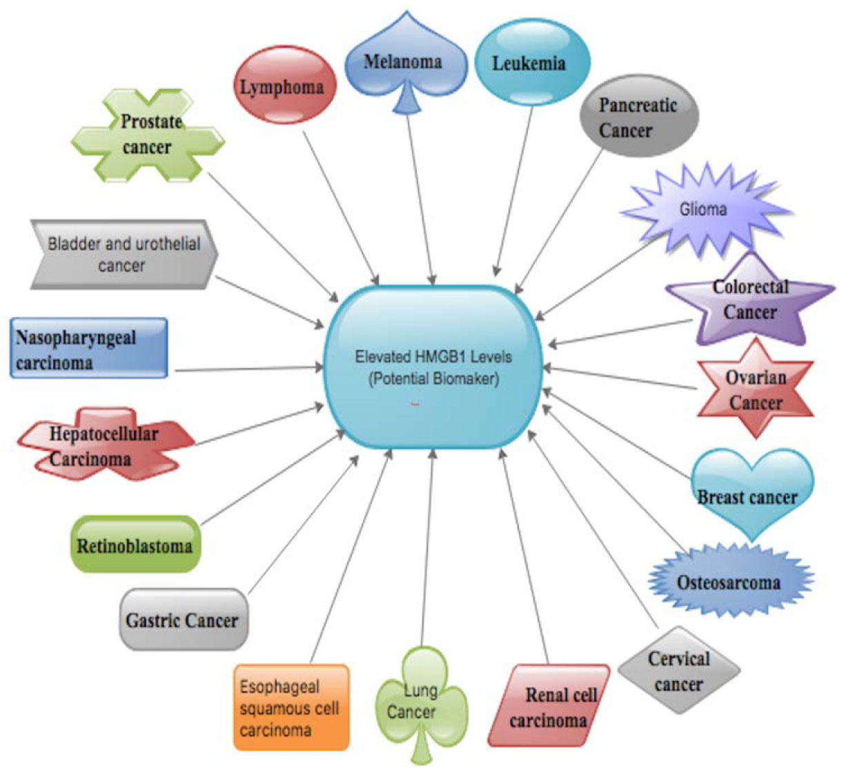
![]() |
1. | Alireza Milani, Kazem Baesi, Elnaz Agi, Azam Bolhassani, Detection of Anti-IgGs against Heat Shock Proteins 27 and 20, HP91 Peptide, and HIV-1 Polypeptides in HIV-Positive and Negative Patients, 2020, 8, 2345-5349, 113, 10.29252/JoMMID.8.3.113 | |
2. | Seidu A. Richard, Elucidating the novel biomarker and therapeutic potentials of High-mobility group box 1 in Subarachnoid hemorrhage: A review, 2019, 6, 2373-7972, 316, 10.3934/Neuroscience.2019.4.316 | |
3. | Worawat Songjang, Chatchai Nensat, Wittawat Jitpewngarm, Arunya Jiraviriyakul, Potential Serum HMGB1, HSP90, and S100A9 as Metastasis Predictive Biomarkers for Cancer Patients and Relevant Cytokines: A Pilot Study, 2024, 25, 1422-0067, 13232, 10.3390/ijms252413232 |
Cancer Type | Mechanism via which HMGB1 function. |
Glioma | HMGB1 facilitates Beclin-1-PI3K-III multifaceted materialization by binding to Beclin-1 via MEK/ERK 1/2 pathway [40,60,61,62]. |
HMGB1 curbed autophagy by inducting the MEK/ERK1/2 pathway, while the genetic suppression of PI3K-III depressed the HMGB1-stimulated phosphorylation of the MEK-ERK1/2 pathway and inhibited autophagic stimulation [40,60]. | |
Dying glioma cells expressed HMGB1 in feedback reaction to the toxins and liquidation with Ad-TK (+GCV) while HMGB1 in turn triggered TLR-2-interventional NF-kB signaling pathway and DC activation [40,64]. | |
Retinoblastoma | Intercommunication between HMGB1 and retinoblastoma protein stimulates HMGB1-mediated ranscriptional repression, cell growth inhibition, G1 cell cycle arrest, apoptosis induction, and cancer growth suppression [65,66]. |
HMGB1 also accelerated cancer metastasis in retinoblastoma by a number of angiogenic factors such as VEGF and cancer necrosis factor [67,68]. | |
The silencing activity of HMGB1 can apparently promote Caspase-3 activation and inhibit the cell cycle and induce apoptosis of cancer cells [67]. | |
Point mutation in the 104LFCSE108 motif of the HMGB1 protein resulted in loss of binding to retinoblastoma, backing the critical role of LXCXE (leucine-X-cysteine-X-glutamic; X = any amino acid) motif in retinoblastoma and HMGB1 interaction [66,69]. | |
Post-transcriptional regulators like microRNAs (miRNAs) have also been implicated to interact with HMGB1 during the pathogenesis of retinoblastoma [70,71]. | |
Melanoma | HMGB1 regulated cell proliferation via interrelating with Sp1 and interfering Sp1-mediated transcription of p21 which is in Coherent with the of function nuclear of HMGB1 [72]. |
UVB radiation on the skin has proven to stimulate HMGB1 secretion by epidermal keratinocytes, leading to neutrophilic inflammatory reaction that trigger angiogenesis and facilitates melanoma metastasis in mice [73,74]. | |
HMGB1 directly triggered the production of IL-10 in TAMs [73]. | |
IL-10, may be produced by melanoma cells51 and cancer-linked myeloid-derived suppressor cells may support immunoregulatory reactions by triggering the downregulation of molecules associated with antigen presentation to CD8+ T cells or by triggering regulatory T cells and/or by suppressing the production of proinflammatory cytokines such as TNFα, IFN-γ and IL-2 by T cells [73,75,76,77,78,79]. | |
HMGB1 1-IL-23-IL-17-IL-6-Stat3 axis plays a role in tumor development in murine models of melanoma [80] | |
Leukemia | HMGB1 is a direct trigger of autophagy in leukemia cells via the stimulation of PI3KC3/MEK/ERK pathway [81,82]. |
HMGB1 regulates autophagy via accumulative transcriptional activities of JNK and ERK in human myeloid leukemia cells [81,83]. | |
HMGB1 secretion by CLL cells, while blockade of the HMGB1-RAGE/TLR-9 signalling pathway inhibited NLC differentiation [84]. | |
Lymphomas | HMGB1 interacts with TLRs and RAGE on the DCs, which are associated with the cross-priming of anti-cancer T lymphocytes in vivo [85,86]. |
Gene polymorphisms in TLR-2 and TLR-4 gene sequences have been postulated as potential facilitates in follicular lymphoma and mucosa-associated lymphoid tissue lymphoma [87,88]. | |
HMGB1 may facilitate lymphoma proliferation by upregulating Survivin secretion [89,90]. | |
Breast cancer | HMGB1 is intricate in E2-mediated pro-autophagy and anti-apoptotic activities though the caveolin-1/HMGB1 pathway [91,92]. |
Caveolin-1/HMGB1 affiliation optimistically synchronizes E2-stimulated cell growth by stimulating autophagy and restraining apoptosis in BT474 cells [91,92,93]. | |
N-glycosylated HMGB1 released from breast cancer cells promoted M-MDSC differentiation from bone marrow through p38/NF-κB/Erk1/2 pathway and also participated in transformation of monocytes into MDSC-like cells [94]. | |
Nasopharyngeal carcinoma (NPC) | HMGB1 was markedly promoted by the EBV infection in NPC CNE-2 cells [95]. |
HMGB1-RAGE signalling triggers activation of key cell signalling pathways, such as MAPK, NF-κB, and Rac/Cdc42 in NPC [96,97]. | |
HMGB1 performances an anti-apoptotic factor role in cancer cells by stimulating the release of Bcl-2 and cIAP2 [97,98,99]. | |
Lung cancer | HMGB1 displays both immune activation and immune-suppressive qualities, depending on receptors, redox state and targeted cells [100,101]. |
HMGB1 has the capability of inducing apoptosis in macrophage-derived DCs in lung cancers, and hence reduction of host anticancer immunity [102]. | |
HMGB1 can stimulate tumor-infiltrating T cells to generate lymphotoxin a1b2 with the resultant recruitment of CD11b+ F4/80+ macrophages into lung cancers [100,103]. | |
Treg, a chemoattractant of HMGB1with a positive feedback mechanism releases HMGB1 receptors TLR-4 and RAGE which in turn stimulates the function of Treg [104]. | |
HMGB1 activates proinflammatory signaling pathways which also stimulate inflammatory reactions, tumor formation and metastasis [100,105]. | |
HMGB1 in necrotic cancer cell lysates amplifies ATP production by delivering a conventional connection between the inflammation and cancer energy metabolism in the lung cancer environment [100,106]. | |
HMGB1 regulates MMP-9 expression and cellular metastatic aptitude in lung cancer cells via active PI3KAkt and NF-κB pathways [107]. | |
HMGB1 and pRb can regulate Topo IIa release as well as genome stability [100,108]. | |
Esophageal squamous cell carcinoma | HMGB1 may have positively influence on the prognosis of patients with ESCC via stimulation of VEGF-C secretion which foster lymph node metastasis, as well as serve as hypothetical focus for antilymphangiogenesis therapy [109]. |
HMGB1 via its receptors can activate numerous intracellular signal transduction pathways such as Ras/MAPK, NF-kB, Rac, and Cdc42, which in turn triggered the secretion of VEGF-C [109]. | |
HMGB1 secreted from radiation-induced cancer cell death may augment engulfment of antigenic constituents by DCs via TRL-4 and mediate cross-presentation of tumor antigens into CD4 and CD8 T cells meritoriously resulting in tumor antigen-specific T-cell responses in murine model [27,86]. | |
HMGB1 and calreticulin cell surface secretions are obligatory for antigen-specific T-cell response in murine model [110,111]. | |
Gastric cancer | HMGB1 facilitates cell migration by epigenetic silencing of semaphorin 3A, reductions in Bax and p53 secretion as well as augments the secretion of Bcl-xL, Bcl-2, CyclinDl and NF-κB via the initiation of FAK/PI3K/mTOR pathway in gastric cancer [112,113]. |
HMGB1 secreted from gastric cancer cells stimulated the release of IL1-beta, IL6 and IL8 in the stromal fibroblasts through the HMGB1-TLR-2/4 signaling pathway [114]. | |
HMGB1 secretion by gastric cancer cells and concurrent nuclear translocation of NF-kB in the fibroblasts could be perceived at the deeper invasion areas [114]. | |
HMGB1, secretion of RAGE is highly associated with the invasive and metastatic activity of gastric cancer by enhancing NF-κB activity [98,107,112,115]. | |
HMGB1-mediated autophagy decreases vincristine-induced apoptosis in gastric cancer partly via upregulation of Mcl-1, a Bcl-2 family member [107]. | |
Hepatocellular carcinoma (HCC) Pancreatic cancer |
HMGB1 secreted from hypoxic tumor microenvironment binds to TLR-4 and RAGE, which in turn mediate HCC invasion and metastasis by triggering inflammasome, NF-κB, and Akt pathways [107,116]. |
P53 may positively regulator HMGB1 secretion during hepatocarcinogenesis [107,117]. | |
HMGB1 interaction with RAGE results in the triggering of the NF-kB, MAPK, and type IV collagenase (MMP-2/MMP-9) signaling pathways, all of which degrade extracellular matrix protein and influence tumor invasion and metastasis [118,119]. | |
HMGB1 or its receptor RAGE by RNAi or antisense nucleotide inhibits pancreatic cancer cell invasion and augmented chemotherapy sensitivity partly by downregulation of autophagy [107,120]. | |
HMGB1-RAGE pathway is a key controller of pancreatic cancer development and remedy [107,121]. | |
HMGB1-RAGE pathway influenced the initiation of autophagy and inhibition of HMGB1-RAGE resulted in amplified apoptosis as well as reduced autophagy in pancreatic cancer cells [122,123]. | |
RAGE and its aptitude to regulate gene secretion via NF-ĸB, basally triggered in pancreatic cancer, was anticipated as one of the likely mechanisms associate with pancreas cancer metastasis [119,122]. | |
RAGE antagonist peptide (RAP) decreased the capability of the ligands to trigger RAGE stimulation of NF-ĸB in cancer cells in vitro and in vivo [122,124]. | |
Renal cell carcinoma | HMGB1Ab triggered a substantial reduction in the proliferation and differentiation of MDSCs in bone marrow cells [125]. |
HMGB1, hence trigger cancer-specific T cell immunity, and stimulate antitumor effect through TLR-4 thereby activating DCs [27,125]. | |
DCs triggered by HMGB1 can activate the initiation and proliferation of T cells as helper T lymphocytes [125,126]. | |
HMGB1 can stimulate the JAK/STAT pathway, reduce cancer cell apoptosis, facilitates cell cycle, and trigger resistance and immune escape in cancer cells [125,127]. | |
HMGB1 could facilitate Treg cells to release IL-10 and dwindle anticancer effect of CD8+ T cells [125]. | |
HMGB1 facilitates the growth and advancement of clear cell renal cell carcinoma via ERK1/2 stimulation, which is partly mediated by RAGE [125]. | |
Prostate cancer | HMGB1 and RAGE are co-secreted in prostate cancer samples and proposed that they may have interrelated roles in the development of prostate cancer [128,129]. |
HMGB1 may regulate androgen receptor either by acting as co-activator of androgen receptor or indirectly connecting with RAGE signaling in prostate oncogenesis [128,129,130]. | |
HMGB1 can augment DNA binding activity of ETS (E-twenty-six) transcription factor in regulating peroxiredoxin-1 and -5 expressions in combating oxidative stress in prostate cancer cells [128,131]. | |
Down-regulation of HMGB1 can lead to apoptosis in LNCaP prostate cancer cells via caspase-3-dependent pathways [128]. | |
HMGB1 plays a key inflammatory role in prostate cancer. Therefore, inflammation may be one mechanism by which HMGB1 may accelerate prostate cancer [128,132]. | |
Bladder and urothelial cancer | HMGB1 can inhibit the secretion of NF-κB/p65 and VEGF-C in 5637 bladder cancer cells and HMGB1 may regulate VEGFC secretion through the NF-κB signaling pathway [133,134]. |
HMGB1 and p53 mediate bladder cancer cell survival via an equilibrium of autophagy and apoptosis [135,136]. | |
HMGB1 facilitates phosphorylation and activation of the ERK1/2 pathway in bladder cancer cell lines and HMGB1 binding to Beclin-1 base on the complex ULK1-mAtg13-FIP200 which results the establishment of the Beclin-1-PI3KC3 complex thus promoting autophagic advancement [61,135,137]. | |
Cervical cancer | HMGB1 gene is a direct target of miR-142 in cervical cancer cells [138]. |
HMGB1 suppresses the human immune function by upregulating Tregs and facilitates IL-10 production [139,140]. | |
HMGB1 inhibits the function of T cells by downregulating NF-κB signaling and polarizing Th1 cells to Th2 cells [139,140,141]. |