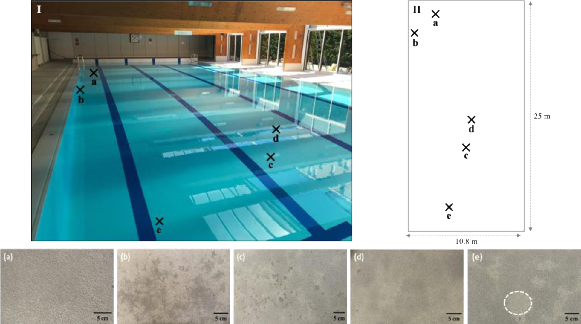
Waterproofing coatings are composite materials made of different layers with complementary functionalities. They may suffer damage that can modify their aesthetic appearance and/or their functionality. In this study, dark stains appearing on a waterproofing coating of a public swimming pool were mapped and characterized at a macroscopic scale through visual observation and by colorimetric analysis, as well as at a microscopic scale with a digital microscope, a confocal laser scanning microscope, and a scanning electron microscope. Five stains were differentiated macroscopically and characterized using colorimetry and principal component analysis. Microscopic observations showed the presence of microorganisms of varied morphology, some filamentous but mostly unicellular. Biofilms consisting of ovoid fluorescent cells with the morphology of Chlorophyta and unicellular cyanobacteria were particularly abundant. The pigmented stains were located at top coat disorders where microbial colonization and biofilm development were observed. The microscopic observations suggested that physical degradation of the surface of the material would have constituted a prerequisite for colonization by pigmented microorganisms which would have led to the development of macroscopically visible pigmented areas. In this case study, the damage remained superficial and did not alter the watertightness of the material so far.
Citation: Clotilde Maestri, Ronan L. Hébert, Patrick Di Martino. Biofilm associated with pigmented areas on a waterproofing coating surface[J]. AIMS Microbiology, 2025, 11(1): 74-86. doi: 10.3934/microbiol.2025005
Waterproofing coatings are composite materials made of different layers with complementary functionalities. They may suffer damage that can modify their aesthetic appearance and/or their functionality. In this study, dark stains appearing on a waterproofing coating of a public swimming pool were mapped and characterized at a macroscopic scale through visual observation and by colorimetric analysis, as well as at a microscopic scale with a digital microscope, a confocal laser scanning microscope, and a scanning electron microscope. Five stains were differentiated macroscopically and characterized using colorimetry and principal component analysis. Microscopic observations showed the presence of microorganisms of varied morphology, some filamentous but mostly unicellular. Biofilms consisting of ovoid fluorescent cells with the morphology of Chlorophyta and unicellular cyanobacteria were particularly abundant. The pigmented stains were located at top coat disorders where microbial colonization and biofilm development were observed. The microscopic observations suggested that physical degradation of the surface of the material would have constituted a prerequisite for colonization by pigmented microorganisms which would have led to the development of macroscopically visible pigmented areas. In this case study, the damage remained superficial and did not alter the watertightness of the material so far.

| [1] | Khanna S, Sah R, Hooda S, et al. (2024) Water proofing system in concrete structures. Int J Res Civ Eng Technol 5: 32-34. https://doi.org/10.48175/ijarsct-17845 |
| [2] | Kavitha R, Ram Vivekananthan M, Dhanagopal K, et al. (2023) An overview of water proofing system in concrete structures. Mater Today Proc . https://doi.org/10.1016/j.matpr.2023.03.515 |
| [3] | Saxena PK, Raut KG, Srinivasan SR, et al. (1991) Polyurethane waterproofing coating for building applications. Constr Build Mater 5: 208-210. https://doi.org/10.1016/0950-0618(91)90052-M |
| [4] | Maestri C, Plancher L, Duthoit A, et al. (2023) Fungal Biodegradation of Polyurethanes. J Fungi (Basel) 9: 760. http://doi.org/10.3390/jof9070760 |
| [5] | Hébert R, Beouch L, Fichet O, et al. (2012) Cracks and stains on façade-cladding made of carbonate rock thin panels. Struct Survey 30: 130-144. https://doi.org/10.1108/02630801211228734 |
| [6] | Newby PT, Mansfield TA, Hamilton RS (1991) Sources and economic implications of building soiling in urban areas. Sci Total Environ 100: 347-365. https://doi.org/10.1016/0048-9697(91)90385-R |
| [7] | Brimblecombe P, Grossi CM (2005) Aesthetic thresholds and blackening of stone buildings. Sci Total Environ 349: 175-189. https://doi.org/10.1016/j.scitotenv.2005.01.009 |
| [8] | Cutler NA, Viles HA, Ahmad S, et al. (2013) Algal ‘greening’ and the conservation of stone heritage structures. Sci Total Environ 442: 152-164. https://doi.org/10.1016/j.scitotenv.2012.10.050 |
| [9] | Di Martino P (2016) What about biofilms on the surface of stone monuments?. Open Conf Proc J 7: 14-28. https://doi.org/10.2174/2210289201607020014 |
| [10] | Liu X, Koestler RJ, Warscheid T, et al. (2020) Microbial deterioration and sustainable conservation of stone monuments and buildings. Nat Sustain 3: 991-1004. https://doi.org/10.1038/s41893-020-00602-5 |
| [11] | Hernández Mariné M, Clavero E, Roldán M (2004) Microscopy methods applied to research on cyanobacteria. Limnetica 23: 179-186. https://doi.org/10.23818/limn.23.16 |
| [12] | Peisker H, Michels J, Gorb S (2013) Evidence for a material gradient in the adhesive tarsal setae of the ladybird beetle Coccinella septempunctata. Nat Commun 4: 1661. https://doi.org/10.1038/ncomms2576 |
| [13] | Hobisch MA, Bossu J, Mandlez D, et al. (2019) Localization of cellulosic fines in paper via fluorescent labeling. Cellulose 26: 6933-6942. https://doi.org/10.1007/s10570-019-02556-0 |
| [14] | Lewin RA (2006) Black algae. J Appl Phycol 18: 699-702. https://doi.org/10.1007/s10811-005-9018-2 |
| [15] | Miller AZ, Laiz L, Gonzalez JM, et al. (2008) Reproducing stone monument photosynthetic-based colonization under laboratory conditions. Sci Total Environ 405: 278-285. https://doi.org/10.1016/j.scitotenv.2008.06.066 |
| [16] | Macedo MF, Miller AZ, Dionísio A, et al. (2009) Biodiversity of cyanobacteria and green algae on monuments in the Mediterranean Basin: an overview. Microbiology (Reading) 155: 3476-3490. https://doi.org/10.1099/mic.0.032508-0 |
| [17] | Li S, Fanesi A, Martin T, et al. (2024) Physiological transition of Chlorella vulgaris from planktonic to immobilized conditions. Algal Res 77: 103354. https://doi.org/10.1016/j.algal.2023.103354 |
| [18] | Garcia-Pichel F, Sherry ND, Castenholtz RW (1992) Evidence for an ultraviolet sunscreen role of the extracellular pigment scytonemin in the terrestrial cyanobacterium Chlorogloeopsis sp. Photochem Photobiol 56: 17-23. https://doi.org/10.1111/j.1751-1097.1992.tb09596.x |
| [19] | Garcia-Pichel F, Castenholz RW (1993) Occurrence of UV-absorbing mycosporine-like compounds among cyanobacterial isolates, and an estimate of their screening capacity. App Env Microbiol 59: 163-169. https://doi.org/10.1128/aem.59.1.163-169.1993 |
| [20] | Proteau PJ, Gerwick WH, Garcia-Pichel F, et al. (1993) The structure of scytonemin, an ultraviolet sunscreen pigment from the sheaths of cyanobacteria. Experientia 49: 825-829. https://doi.org/10.1007/BF01923559 |
| [21] | Gaylarde CC, Gaylarde PM (2005) A comparative study of the major microbial biomass of biofilms on exteriors of buildings in Europe and Latin America. Int Biodeterior Biodegrad 55: 131-139. https://doi.org/10.1016/j.ibiod.2004.10.001 |
| [22] | Bruno L, Valle V (2017) Effect of white and monochromatic lights on cyanobacteria and biofilms from Roman Catacombs. Int Biodeterior Biodegrad 123: 286-295. https://doi.org/10.1016/j.ibiod.2017.07.013 |
| [23] | Komar M, Nowicka-Krawczyk P, Ruman T, et al. (2022) Metabolomic analysis of photosynthetic biofilms on building façades in temperate climate zones. Int Biodeterior Biodegrad 169: 105374. https://doi.org/10.1016/j.ibiod.2022.105374 |
| [24] | Nowicka-Krawczyk P, Komar M, Gutarowska B (2022) Towards understanding the link between the deterioration of building materials and the nature of aerophytic green algae. Sci Total Environ 802: 149856. https://doi.org/10.1016/j.scitotenv.2021.149856 |
| [25] | Viles HA, Taylor MP, Yates TJS, et al. (2002) Soiling and decay of N.M.E.P. limestone tablets. Sci Total Environ 292: 215-229. https://doi.org/10.1016/S0048-9697(01)01124-X |
| [26] | Viles HA, Gorbushina AA (2003) Soiling and microbial colonisation on urban roadside limestone: a three-year study in Oxford, England. Build Environ 38: 1217-1224. https://doi.org/10.1016/S0360-1323(03)00078-7 |
| [27] | Rajkowska K, Otlewska A, Kozirog A, et al. (2014) Assessment of biological colonization of historic buildings in the former Auschwitz II-Birkenau concentration camp. Ann Microbiol 64: 799-808. https://doi.org/10.1007/s13213-013-0716-8 |
| [28] | Komar M, Nowicka-Krawczyk P, Ruman T, et al. (2023) Biodeterioration potential of algae on building materials-Model study. Int Biodeterior Biodegrad 180: 105593. https://doi.org/10.1016/j.ibiod.2023.105593 |
| [29] | Saiz-Jimenez C (1999) Biogeochemistry of weathering processes in Monuments. Geomicrobiol J 16: 27-37. https://doi.org/10.1080/014904599270721 |
| [30] | Błaszczyński T, Łowińska-Kluge A (2007) Experimental investigation and assessment of damage in the case of swimming-pool repairs. Archiv Civ Mech Eng 7: 5-20. https://doi.org/10.1016/S1644-9665(12)60001-6 |
| [31] | Maj M, Ubysz A (2022) The reasons for the loss of polyurea coatings adhesion to the concrete substrate in chemically aggressive water tanks. Eng Fail Anal 142: 106774. https://doi.org/10.1016/j.engfailanal.2022.106774 |
| [32] | Almusallam AA, Khan FM, Dulaijan SU, et al. (2003) Effectiveness of surface coatings in improving concrete durability. Cem Concr Compos 25: 473-481. https://doi.org/10.1016/S0958-9465(02)00087-2 |
| [33] | Tsukagoshi M, Miyauchi H, Tanaka K (2012) Protective performance of polyurethane waterproofing membrane against carbonation in cracked areas of mortar substrate. Constr Build Mater 36: 895-905. https://doi.org/10.1016/j.conbuildmat.2012.06.072 |
| [34] | Somarathna HMCC, Raman SN, Mohotti D, et al. (2018) The use of polyurethane for structural and infrastructural engineering applications: A state-of-the-art review. Constr Build Mater 190: 995-1014. https://doi.org/10.1016/j.conbuildmat.2018.09.166 |
| [35] | Khatua S, Hsieh YL (1997) Chlorine degradation of polyether-based polyurethane. J Polym Sci A: Polym Chem 35: 3263-3273. https://doi.org/10.1002/(SICI)1099-0518(19971115)35:15%3C3263::AID-POLA20%3E3.0.CO;2-8 |


Figures(5) / Tables(1)

Clotilde Maestri, Ronan L. Hébert, Patrick Di Martino. Biofilm associated with pigmented areas on a waterproofing coating surface[J]. AIMS Microbiology, 2025, 11(1): 74-86. doi: 10.3934/microbiol.2025005


 DownLoad: