Tumor Treating Fields (TTFs) is a new tumor electric therapy that has success with many tumor types. This modality is very low-intensity alternating current (AC) fields (1–3 V/cm) and intermediate frequency (100–300 kHz). The delivery of TTFs was applied only through completely insulated electrodes by using external high voltage to avoid any reactions that may occur through using conductive electrodes, specially the generation of oxidative reactions. In order to achieve the required effect, the patient can be exposed to the technique for continuous three days, which may lead to long-term side effects. The adoption of the idea that TTFs should be applied through insulated electrodes did not give the chance to evaluate the uninsulated electrodes to conduct TTFs. This study aims to evaluate the possibility of using uninsulated electrodes, find the optimum exposure time, and determine the accompanying oxidative stress on human non-small cell lung carcinoma (NSCLC) cells. Here we found that applying TTFs through uninsulated electrodes produced 18–29% tumor growth inhibition for exposure time of 2.5 min to 40 min without significant oxidative stress. These findings will lead to the possibility of using the uninsulated electrodes TTFs in simple technology. The treatment quality will be improved by highly decreasing the exposure time.
Citation: Mamdouh M. Shawki, Seham Elabd. Tumor treating fields (TTFs) using uninsulated electrodes induce cell death in human non-small cell lung carcinoma (NSCLC) cells[J]. AIMS Biophysics, 2021, 8(2): 143-156. doi: 10.3934/biophy.2021011
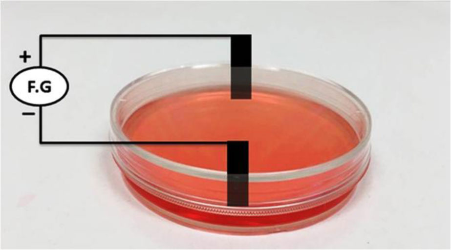
Tumor Treating Fields (TTFs) is a new tumor electric therapy that has success with many tumor types. This modality is very low-intensity alternating current (AC) fields (1–3 V/cm) and intermediate frequency (100–300 kHz). The delivery of TTFs was applied only through completely insulated electrodes by using external high voltage to avoid any reactions that may occur through using conductive electrodes, specially the generation of oxidative reactions. In order to achieve the required effect, the patient can be exposed to the technique for continuous three days, which may lead to long-term side effects. The adoption of the idea that TTFs should be applied through insulated electrodes did not give the chance to evaluate the uninsulated electrodes to conduct TTFs. This study aims to evaluate the possibility of using uninsulated electrodes, find the optimum exposure time, and determine the accompanying oxidative stress on human non-small cell lung carcinoma (NSCLC) cells. Here we found that applying TTFs through uninsulated electrodes produced 18–29% tumor growth inhibition for exposure time of 2.5 min to 40 min without significant oxidative stress. These findings will lead to the possibility of using the uninsulated electrodes TTFs in simple technology. The treatment quality will be improved by highly decreasing the exposure time.

| [1] |
De Groot P, Wu CC, Carter BW, et al. (2018) The epidemiology of lung cancer. Transl Lung Cancer Res 7: 220-233. doi: 10.21037/tlcr.2018.05.06

|
| [2] |
Oser MG, Niederst MJ, Sequist LV, et al. (2015) Transformation from non-small-cell lung cancer to small-cell lung cancer: molecular drivers and cells of origin. Lancet Oncol 16: e165-e172. doi: 10.1016/S1470-2045(14)71180-5
|
| [3] | Lemjabbar-Alaoui H, Hassan OUI, Yang YW, et al. (2015) Lung cancer: biology and treatment options. BBA-Rev Cancer 1856: 189-210. |
| [4] |
Rubinsky B (2007) Irreversible electroporation in medicine. Technol Cancer Res Treat 6: 255-259. doi: 10.1177/153303460700600401
|
| [5] |
Sersa G, Miklavcic D, Cemazar M, et al. (2008) Electrochemotherapy in treatment of tumours. Eur J Surg Oncol 34: 232-240. doi: 10.1016/j.ejso.2007.05.016
|
| [6] |
Kirson ED, Dbalý V, Tovaryš F, et al. (2007) Alternating electric fields arrest cell proliferation in animal tumor models and human brain tumors. Proc Natl Acad Sci 104: 10152-10157. doi: 10.1073/pnas.0702916104
|
| [7] |
Berkelmann L, Bader A, Meshksar S, et al. (2019) Tumour-treating fields (TTFields): Investigations on the mechanism of action by electromagnetic exposure of cells in telophase/cytokinesis. Sci Rep 9: 7362. doi: 10.1038/s41598-019-43621-9
|
| [8] |
Giladi M, Weinberg U, Schneiderman RS, et al. (2014) Alternating electric fields (tumor-treating fields therapy) can improve chemotherapy treatment efficacy in non-small cell lung cancer both in vitro and in vivo. Semin Oncol 41: S35-S41. doi: 10.1053/j.seminoncol.2014.09.006
|
| [9] | Porat Y, Giladi M, Schneiderman RS, et al. (2017) Determining the optimal inhibitory frequency for cancerous cells using tumor treating fields (TTFields). J Vis Exp 123: 55820. |
| [10] |
Spinelli E, Haberman M (2010) Insulating electrodes: a review on biopotential front ends for dielectric skin–electrode interfaces. Physiol Meas 31: S183-S198. doi: 10.1088/0967-3334/31/10/S03
|
| [11] |
Wenger C, Miranda P, Salvador R, et al. (2018) A review on tumor-treating fields (TTFields): clinical implications inferred from computational modeling. IEEE Rev Biomed Eng 11: 195-207. doi: 10.1109/RBME.2017.2765282
|
| [12] |
Kanner AA, Wong ET, Villano JL, et al. (2014) Post hoc analyses of intention-to-treat population in phase III comparison of NovoTTF-100ATM system versus best physician's choice chemotherapy. Semin Oncol 41: S25-S34. doi: 10.1053/j.seminoncol.2014.09.008
|
| [13] |
Jeong J, Kimb C, Yoon J (2009) The effect of electrode material on the generation of oxidants and microbial inactivation in the electrochemical disinfection processes. Water Res 43: 895-901. doi: 10.1016/j.watres.2008.11.033
|
| [14] |
Sung J, Seo J, Jo Y, et al. (2018) Development of a method for improving the electric field distribution in patients undergoing tumor-treating fields therapy. J Korean Phys Soc 73: 1577-1583. doi: 10.3938/jkps.73.1577
|
| [15] |
Franken NAP, Rodermond HM, Stap J, et al. (2006) Clonogenic assay of cells in vitro. Nat Protoc 1: 2315-2319. doi: 10.1038/nprot.2006.339
|
| [16] |
Wlodkowic D, Skommer J, Darzynkiewicz Z (2009) Flow cytometry-based apoptosis detection. Apoptosis Totowa: Humana Press, 19-32. doi: 10.1007/978-1-60327-017-5_2
|
| [17] |
Pozarowski P, Darzynkiewicz Z (2004) Analysis of cell cycle by flow cytometry. Checkpoint Controls and Cancer Humana Press, 301-311. doi: 10.1385/1-59259-811-0:301
|
| [18] |
Bradford MM (1976) A rapid and sensitive method for the quantitation of microgram quantities of protein utilizing the principle of protein-dye binding. Anal Biochem 72: 248-254. doi: 10.1016/0003-2697(76)90527-3
|
| [19] |
Marques SS, Magalhães LM, Tóth IV, et al. (2014) Insights on antioxidant assays for biological samples based on the reduction of copper complexes-the importance of analytical conditions. Int J Mol Sci 15: 11387-11402. doi: 10.3390/ijms150711387
|
| [20] |
Baker MA, Cerniglia GJ, Zaman A (1990) Microtiter plate assay for the measurement of glutathione and glutathione disulfide in large numbers of biological samples. Anal Biochem 190: 360-365. doi: 10.1016/0003-2697(90)90208-Q
|
| [21] |
Eyer P, Podhradsky D (1986) Evaluation of the micromethod for determination of glutathione using enzymatic cycling and Ellman's reagent. Anal Biochem 153: 57-66. doi: 10.1016/0003-2697(86)90061-8
|
| [22] |
Shah PP, White T, Khalafallah AM, et al. (2020) A systematic review of tumor treating fields therapy for high grade gliomas. J Neuro-oncol 148: 433-443. doi: 10.1007/s11060-020-03563-z
|
| [23] |
Cifra M, Fields JZ, Farhadi A (2011) Electromagnetic cellular interactions. Prog Biophys Mol Biol 105: 223-246. doi: 10.1016/j.pbiomolbio.2010.07.003
|
| [24] |
Kim CY, Paek SH, Nam D, et al. (2020) Tumor treating fields plus temozolomide for newly diagnosed glioblastoma: a sub-group analysis of Korean patients in the EF-14 phase 3 trial. J Neuro-oncol 146: 399-406. doi: 10.1007/s11060-019-03361-2
|
| [25] |
Mun EJ, Babiker HM, Weinberg U, et al. (2018) Tumor-treating fields: A fourth modality in cancer treatment. Clin Cancer Res 24: 266-275. doi: 10.1158/1078-0432.CCR-17-1117
|
| [26] |
Tuszynski JA, Wenger C, Friesen DE, et al. (2016) An overview of sub-cellular mechanisms involved in the action of TTFields. Int J Environ Res Public Health 13: 1128. doi: 10.3390/ijerph13111128
|
| [27] |
Li X, Yang F, Rubinsky B (2020) A theoretical study on the biophysical mechanisms by which tumor treating fields affect tumor cells during mitosis. IEEE T Biomed Eng 67: 2594-2602. doi: 10.1109/TBME.2020.2965883
|
| [28] |
Voloshin T, Kaynan N, Davidi S, et al. (2020) Tumor-treating fields (TTFields) induce immunogenic cell death resulting in enhanced antitumor efficacy when combined with anti-PD-1 therapy. Cancer Immunol Immunother 69: 1191-1204. doi: 10.1007/s00262-020-02534-7
|
| [29] |
Wenger C, Salvador R, Basser PJ, et al. (2016) Improving tumor treating fields treatment efficacy in patients with glioblastoma using personalized array layouts. Int J Radiation Oncol Biol Phys 94: 1137-1143. doi: 10.1016/j.ijrobp.2015.11.042
|
| [30] |
Davies AM, Weinberg U, Palti Y (2013) Tumor treating fields: A new frontier in cancer therapy. Ann NY Acad Sci 1291: 86-95. doi: 10.1111/nyas.12112
|
| [31] | Puente C, López I (2018) Direct electrochemical synthesis of metal complexes. Direct Synthesis of Metal Complexes Elsevier, 87-141. |


Figures(5) / Tables(1)

Mamdouh M. Shawki, Seham Elabd. Tumor treating fields (TTFs) using uninsulated electrodes induce cell death in human non-small cell lung carcinoma (NSCLC) cells[J]. AIMS Biophysics, 2021, 8(2): 143-156. doi: 10.3934/biophy.2021011


 DownLoad: